Case 7243
Sex Cord Stromal Tumor in a 17 Year Old Girl
C. Bahia1, M. O. Castro2, G. Bastos1, J. Parra1, E. Vieira1, T. M. Cunha3, F. Aleixo1
1Centro Hospitalar do Barlavento Algarvio; 2Centro Hospitalar do Baixo Alentejo; 3Instituto Português
de Oncologia Francisco Gentil, Lisboa
Portugal
Section: Genital (Female) Imaging Published: 2009, Feb. 2
Patient: 17 year(s), female
Clinical Summary
A 17 year old girl was presented with painless abdominal distention. Physical examination revealed a
palpable abdominal mass. Laboratory tests were normal.
Clinical History and Imaging Procedures
The patient was admitted to the hospital with painless abdominal distension and underwent
ultrasonographic evaluation, which revealed a large left abdominal mass. The lesion was heterogeneous,
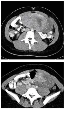
mostly solid with a small cystic component, demonstrating prominent vascularity. A CT scan was done,
revealing a well circumscribed mass, with 18 x 17 cm, localized predominantly in the left abdomen,
originating from the right ovary. This mass was heterogeneous with an irregular shaped central cystic
area, a lobulated soft tissue density component and a peripheral left hypodense area. After contrast
administration there was enhancement of the central mass (Fig. 1a) and prominent vessels converging
to the hilum were evident (Fig. 1b). Increased caliber of right ovarian vessels was also noted (Fig. 1b).
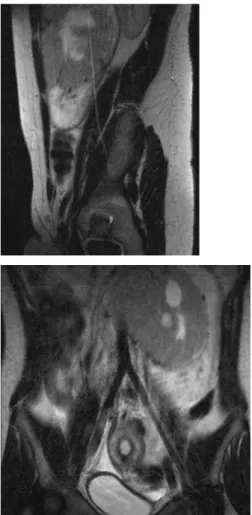
On MRI the central mass was moderately hypointense on T1WI (Fig. 2), of intermediate signal intensity
on T2WI (Fig. 3 a,b), with an irregular central cystic area. It was surrounded by a smoothly marginated
solid component with hypointense capsule on T1WI and T2WI, crossed by several vessels. There was
enhancement of the central mass and of the tumour capsule after gadolinium administration (Fig. 4).
Surgery was done, with total resection of the mass and partial resection of the right fallopian tube (Fig.
5).
consisting on two distinct components, one external corresponding to a fibrothecoma, and one internal of
stromal origin with an undetermined pattern (Fig. 6,7).
Discussion
Sex cord stromal tumours derive from coelomic epithelium or mesenchymal cells of the embryonic
gonads. This group of tumours represents approximately 8% of ovarian neoplasms and affects all age
groups. The most common types are granulosa cell tumours, fibrothecomas and Sertoli-Leydig cell
tumours. Fibromas and fibrothecomas belong to a spectrum of benign tumours. Lipid-rich thecoma
demonstrates estrogenic activity and few fibroblasts. Fibroma is composed of spindle cells that form
collagen, has no thecal cells and demonstrates no estrogenic activity.
A broad spectrum of ultrasonographic features is seen and in most cases the appearance of the tumour
is non-specific. CT shows a homogeneous solid tumour with delayed enhancement.
Ovarian fibromas usually display low signal intensity on both T1WI and T2WI. High signal intensity on
T2WI corresponds to regions of hyalinization and myxomatous changes. Intratumoral oedema is also a
feature of larger fibromas / fibrothecomas.
Differential diagnosis in this age group should include sclerosing stromal tumour (SST). SST occur
predominantly in the second and third decades of life in 80% of cases, usually appearing as a large
mass with hyperintense cystic components and a heterogeneous solid component with intermediate to
high signal intensity on T2WI and early peripheral enhancement with centripetal progression.
Dysgerminoma is another entity to be considered. Characteristic imaging findings include a
multilobulated solid mass with prominent fibrovascular septa and areas of necrosis and haemorrhage.
Knowledge of key imaging features of sex cord stromal tumours may provide criteria to narrow the
differential diagnosis of adnexal masses.
Final Diagnosis
Sex Cord Stromal Tumor of Undetermined Pattern
Figure 1 Axial CT after contrast administration
Enhancement of the central mass and capsule with
prominent vascularity
Increased caliber of right ovarian vessels converging to
the hilum
Figure 2 Axial T1WI
Figure 3 T2WI
Sagittal T2WI showing a mass of intermediate signal
with irregular central cystic area, surrounded by a
hyperintense component, traversed by several vessels.
This mass was delineated by a hypointense capsule.
Coronal T2WI showing a mass of intermediate signal
with irregular central cystic area, surrounded by a
hyperintense component, traversed by several vessels.
This mass was delineated by a hypointense capsule.
Figure 4 Axial fat-suppressed T1WI GRE after gadolinium administration
Enhancement of the central mass and of the tumor
capsule.
Figure 5 Surgical specimen
Figure 6 Photomicrograph
Peripheral component: collagen fibres with fibroblasts
and theca cells characteristic of fibrothecoma.
Figure 7 Photomicrograph
Central mass: monomorphic cell population with
moderate atypia forming trabecules. Some cells show
mitotic activity.
MeSH
Ovary [A05.360.319.114.630]
The reproductive organ (GONADS) in female animals. In vertebrates, the ovary contains two functional
parts: the OVARIAN FOLLICLE for the production of female germ cells (OOGENESIS); and the
endocrine cells (GRANULOSA CELLS, THECA CELLS, and LUTEAL CELLS) for the production of
ESTROGENS and PROGESTERONE.
Sex Cord-Stromal Tumor [C04.557.475.750]
A malignant neoplasm of the ovary or testis. These tumors differentiate toward sex cords (in embryonic
gonads) in the form of female (i.e., granulosa and theca) cells, male (i.e., Sertoli and Leydig) cells, or
indifferent elements. In the ovary, sex cord-stromal tumors comprise 5% of all ovarian neoplasms. In the
testes, Leydig and Sertoli cell tumors comprise about 5% of all testicular neoplasms, 10% of which
behave in a malignant fashion. (From Segen, Dictionary of Modern Medicine, 1992)
References
[1] Jung SE, Lee JM, Rha SE, Byun JY, Jung JI, Hahn ST (2002) CT and MR Imaging of Ovarian
Tumors with Emphasis on Differential Diagnosis. Radiographics 22:1305-1325.
Solid and Cystic Components That Mimic Malignancy. AJR Am J Roentgenol 182:1259-1265
[3] Tamai K, Koyama T, Saga T, Kido A, Kataoka M, Umeoka S, Fujii S, Togashi K (2006) MR features
of physiologic and benign conditions of the ovary. Eur Radiol 16: 2700-2711
[4] Diagnostic Imaging: Gynecology, Hedvig Hricak, Saunders, 2008
[5] MRI and CT of the Female Pelvis, Bernd Hamm, Rosemarie Forstner, Springer, 1st edition, 2007
Citation
C. Bahia1, M. O. Castro2, G. Bastos1, J. Parra1, E. Vieira1, T. M. Cunha3, F. Aleixo1
1Centro Hospitalar do Barlavento Algarvio; 2Centro Hospitalar do Baixo Alentejo; 3Instituto Português
de Oncologia Francisco Gentil, Lisboa
Portugal (2009, Feb. 2)