online | memorias.ioc.fiocruz.br
Validation of the Dual-path Platform chromatographic immunoassay
(DPP
®CVL rapid test) for the serodiagnosis of canine
visceral leishmaniasis
Fabiano Borges Figueiredo1, Tassia Cristina Bello de Vasconcelos2/+,
Maria de Fátima Madeira3, Rodrigo Caldas Menezes4, Ana Nilce Silveira Maia-Elkhoury5, Andreza Pain Marcelino6, Guilherme L Werneck7
1Fundação Oswaldo Cruz-Fiocruz, Instituto Carlos Chagas, Laboratório de Biologia Celular, Curitiba, PR, Brasil 2Prefeitura Municipal de Resende, Vigilância em Saúde, Centro de Controle de Zoonoses, Resende, RJ, Brasil
3Fundação Oswaldo Cruz-Fiocruz, Instituto Nacional de Infectologia, Laboratório de Vigilância em Leishmanioses, Rio de Janeiro, RJ, Brasil 4Fundação Oswaldo Cruz-Fiocruz, Instituto Nacional de infectologia, Laboratório de Pesquisa Clínica em Dermatozoonoses em Animais
Domésticos, Rio de Janeiro, RJ, Brasil
5Organização Pan-Americana de Saúde, Doenças Negligenciadas, Tropicais e Transmitidas por Vetores, Doenças Transmissíveis e Determinantes Ambientais de Saúde, Duque de Caxias, RJ, Brasil
6Fundação Ezequiel Dias, Instituto Otávio Magalhães, Belo Horizonte, MG, Brasil
7Universidade do Estado do Rio de Janeiro, Instituto de Medicina Social, Departamento de Epidemiologia, Rio de Janeiro, RJ, Brasil
BACKGROUND Visceral leishmaniasis is a major public health challenge in South America, and dogs are its main urban reservoir.
OBJECTIVE Validation of the canine Dual-path Platform immunoassay for canine visceral leishmaniasis (DPP® CVL) for a sample
set composed of 1446 dogs from different Brazilian endemic areas.
METHODS A well-defined reference standard by means of parasitological culture, immunohistochemistry, and histopathology was used. Animals were classified as asymptomatic, oligosymptomatic, or symptomatic. Sensitivity and specificity were assessed
as a single set and in clinical groups. A reproducibility assessment of the tests was conducted using the Kappa (κ) index at three
different laboratories (A, B, and C).
FINDINGS Overall, 89% sensitivity and 70% specificity were obtained for the entire sample set. Analysis of the clinical groups showed a gradual decrease in the sensitivity and an increase in the specificity with the reduction of clinical signs in the dogs that were assessed, reaching a sensitivity of 75% (42.8-94.5%) among asymptomatic dogs and lower specificity of 56% (46.2-66.3%)
among symptomatic dogs. Inter-laboratory agreement was substantial (κAB= 0.778; κAC= 0.645; κCB= 0.711).
MAIN CONCLUSIONS The test performance is somewhat dependent on canine symptomatology, but such influence was less evident than in previous studies. Favourable results for sensitivity and specificity can be obtained even in asymptomatic animals; however, caution is needed in these evaluations, and the results suggest that the immunochromatographic test may be further improved for better investigation in asymptomatic dogs. The results obtained confirm the usefulness of DPP® CVL for application
in serological surveys.
Key words: Dual-path Platform CVL rapid test - visceral leishmaniasis - dog - diagnosis
Visceral leishmaniasis (VL) is typically a zoonosis that affects humans and other species of domestic and wild animals, but anthroponotic transmission predomi-nates on the Indian subcontinent and in parts of Africa. (1,2) On the American continents, this disease is caused
by Leishmania infantum (sin. Leishmania chagasi),and
sand flies of the genus Lutzomyia are the vectors in-volved in its transmission.(2,3,4,5)
doi: 10.1590/0074-02760180260
Financial support: FAPERJ (Projects Jovem Cientista do Nosso Estado, Cientista do Nosso Estado and CNPq).
GLW and FBF hold a CNPq grant for productivity in research. + Corresponding author: [email protected] / [email protected]
Received 23 May 2018 Accepted 25 September 2018
In South America, VL is expanding geographically and is a great challenge to public health.(2,5,6,7,8,9) Human and canine cases have been reported in both rural and urban areas,(2,10,11) and Brazil is among the top four coun-tries in the world with the largest numbers of cases of this disease.(12)
In Brazil, where the transmission cycle of VL is pre-dominantly zoonotic, dogs are the main urban reservoir. (5) Diagnosis in this host is complex and can be conducted
by means of serological, molecular and parasitological methods.(13) Parasitological techniques are considered the reference standard,(14) but, in endemic areas, serolog-ical tests are used as a tool in epidemiologserolog-ical surveys to facilitate diagnosis and decision-making.(2,15)
combining antigens K9, K26 and K39) of L. infantum.(16) Despite such characteristics and the ease of application, discussion on the accuracy of DPP persists, especially regarding its sensitivity for detection of infected asymp-tomatic animals.(17)
In this context, there is a need to conduct a study that addresses a representative sample of dogs from an en-demic area, that uses a well-defined reference standard and blind analysis, and that follows the recommended methodological principles for the preparation and report of diagnostic accuracy studies.(18) With this perspective, this study aimed to validate and assess the inter-labo-ratory concordance of the DPP® CVL chromatographic immunoassay by applying it to samples of animals from different Brazilian endemic areas.
MATERIALS AND METHODS
The present study conducted the validation of a mul-ticentric, blind, diagnostic test of a sample set composed of 1446 dogs that were systematically selected in four municipalities located in different regions of Brazil in which VL is endemic. Sample size calculations were based on an estimated 10% CVL prevalence, 90% test sensitivity, 80% test specificity, and 5% error.
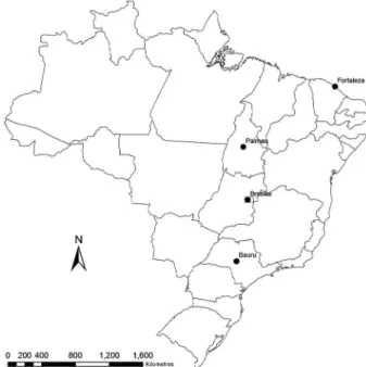
Participating municipalities and their respective states and regions were as follows: Bauru, São Paulo state, Southeast region; Brasília, Federal District, Mid-West region; Palmas, Tocantins state, North region; Fortaleza, Ceará state, Northeast region (Fig. 1). In each municipality, three distinct, non-continuous neighbour-hoods with the highest prevalence of CVL were selected. The dogs were selected through an active, door-to-door search. The animal selection process for the study fol-lowed a systematic sampling procedure beginning with
a randomly selected house, in which at least one dog was present. The subsequent house was passed, and the next one was visited until another dog was found; this process continued until there were no more houses available in each neighbourhood or until the sample size calculated for the municipality was complete.
All animals from the chosen households were evalu-ated and selected according to the following inclusion cri-teria: dogs whose owner had resided in the study region for at least six months; dogs whose owner was of legal age and qualified to sign an informed consent form; dogs aged ≥ 8 months; dogs amenable to sedation; and dogs without previous clinical assessment or laboratorial diagnosis for CVL. Exclusion criteria were as follows: pregnant bitches; aggressive dogs that could not be managed by the field team; dogs without an owner; or dogs undergoing vaccina-tion or any anti-Leishmania chemotherapeutic treatment.
Clinical evaluation was performed by veterinarians on the research team, and animals were classified ac-cording to the presence of clinical signs suggestive of CVL. To this end, despite the LeishVet guidelines for classification of CVL, which consider both clinical signs and clinicopathological abnormalities,(13) dogs enrolled in this study were evaluated exclusively by the clinical criteria due to the operational impossibility of perform-ing pathological analyses for all animals in such a large sample set. The main signs of CVL considered were ony-chogryphosis, ophthalmologic abnormalities, adenitis, cachexia, hepatosplenomegaly, desquamation, and crust -ed ulcers; dogs were classifi-ed as asymptomatic (the ab-sence of clinical signs), oligosymptomatic (the preab-sence of one to three clinical signs), or symptomatic (the presence of more than three clinical signs according to the criterion adapted from Mancianti et al.).(19)
The samples were collected with the aim of building the National Serological Panel of Canine Visceral Leish-maniasis in Brazil during the period of 2008 to 2009. For this collection, dogs were gagged, mechanically con-tained, and sedated using ketamine hydrochloride (10 mg/kg) with acepromazine maleate (0.2 mg/kg). Subse-quently, blood samples were collected from the jugular vein for serological evaluation. Fragments of healthy skin and, when present, of skin lesions were collected for parasitological culture, immunohistochemistry, and histopathology. Trichotomy using disposable stainless-steel blades, antisepsis, and 2% lidocaine as a local an-aesthesia was performed prior to biopsy and collect cu-taneous fragments. Four fragments of healthy skin were collected from the scapular region of each animal using a 3 mm punch. Two of these skin fragments were stored in sterile saline solution with antifungals and antibiot-ics for the isolation of the parasite in culture medium, according to the protocol by Madeira et al.(20) The other two fragments were stored in 10% buffered formalin for histopathology (HP) and immunohistochemistry (IHC) according to Menezes et al.(21)
After sample collection and clinical evaluation in the field, the samples were immediately sent to our collabo-rating laboratories for the proposed analyses to be done within similar timeframes while respecting the work dy-namics of each laboratory.
The parasites that were isolated in culture were char-acterised by isoenzymes using five enzymatic systems based on protocols previously defined by Cupolillo et al.: 6PGDH, GPI, NH, G6PDH, and PGM.(22) The characteri-sation was performed to determine the species of CVL in each case and to positively identify cases of L. infantum.
Serological immunoassays were performed using DPP® CVL kits according to the manufacturer’s rec-ommendations.
Collected samples were taken to reference laboratory A, where the parasitological examinations were processed. Aliquots of the serum samples were prepared, stored at -70ºC, and then sent to three different laboratories: na-tional reference laboratory A, state reference laboratory B, and municipal reference laboratory C. The samples were processed without noting the results of the parasi-tological tests to allow a blind analysis to be performed. The results of the clinical assessments and serological and parasitological examinations were statistically analysed independently in an epidemiology reference laboratory.
The results obtained were entered into a Microsoft Excel-Office® spreadsheet. Based on the cross-distri-bution of positive and negative results in a 2x2 contin-gency table, sensitivity, specificity, accuracy, positive and negative predictive values, and the respective 95% confidence intervals (95% CI) were calculated with reference to the parasitological culture techniques, HP, and IHC. The sensitivity and specificity of DPP® CVL were also analysed separating the animals into three groups based on their symptoms: asymptomatic, oligo-symptomatic, and symptomatic.
As a reference standard for validation of the serologi-cal tests, dogs with at least one positive result in any of the three parasitological diagnostic tests were considered “cases” of Leishmania infection, whereas dogs with nega-tive results for the three tests were considered “non-cases”.
Ethics - Study procedures were approved by the
Eth-ics Committee on Animal Use (CEUA-FIOCRUZ) under license no. L-038/08.
RESULTS
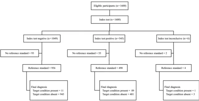
Fig. 2 shows the flow chart of the study participants with detailed information on the index and reference standard results. Table I presents in detail the infection prevalence, sensitivity, and specificity, as well as the positive and negative predictive values of the DPP® im-munoassays for all 1446 dogs in the sample set, both for the total canine population and the clinical subgroups (asymptomatic, oligosymptomatic, and symptomatic). The global prevalence of infection based on the refer-ence standard was 6.9%, which increased with the pres-ence of clinical signs of CVL. Positivity in the different parasitological tests was 4.0%, 3.8% and 5.5% for para-sitological culture, histopathology, and immunohisto-chemistry, respectively. High overall sensitivity (89%) was observed. Sensitivity gradually decreased with the reduction of symptomatology in the animals and reached the lowest level (75%) in asymptomatic dogs. General specificity was 70%. Specificity gradually decreased with an increase in signs and symptoms in the dogs and reached the lowest level in symptomatic animals (56%).
As for inter-laboratory agreement, the Kappa (κ) in -dices obtained from the comparisons between the three participating laboratories (A, B, and C) were κAB=0.778, κAC=0.645 and κCB=0.711; the concordance was substan-tial according to the classification by Landis and Koch.(23) Table II shows the prevalence of infection, sensitivity, specificity, and positive and negative predictive values for the total canine population and clinical subgroups (asymp-tomatic, oligosymp(asymp-tomatic, and symptomatic) by munici-pality investigated. In general, consistent sensitivity and specificity results were observed between the
ties, particularly with respect to increased sensitivity and decreased specificity as the analysed dogs presented more symptoms. The only anomaly was observed in Brasília, where sensitivity decreased as the symptoms increased. Nevertheless, the confidence intervals are quite broad, in-dicating a low precision in the estimates owing mainly to the small number of dogs with asymptomatic infection. In addition, as expected, the predictive values vary widely between municipalities because they depend directly on the prevalence values of canine infection.
DISCUSSION
The high overall sensitivity observed confirms the re-sults of previous studies conducted with smaller sample sizes.(24,25) In fact, the DPP® CVL test was developed for joint detection of antibodies against K26 and K39 anti-gens,(26) and historically, studies of the anti-Leishmania canine chromatographic immunoassay formulation have indicated an increased sensitivity when using both anti-gens together, while the use of k39 or rk39 in isolation has resulted in lower sensitivities.(27,28) Subsequently, al-though Otranto et al.(29) reached high sensitivity with the use of the rk39 antigen alone, other studies have suggested that the combined use of different antigens is associated with increased sensitivity in immunochromatographic tests;(17,25,30) Souza Filho et al.(31) demonstrated high sen-sitivity with the use of the AlereTM test, which also uti-lises chimaera rK28. High sensitivity is a characteristic required when using diagnostic tests as a screening tool for the Visceral Leishmaniasis Control Programme.(32)
The parasitological methods used in this study are considered the gold standards for leishmaniasis diag-nosis.(14) Despite some limitations, such as the need for pathologist expertise in microscopic amastigote detec -tion(33) and its susceptibility to contamination, parasito-logical diagnosis is still considered the best method for diagnosis because of its high specificity.(14) However, even considering that we used three different parasito-logical tests to build our reference standard, one should keep in mind that the results of the study might be slightly biased due to the well-known imperfections in the sensitivity of such tests.
The results of the clinical group evaluations showed a gradual decrease in test sensitivity accompanying the reduction of clinical signs in the dogs. Indeed, studies conducted using methodologies based on detection of serological response present high sensitivity and speci-ficity in symptomatic dogs.(34) However, it is worth not-ing that the mean sensitivity found in asymptomatic animals is still considerably higher than that observed in asymptomatic dogs in studies with smaller sample sizes, in which sensitivity was close to 50% as determined by the DPP® CVL test or similar immunoassays using rk39 antigen;(17,35) the assessment of this study also showed high negative predictive values. Such variation, in com-parison with symptomatic dogs, is probably explained by the fact that the latter present high levels of non-protective antibodies,(36,37) which would facilitate their detection, whereas the lower antibody levels detected in asymptomatic animals influence the accuracy of the serological methodologies.(34) It should be noted that the sensitivity of chromatographic immunoassays may vary according to the course of infection.(38)
Thus, the large-scale assessment performed in this study demonstrates that symptomatology affects test performance but suggests that such influence occurs in a smaller proportion of tests than previously observed. In-deed, favourable results can also be found in asymptom-atic animals. Recently, Larson et al.(39) demonstrated that most animals, whether symptomatic or asymptomatic, tested positive in less than 3 min when the response time of the DPP® CVL test was measured. Laurenti et al.(25) de-tected infection of both symptomatic and asymptomatic animals in equal proportions. However, such findings do not eliminate the need for caution when assessing asymp-tomatic dogs, and the results of rapid tests, especially neg-ative ones, should generally be evaluated with caution.(26) Accordingly, the use in parallel (jointly) of enzyme-linked immunosorbent assay (ELISA) can increase the sensitivity of the assessment.(24) This assay is already used serially as a confirmatory test for CVL according to the Brazilian Ministry of Health protocol.(15) Regard-ing the application of this protocol, Coura-Vital et al.(40) demonstrated an increase in CVL detection, in relation TABLE I
Prevalence of infection, sensitivity, specificity, and positive/negative predictive values regarding
the Dual-path Platform Chromatographic Immunoassay (DPP® CVL) in a sample composed of 1446 dogs from areas endemic
for canine visceral leishmaniasis assessed in single or clinical groups (asymptomatic, oligosymptomatic, and symptomatic)
Clinical condition
Number of dogs*
Prevalence of infection (95% CI)
(%)
Sensitivity (95% CI)
(%)
Specificity (95% CI)
(%)
Positive predictive value
(95% CI) (%)
Negative predictive value
(95% CI) (%)
All (single group) 1446 6.9 (5.7-8.4) 89.0 (81.2-94.4) 70.2 (67.7-72.6) 18.2 (14.8-21.9) 98.8 (98.0-99.4) Asymptomatic 448 2.7 (1.4-4.6) 75.0 (42.8-94.5) 72.9 (68.5-77.1) 7.1 (3.29-13.0) 99.1 (97.3-99.8) Oligosymptomatic 721 5.1 (3.6-7.0) 89.2 (74.6-97.0) 70.3 (66.7-73.7) 14.0 (9.8-19.1) 99.2 (97.9-99.8) Symptomatic 149 32.0 (25.0-40.4) 93.8 (82.8-98.7) 56.4 (46.2-66.3) 50.6 (39.8-61.3) 95.0 (86.1-99.0)
to prevalence and incidence measurements, when DPP® CVL was utilised jointly with ELISAs as opposed to the previously used immunofluorescence technique. Nev-ertheless, there is discussion of reversing the protocol order, especially in locations with great diagnostic de-mand; such discussion suggests the use of ELISAs as a screening method and DPP® CVL for confirmation because of the high specificity and positive predictive value previously reported for DPP® CVL and aims to re-duce the costs and increase the quality control of evalu-ation.(25,40) However, the results of this survey showed a relative reduction in specificity, as well as in positive predictive value, when compared with studies conducted with smaller sample sets,(24,25) which indicates a need for caution in the face of such propositions.
It is also important to highlight that the DPP® se-quence as a screening method with ELISA as a con-firmatory test is appropriate to the reality of small municipalities unable to maintain a laboratory for the performance of ELISAs, which can only be performed in central laboratories to confirm the diagnosis.(40) In this context, the DPP® CVL test is a screening tool that is easy to store, transport, and use and is able to achieve simple and fast results without the need of specialised laboratories.(24) In addition, the substantial agreement
between the three participating laboratories in a large-scale blind analysis demonstrates the reproducibility of the results and confirms the ease of use of the DPP® CVL assay, which decreased execution errors.
Another aspect worth mentioning is the necessity to verify, prior to the diagnostic test, any possible anti-
Leish-mania vaccination of the dogs, considering that
serologi-cal tests may not distinguish between infected and vac-cinated animals.(41) Studies have diverged with regard to the results obtained on cross-reactivity: Campos et al.(42) recently demonstrated no cross-reactivity of DPP® CVL for up to 12 months after vaccination of animals in a non-endemic area, whereas Marcondes et al.(43) reported that the test can cross-react with vaccine antibodies for up to six months after vaccination. Therefore, such information must be considered before the interpretation of test results.
A comprehensive assessment of possible cross-reac-tivity, which the method is subject to, is also suggested. (34) The results in the literature are still contradictory,
pre-senting studies that did not observe cross-reactivity(24,44) as well as surveys that demonstrated cross-reactivity with canine babesiosis(25) and Leishmania braziliensis.(17)
Ultimately, Schubach et al.(32) used data from one of the cities enrolled in our four-city study (namely, For-taleza) to evaluate the performance of the rapid test and TABLE II
Prevalence of infection, sensitivity, specificity, and positive/negative predictive values regarding
the Dual-path Platform Chromatographic Immunoassay (DPP® CVL) in a sample of 1446 dogs from areas endemic
for canine visceral leishmaniasis assessed in single or clinical groups (asymptomatic, oligosymptomatic, and symptomatic) according to the municipality investigated
Mu
n
ic
ip
al
it
y
Clinical condition
Number of dogs*
Prevalence of infection
(95% CI) (%)
Sensitivity (95% CI)
(%)
Specificity (95% CI)
(%)
Positive predictive value
(95% CI) (%)
Negative predictive value
(95% CI) (%)
F
o
rt
a
le
z
a All (single group) 333 7.5 (4.9-10.9) 92.0 (74.0-99.0) 71.8 (66.4-76.7) 20.9 (13.7-29.7) 99.1 (96.8-99.9)
Asymptomatic 134 3.7 (1.2-8.5) 60.0 (14.7-94.7) 72.9 (64.3-80.3) 7.9 (1.7-21.4) 97.9 (92.7-99.7) Oligosymptomatic 132 3.0 (0.8-7.6) 100.0 (39.8-100.0) 71.9 (63.2-79.5) 10.0 (2.8-23.7) 100.0 (96.1-100.0)
Symptomatic 36 39.0 (23.0-56.5) 100.0 (76.8-100.0) 45.5 (24.4-67.8) 53.8 (33.4-73.4) 100.0 (69.2-100.0)
Pa
lm
a
s All (single group) 377 2.7 (1.3-4.8) 100.0 (69.0-100.0) 59.4 (54.2-64.5) 6.3 (3.1-11.3) 100.0 (98.3-100.0)
Asymptomatic 129 1.6 (0.2-5.5) 100.0 (15.8-100.0) 64.6 (55.6-72.8) 4.3 (0.5-14.5) 100.0 (95.6-100.0) Oligosymptomatic 179 2.2 (0.6-5.6) 100.0 (39.8-100.0) 57.1 (49.5-64.6) 5.1 (1.4-12.5) 100.0 (96.4-100.0) Symptomatic 16 25.0 (7.3-52.4) 100.0 (39.8-100.0) 33.3 (9.9-65.1) 33.3 (9.9-65.1) 100.0 (39.8-100.0)
Ba
u
ru
All (single group) 379 11.0 (8.3-15.0) 83.7 (69.3-93.2) 70.5 (65.3-75.4) 26.7 (19.4-35.0) 97.1 (94.2-98.8) Asymptomatic 76 3.9 (0.8-11.1) 66.7 (9.4-99.2) 69.9 (58.0-80.1) 8.3 (1.0-27.0) 98.1 (89.7-100.0) Oligosymptomatic 220 7.3 (4.2-11.5) 81.3 (54.4-96.0) 72.5 (65.9-78.5) 18.8 (10.4-30.1) 98.0 (94.3-99.6) Symptomatic 66 35.0 (24.0-47.6) 91.3 (72.0-98.9) 67.4 (51.5-80.9) 60.0 (42.1-76.1) 93.5 (78.6-99.2)
B
ras
íl
ia All (single group) 357 6.2 (3.9-9.2) 90.9 (70.8-98.9) 80.3 (75.6-84.4) 23.3 (14.8-33.6) 99.3 (97.4-99.9)
Asymptomatic 109 1.8 (0.2-6.5) 100.0 (15.8-100.0) 85.0 (76.9-91.2) 11.1 (1.4-34.7) 100.0 (96.0-100.0) Oligosymptomatic 190 6.8 (3.7-11.4) 92.3 (84.0-99.8) 79.7 (73.0-85.3) 25.0 (13.6-39.6) 99.3 (96.1-100.0)
Symptomatic 31 23.0 (9.6-41.1) 85.7 (42.1-99.6) 58.3 (36.6-77.9) 37.5 (15.2-64.6) 93.3 (68.1-99.8)
found comparable accuracy values using whole blood and serum samples through electronic or visual read-ings. Although they used some of the data from our study, it should be noted that our study does not focus on the stability of the results between types of samples. We used a much larger sample to evaluate accuracy and reli-ability of the test, as well as how this relates to the pres-ence of CVL clinical signs. It is strongly recommended, however, that future systematic reviews in this field do not include both papers as if they used completely dif-ferent sample sets.
In conclusion, DPP® CVL performance is altered ac-cording to canine symptomatology, but such influence was less evident than in previous studies. Favourable re-sults for sensitivity and specificity can be obtained even in asymptomatic animals; however, caution is needed in these evaluations, and the results suggest that immuno-chromatographic assays may be further improved for better investigation in asymptomatic dogs. However, the results obtained confirm the usefulness of DPP® CVL for application in serological surveys.
AUTHORS’ CONTRIBUTION
FBF - Conceptualisation, sample collection, parasitologi-cal methods, data analysis, and manuscript writing and view; TCBV - data analysis and manuscript writing and re-view; MFM - parasitological methods and manuscript writing and review; RCM - histopathology methods and manuscript writing and review; ANSME - conceptualisation and manu-script writing and review; APM - serological methods and manuscript writing and review; GLW - conceptualisation, data analysis and manuscript writing and review.
REFERENCES
1. Ready PD. Epidemiology of visceral leishmaniasis. Clin Epidemiol. 2014; 6: 147-54.
2. MS/SVS/DVE - Ministério da Saúde/Secretaria de Vigilância em Saúde/Departamento de Vigilância Epidemiológica. Manual de vigilância e controle da leishmaniose visceral. Brasília: Ministé-rio da Saúde; 2006. 120 pp.
3. Danta-Torres F. Canine leishmaniosis in South America. Parasit Vectors. 2009; 2(Suppl. 1): S1.
4. PAHO - Pan American Health Organization. Leishmaniasis: epi-demiological report of the Americas. Nº 1. 2013. Available from:
http://www.paho.org/hq/index.php?option=com_topics&view=re adall&cid=6721&Itemid=40754&lang=pt.
5. Werneck GL. Visceral leishmaniasis in Brazil: rationale and concerns related to reservoir control. Rev Saude Publica. 2014; 48(5): 851-5.
6. Werneck GL. Expansão geográfica da leishmaniose visceral no
Brasil. Cad Saude Publica. 2008; 26(4): 644-5.
7. WHO - World Health Organization. Leishmaniasis. Control of the
leishmaniasis: report of a meeting of the WHO Expert Commit -tee on the Control of Leishmaniases. 2010. Available from: http://
whqlibdoc.who.int/trs/WHo_TRs_949_eng.pdf.
8. MS/SVS - Ministério da Saúde/Secretaria de Vigilância em Saúde. Guia de vigilância em saúde. Brasília: Ministério da Saúde; 2014. 812 pp.
9. PAHO - Pan American Health Organization. Leishmaniasis. Eepide-miological Report of the Americas. Report 6. 2018. Available from:
http://iris.paho.org/xmlui/bitstream/handle/123456789/34856/ LeishReport6_eng.pdf?sequence=1&isAllowed=y.
10. Bevilacqua PD, Paixão HH, Modena CM, Castro MCPS.
Urbani-zação da leishmaniose visceral em Belo Horizonte. Arq Bras Med Vet Zootec. 2001; 53(1): 1-8.
11. Gontijo CMF, Melo MN. Leishmaniose visceral no Brasil: quadro atual, desafios e perspectivas. Rev Bras Epidemiol. 2004;7: 338-49.
12. WHO - World Health Organization. Weekly epidemiological re-cord. 2017. Available ifrom: http://apps.who.int/iris/bitstream/ handle/10665/258973/WER9238.pdf;jsessionid=1D87FB89DFEE
5A59D71812B6E6651D27?sequence=1.
13. Solano-Gallego L, Miró G, Koutinas A, Cardoso L, Pennisi MG, Ferrer L, et al. LeishVet guidelines for the practical management of canine leishmaniosis. Parasit Vectors. 2011; 4: 86.
14. De Vries HJ, Reedijk SH, Schallig HD. Cutaneous leishmaniasis: recent developments in diagnosis and management. Am J Clin Dermatol. 2015; 16(2): 99-109.
15. MS/SVS/DVIT - Ministério da Saúde/Secretaria de Vigilância em Saúde/Departamento de Vigilância de Doenças Transmissíveis. Nota Técnica Conjunta 01/2011. Brasília: CGDT/CGLAB/DVIT/ SVS/MS; 2011.
16. Pattabhi S, Whittle J, Mohamath R, El-Safi S, Moulton GG, Gude-rian JA, et al. Design, development and evaluation of rK28-based point-of-care tests for improving rapid diagnosis of visceral leish-maniasis. PLoS Negl Trop Dis. 2010; 4(9): e822.
17. Grimaldi G, Teva A, Ferreira AL, dos Santos CB, Pinto IS, de-Azevedo CT, et al. Evaluation of a novel chromatographic immu-noassay based on Dual-Path Platform technology (DPP(R) CVL rapid test) for the serodiagnosis of canine visceral leishmaniasis. Trans R Soc Trop Med Hyg. 2012;106: 54-9.
18. Whiting PF, Rutjes AW, Westwood ME, Mallett S, Deeks JJ, Reitsma JB, et al. QUADAS-2: a revised tool for the quality as-sessment of diagnostic accuracy studies. Ann Intern Med. 2011; 155(8): 529-36.
19. Mancianti F, Gramiccia M, Gradoni L, Pieri S. Studies on canine leishmaniasis control. 1. Evolution of infection of different clini-cal forms of canine leishmaniasis following antimonial treatment. Trans R Soc Trop Med Hyg. 1988;82(4): 566-7.
20. Madeira MF, Schubach AO, Schubach TM, Pereira SA, Figueire-do FB, Baptista C, et al. Post mortem parasitological evaluation of dogs seroreactive for Leishmania from Rio de Janeiro, Brazil. Vet Parasitol. 2006;138(3-4): 366-70.
21. Menezes RC, Figueiredo FB, Wise AG, Madeira MF, Oliveira RV, Schubach TM, et al. Sensitivity and specificity of in situ hybrid-ization for diagnosis of cutaneous infection by Leishmania infan-tum in dogs. J Clin Microbiol. 2013;51: 206-11.
22. Cupolillo E, Grimaldi JG, Momen H. A general classification of new world Leishmania using numeral zymotaxomomy. Am J Trop
Med Hyg. 1994; 50(3): 296-311.
23. Landis JR, Koch GG. The measurement of observer agreement for categorical data. Biometrics. 1977; 33(1): 159-74.
24. da Silva DA, Madeira MF, Abrantes TR, Barbosa Filho CJL, Figueiredo FB. Assessment of serological tests for the diagnosis of canine visceral leishmaniasis. Vet J. 2013; 195(2): 252-3.
25. Laurenti MD, Leandro MVS, Tomokane TY, De Lucca HRL, As-char M, Souza CSF, et al. Comparative evaluation of the DPP® CVL rapid test for canine serodiagnosis in area of visceral leish-maniasis. Vet Parasitol. 2014; 205(3): 444-50.
26. Pinto AJW, Ribeiro VM, Tafuri WL. The immunochromatogra-phy use in canine visceral leishmaniasis in Brazil: A “quick
27. Reithinger R, Quinnell RJ, Alexander B, Davies CR. Rapid detec -tion of Leishmania infantum infection in dogs: comparative study using an immunochromatographic dipstick test, enzyme-linked im-munosorbent assay, and PCR. J Clin Microbiol. 2002; 40(7): 2352-6.
28. da Costa RT, França JC, Mayrink W, Nascimento E, Genaro O, Campos-Neto A. Standardization of a rapid immunochromato-graphic test with the recombinant antigens K39 and K26 for the diagnosis of canine visceral leishmaniasis. Trans R Soc Trop Med Hyg. 2003; 97(6): 678-82.
29. Otranto D, Paradies P, Sasanelli M, Spinelli R, Brandonisio O. Rapid immunochromatographic test for serodiagnosis of canine leishmaniasis. J Clin Microbiol. 2004; 42(6): 2769-70.
30. Fraga DBM, da Silva ED, Pacheco LV, Borja LS, de Oliveira IQ, Coura-Vital W, et al. A multicentric evaluation of the recombinant
Leishmania infantum antigen-based immunochromatographic as-say for the serodiagnosis of canine visceral leishmaniasis. Parasit Vectors. 2014; 7(1): 136.
31. Souza Filho JA, Barbosa JR, Figueiredo FB, Mendes Jr AA, Silva SR, Coelho GL, et al. Performance of Alere™ immunochromatho-graphic test for the diagnosis of canine visceral leishmaniasis. Vet Parasitol. 2016; 225: 114-6.
32. Schubach EY, Figueiredo FB, Romero GA. Accuracy and repro-ducibility of a rapid chromatographic immunoassay for the diag-nosis of canine visceral leishmaniasis in Brazil. Trans R Soc Trop Med Hyg. 2014; 108(9): 568-74.
33. Boelaert M, Bhattacharya S, Chappuis F, El Safi SH, Hailu A, Mondal D, et al. Evaluation of rapid diagnostic tests: visceral leishmaniasis. Nat Rev Microbiol. 2007; 5: S30-S39.
34. Ferrer L, Aisa MJ, Roura X, Portus M. Serological diagnosis and treatment of canine leishmaniasis. Vet Rec. 1995;136(2): 514-16.
35. Mettler M, Grimm F, Capelli G, Camp H, Deplazes P. Evaluation of enzyme-linked immunosorbent assays, an immunofluorescent-antibody test, and two rapid tests (immunochromatographic-dip-stick and gel tests) for serological diagnosis of symptomatic and asymptomatic Leishmania infections in dogs. J Clin Microbiol. 2005; 43(11): 5515-9.
36. Pinelli E, Killick-Kendrick R, Wagenaar J, Bernadina W, Del Real G, Ruitenberg J. Cellular and humoral immune responses in dogs
experimentally and naturally infected with Leishmania infantum. Infect Immun. 1994; 62(1): 229-35.
37. Barbiéri CL. Immunology of canine leishmaniasis. Parasite Im-munol. 2006; 28(7): 329-37.
38. Quinnell RJ, Carson C, Reithinger R, Garcez LM, Courtenay O. Evaluation of rK39 rapid diagnostic tests for canine visceral leish-maniasis: longitudinal study and meta-analysis. PLoS Negl Trop Dis. 2013; 7(1): e1992.
39. Larson M, Toepp A, Scott B, Kurtz M, Fowler H, Esfandiari J, et al. Semi-quantitative measurement of asymptomatic L. infantum
infection and symptomatic visceral leishmaniasis in dogs using Dual-Path Platform® CVL. Appl Microbiol Biotechnol. 2017; 101(1): 381-90.
40. Coura-Vital W, Ker HG, Roatt BM, Aguiar-Soares RDO, Leal GGdA, Moreira N, et al. Evaluation of change in canine diag-nosis protocol adopted by the visceral leishmaniasis control program in Brazil and a new proposal for diagnosis. PLoS One. 2014; 9(3): e91009.
41. Solano-Gallego L, Cardoso L, Pennisi MG, Petersen C, Bourdeau P, Oliva G, et al. Diagnostic challenges in the era of canine Leish-mania infantum vaccines. Trends Parasitol. 2017; 33(9): 706-17.
42. Campos MPD, Luca PMD, Renzetti ARDS, Souza SMMD, Mendes Jr AAV, Barros RS, et al. Can vaccines against canine visceral leishmaniasis interfere with the serological diagnostics
recommended by the Brazilian Ministry of Health? Cienc Rural.
2017; 47(4): e20160846.
43. Marcondes M, de Lima VMF, de Araújo MDFL, Hiramoto RM, Tolezano JE, Vieira RF, et al. Longitudinal analysis of serological tests officially adopted by the Brazilian Ministry of Health for the diagnosis of canine visceral leishmaniasis in dogs vaccinated with Leishmune®. Vet Parasitol. 2013; 197(3): 649-52.
44. Krawczak FDS, Reis IA, Silveira JAD, Avelar DM, Marcelino AP, Werneck GL, et al. Leishmania, Babesia and Ehrlichia in urban
pet dogs: co-infection or cross-reaction in serological methods?