351
Optimization of mammographic images
Radiol Bras 2006;39(5):351–354 Original Article
OPTIMIZATION OF MAMMOGRAPHIC IMAGES*
Diana Rodrigues de Pina1
, José Morceli1
, Sérgio Barbosa Duarte2
, Thomaz Ghilardi Netto3
OBJECTIVE: The aim of this study is the optimization of mammographic images with a considerable radia-tion dose reducradia-tion. MATERIALS AND METHODS: In the present study the X-ray beam was calibrated for each tension (kVp), aiming at determining the best combination between kVp and mAs, resulting in optical densities of about 1.0 above the base-plus-fog density. RESULTS: This study will bring into question the methods for X-ray beam calibration, the choice of the best image by means of visual grading analysis, com-parisons between doses and tube load (kVp × mAs) delivered by the techniques described in this study and by those adopted in the clinical routine at Service of Diagnostic Imaging of Faculdade de Medicina de Botucatu Clinics Hospital, Botucatu, SP, Brazil. Excellent breast radiographic images have been obtained with doses and tube loads reduction of respectively 36.8% and 46.2% in comparison to those employed in our institu-tions’ clinical routine. CONCLUSION: This study is a contribution to the optimization of the risk-benefit ratio for patients and cost-benefit ratio for the institution.
Keywords: Mammography; Homogeneous phantom; Quality control; Dose reduction.
Otimização de imagens mamográficas.
OBJETIVO: Este trabalho tem como objetivo a otimização de imagens mamográficas, com consideráveis reduções de doses. MATERIAIS E MÉTODOS: Neste estudo o feixe de raios-X foi calibrado para cada tensão (kVp), de modo a determinar a melhor combinação de kVp e mAs que irá proporcionar uma densidade ótica (DO) em torno de 1.0 acima da base mais véu do filme utilizado. RESULTADOS: Serão discutidas questões sobre os métodos empregados para a seleção de parâmetros de exposição do feixe de raios-X, seleção da melhor imagem utilizando o método de avaliação gradativa visual, comparações entre as doses e carga do tubo (kVp × mAs) proporcionadas pelas técnicas determinadas neste estudo e pelas utilizadas na rotina clí-nica do Serviço de Diagnóstico por Imagem do Hospital das Clíclí-nicas da Faculdade de Medicina de Botucatu. Neste estudo foram obtidas imagens radiográficas de mama de excelente qualidade, com redução de dose e carga de tubo, respectivamente, de 36,8% e 46,2%, quando comparadas com a técnica utilizada pela rotina clínica da instituição. CONCLUSÃO: Esta pesquisa vem contribuir com a otimização da relação risco-benefício para o paciente e custo-risco-benefício para a instituição.
Unitermos: Mamografia; Fantoma homogêneo; Controle de qualidade; Redução de dose.
Abstract
Resumo
* Study developed at Hospital das Clínicas da Faculdade de Medicina de Botucatu – Universidade Estadual Paulista “Júlio de Mesquita Filho” (Unesp), Botucatu, SP, Brazil.
1. Assistant Doctor-Professors at Department of Tropical Dis-eases and Diagnostic Imaging of Faculdade de Medicina de Botucatu, Unesp.
2. Researcher at Centro Brasileiro de Pesquisas Físicas, Rio de Janeiro, RJ.
3. Titular Professor at Faculdade de Filosofia, Ciências e Letras de Ribeirão Preto – Universidade de São Paulo.
Mailing address: Profa. Dra. Diana Rodrigues de Pina. Depar-tamento de Doenças Tropicais e Diagnóstico por Imagem da Faculdade de Medicina de Botucatu, Unesp. Distrito de Rubião Junior, s/nº. Botucatu, SP, Brazil 18618-970. E-mail: drpina@ fmb.unesp.br / [email protected]
Received March 23,2005. Accepted after revision February 10, 2006.
INTRODUCTION
In Brazil, deaths caused by breast can-cer represent 16% of the mortality related to malignant neoplasms among women(1).
The early detection or screening is the pro-cess aimed at detecting a determined type of cancer in its early phase, before the ap-pearance of any symptom(2). In this case,
the mammography is the method of choice, since it may detect up to 90% of breast cancers. However, neoplasm detec-tion by means of a mammogram depends on factors associated with parameters like screen-film system, radiographic tech-niques, an adequate angulation of the X-ray tube, correct positioning of the patient, satisfactory compression and exposure of the breast(2–3).
Presently, in Brazil, there are more than 2,700 mammographs, corresponding to an average of one for each 32 thousand inhab-itants(4). An alarming data is that more than
60% of these mammographs are not sub-mitted to a quality control process(4).
Typi-cally, mammographic images acquired in services of diagnostic imaging present a very low quality level, likely to generate a significant rate of negative or false-positive results in the clinical routine(4). It
is important to mention that the image
qual-ity depends on optical densities (OD) sat-isfactory to the physiological response of the human eye, besides a good visualiza-tion of tissues relevant for a safe medical diagnosis. The breast tissue presents simi-lar density and effective atomic number, so the contrast produced by interaction be-tween radiation and this tissue is low(5).
Therefore, the mammographic examination requires the highest technical standard be-cause of the breast tissue structure itself and the very particular geometry of breast ra-diography(5–7).
In the present study, we propose a pro-cedure for selection of exposure parameters appropriate for any screen-film system, aiming at achieving radiographic tech-niques which are able to deliver images of a typical 3 cm thickness compressed breast, with higher acceptance rates in tests of gradual visual evaluation(8.9), and
352
Pina DR et al.
Radiol Bras 2006;39(5):351–354 MATERIALS AND METHODS
In the present study, we have utilized a phantom approved by Colégio Brasileiro de Radiologia (Brazilian College of Radi-ology) for accreditation in mammography. The homogeneous portion of this phantom and the portion including analytical details were utilized in a way to simulate a 3 cm-thickness compressed breast with an aver-age composition (50% glandular tissue and 50% fat tissue).
The homogeneous portion of this phan-tom was utilized for selection of X-ray beam exposure parameters(10,11), in a way to
obtain radiographic techniques responsible for the yielding of an OD satisfactory to the human eye. The optical densities originated from sensitometric curves produced by the time scale sensitometric method(10–12). This
is a very efficient method for experiments in radiodiagnosis(10–13). Figure 1 presents an
experimental arrangement utilizing the sen-sitometric method adopted in this study where a phantom can be observed (a) po-sitioned on a shield system; (b) mounted on the screen-film bucky tray (c). In this pro-cedure, a manual exposure control was adopted in a way to expose, on the screen-film system, only previously established regions, varying the exposure time and maintaining a constant kVp. The result of this procedure is presented on Figure 2 showing different ranges of optical density on the film as a result of the variation of the exposure time.
Figure 1. Experimental arrangement of the time scale sensitometric method.
Figure 2. Optical density ranges obtained through the time scale sensitometric method.
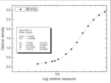
Figure 3. Typical curve obtained through the time scale sensitometric method utilizing a 3 cm homogeneous acrylic phantom with 28 kVp.
the production of images with different ODs, but with a same radiographic contrast because of the constant kVp. Such images were subjectively analyzed by 11 special-ists in Radiology, utilizing the method of gradual visual evaluation(8,9). These
profes-sionals identified the more appropriate gray tone for breast images production. This procedure was performed for a kVP range between 23 and 31, 1 kVP step, so for each kVP value we have an image selected with the best gray tone (OD) for the physiologi-cal response of the human eye. These radi-ographic techniques are shown in Table 1. It is important to note that all the images produced according the techniques in-cluded in Table 1 present a same gray tone, however, one differs from each other in terms of radiographic contrast, since the images are produced with different kVp. Once again, the method of gradual visual evaluation was utilized to identify the im-age providing the best visualization of the phantom structures, i.e., the best radio-graphic contrast for a typical 3 cm-thick-ness compressed breast.
The radiographic technique illustrated in Table 1, capable of providing the best vi-sualization of the phantom structures (28 kVp and 25 mAs), should be associated with the automatic exposure control in or-der to achieve a same exposure rate (con-stant kVp) in the screen-film system. This procedure is compatible with the necessity of an automatic exposure control in mam-mographic examinations(7,14).
The relation between the logarithm of relative exposure (LRE) and the DO yields a response curve or sensitometric curve of the screen-film system for a determined kVp(10–13)), as per Figure 3. The best OD
yielded from the linear region of sensito-metric curve. In this procedure, the mAs corresponding to the OD of the linear re-gion of the curve were combined with the selected kVp , generating a set of seven radiographic techniques. These techniques were utilized for obtaining images from the phantom with analytical details, leading to
Op
ti
c
a
l
d
e
n
s
it
y
353
Optimization of mammographic images
Radiol Bras 2006;39(5):351–354 The best image obtained in the present study was compared, by means of gradual visual evaluation, with those images pro-duced in the clinical routine of Faculdade de Medicina de Botucatu – Universidade Estadual Paulista (HCFMB-Unesp) Clinics Hospital. The clinical routine image was determined by a technician in Radiology duly instructed to consider the phantom as a typical 3 cm-thickness compressed breast. Thus, the technician selected three kVp and utilized the mammograph´s automatic ex-posure control in a way to produce three images of the analytical phantom. One image was selected by means of gradual visual evaluation, as the best in a set of images produced with the clinical routine techniques.
Finally, with the aid of lithium fluoride (LIF) termoluminescent dosemeters(15),
ra-diation doses were monitored on the phan-tom entrance surface, utilizing the opti-mized radiographic techniques found in the present study and those utilized in the HCFMB-Unesp service routine.
In the present research, we have utilized a General Electric Senographe 600T – Se-nix H.F mammograph, with Mo/Mo target/ filter combination, IBF-Medix (18 × 24) cm film in combination with a Kodak Min-R intensifying screen, a standalone Momo-ray Detail HT-300 (Agfa) automatic pro-cessor, MRA densitometer and sensitom-eter. The possible processor variations were evaluated with a Victoreen 21-step dual color model 07-417 and a MRA digital densitometer. It is important to note that the clinical imaging diagnostic routine utilizes the above mentioned screen-film system.
RESULTS
The Table 1 presents the kVp evaluated in the present study with the respective mAs which produced ODs satisfactory to the human eye, obtained with the homoge-neous phantom utilizing the time scale sen-sitometric method. It is important to note that the base-plus-fog-density value uti-lized is 0.20.
According to data shown Table 1, the OD that is most satisfactory to the human eye for visualizing the breast structures is around 1.27. The kVp/mAs combination (in bold types) that has provided the best
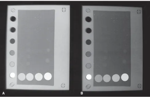
visualization of the analytical phantom structures corresponds to 28 kVp/ 25 mAs. The image that corresponds to the radio-graphic technique found in this study is presented in Figure 4A. The Figure 4B pre-sents the best image produced by the radio-graphic technique utilized by the clinical
routine, simulating a typical 3 cm-thickness compressed breast. The image produced by the clinical routine (Figure 4B) presents a decrease in radiographic contrast with a gray tone around 1.5, difficulting the visu-alization of structures relevant for a safe medical diagnosis.
The Table 2 includes the radiographic technique determined by this research as well as those utilized in the service routine with their respective doses (in mGy) and tube loads, simulating the exposure of a typical 3 cm-thickness compressed breast.
DISCUSSION
The time scale sensitometry method adopted in the present research allows evaluating the exposures of the sensitomet-ric curve linear region for a given kVp. Therefore it is possible to define the best gray tone for a radiographic image associ-ating it (through exposure evaluation) with
Table 1 kVp and mAs combinations utilized for obtaining optical densities more satisfactory to the human eye physiological response, according spe-cialists in Radiology of HCFMB-Unesp, utilizing a homogeneous breast phantom.
kVp ± 0.01
23.00
24.00
25.00
26.00
27.00
28.00
29.00
30.00
31.00
mAs ± 0.01
80.00
63.00
50.00
40.00
32.00
25.00
20.00
16.00
16.00
OD ± 0.01
1.26
1.27
1.26
1.26
1.27
1.27
1.24
1.28
1.27
Figure 4. Images from an analytical phantom simulating a typical 3 cm-thickness compressed breast. A:
Image produced with the radiographic technique obtained in the present study; B: Image produced with
the radiographic technique utilized in the HCFMB-Unesp clinical routine.
A B
D(mGy), dose (in milligray) obtained on the phantom entrance surface; CT(J), tube load (kVp × mAs) with units in joules.
Table 2 Comparison between doses and tube load (kVp x mAs) produced by radiographic techniques obtained in this study and by HCFMB-Unesp clinical routine.
Radiographic technique
kVp ± 0.01
mAs ± 0.01 D (mGy) ± 0.001
CT (J) ± 0.1
This study
28.00
25.00 2.133
560.0
Clinical routine HCFMB-Unesp
26.00
40.00 3.688
354
Pina DR et al.
Radiol Bras 2006;39(5):351–354 the automatic exposure control for daily
procedures. This method may be employed every time any sensitometric factor (screen-film system and automatic processor) pre-sents considerable variations in order to change the image quality. Also, with this method, the best radiographic contrast of the simulated breast can be determined. This procedure must be based on evalua-tions of kVp that should be combined with mAs, yielding a gray tone adequate to the human eye physiological response.
The kVp and OD values reported in the present study are in compliance with those suggested by American College of Radiol-ogy(9), i.e., 28 kVp and an OD above the
base-plus-fog density of the film utilized. It is important to note that the observers involved in the evaluation of the mammo-graphic images, were not familiar with the radiographic techniques utilized for the images acquisition.
Another important detail to be taken into consideration is that the automatic ex-posure control utilized by the sector of di-agnostic imaging where this research was developed, was duly calibrated for a more sensitive screen-film system. This explains the gray tone (1.50) in the image produced by the clinical routine (Figure 4B) when compared with those obtained by the present study (Figure 4A). Additionally, the Figure 4B, presents considerable decrease in radiographic contrast as a result of an inadequate kVp value when compared with the image presented in Figure 4A.
Besides the optimization of the image quality, the present study also provides a dose reduction and tube load of respec-tively 36.8% and 46.2%, when compared
with the technique utilized by the HCFMB-Unesp clinical routine, simulating an expo-sure of a typical 3 cm-thickness com-pressed breast.
The results of the present study drew the attention of the HCFMB-Unesp clinical staff and immediately the necessary steps were taken to improve the quality of mam-mographic images in that service. All the screen-film systems were replaced, and the most appropriate processing chemicals for the new screen film system started to be utilized. Also, it is important to note that, currently, the mammography processor undergoes daily quality control tests, a pro-cess that contributes to assure the quality of mammograms (about 460 per month) performed in HCFMB-Unesp. Finally, as a result of this research, the institution was granted the seal of quality in mammogra-phy by Colégio Brasileiro de Radiologia (Brazilian College of Radiology).
Acknowledgements
The authors would like to express their gratitude for the Conselho Nacional de Desenvolvimento Cientifico e Tecnológico (CNPq) financial support, for the coopera-tion from the whole technical and clinical staff of the Sector of Diagnostic Imaging, to Kodak, particularly to the physicist Fran-cisco Carrieri, for supplying the chassis utilized in the present study, and to Murilo Stelzer for designing the figures.
REFERENCES
1. Câncer de mama. Disponível em: http://www. inca.gov.br/conteudo_view.asp?id=336. Aces-sado em: 10/2/2006.
2. Guia Europeu para a Garantia de Qualidade no Rastreio por Mamografia CEC. Programa
Euro-pa contra o cancro Acções de proteção contra ra-diações, 1992.
3. Ghilardi Netto T, Trad CS. Princípios físicos e o controle de qualidade da imagem e da exposição em mamografia. Radiol Bras 1983;16:125–130. 4. Programa de Qualidade em Mamografia. Dispo-nível em: http://www.hospitalar.com.br/noticias/ not1634.html. Acessado em: 10/2/2006. 5. Barnes GY, Donald Frey G. Screen film
mammog-raphy. Madison, WI: Medical Physics Publishing, 1991;159–175.
6. Magalhães LAG, Azevedo ACP, Carvalho ACP. A importância do controle de qualidade de proces-sadoras automáticas. Radiol Bras 2002;35:357– 363.
7. American College of Radiology Committee on Quality Assurance in Mammography. Quality Control Medical Physicist’s Manual, 1999. 8. Sund P, Bath M, Kheddache S, Mansson LG.
Comparison of visual grading analysis and deter-mination of detective quantum efficiency for evaluating system performance in digital chest ra-diography. Eur Radiol 2004;14:48–58. 9. Tuczek HV, Fritz P, Schwarzmann P, Wu X,
Mahner G. Breast carcinoma. Correlations be-tween visual diagnostic criteria for histologic grading and features of image analysis.Anal Quant Cytol Histol 1996;18:481–493. 10. Pina DR, Ghilardi Netto T, Rocha SL, Brochi
MAC, Trad CS. Construção de um fantoma ho-mogêneo para padronização de imagens radiográ-ficas. Radiol Bras 2000;33:41–44.
11. Pina DR, Duarte SB, Ghilardi Netto T, Trad CS, Brochi MAC, Oliveira SC. Optimization of standard patient radiographic images for chest, skull and pelvis exams in conventional x-ray equipment. Phys Med Biol 2004;49:N215–226. 12. Góes EG, Pela CA, Ghilardi Netto T. A time-scale sensitometric method for evaluating screen-film systems. Phys Med Biol 1997;42:1939–1946. 13. Haus AG. Screen-film image receptors and film
processing. Syllabus: A categorical course in physics-technical aspects of breast imaging. 2nd ed. Oak Brook, IL: RSNA, 1993;69–83. 14. Ministério da Saúde, Secretaria da Vigilância
Sanitária. Diretrizes de proteção radiológica em diagnóstico médico e odontológico. Portaria nº
453, 1998.