4 artigo 360
oRIgINAL ARTICLE
The authors declare that they did not have any conflict of interests in producing this article.
1 - Trainee Physician specializing in knee surgery and lower-limb trauma at Hospital das Clínicas, Ribeirão Preto School of Medicine, University of São Paulo (HCFMRP-USP). 2 - Attending Physician in the Discipline of Knee Surgery and Lower-limb Trauma, HCFMRP-USP.
3 - PhD. Professor in the Department of Biomechanics, Medicine and Rehabilitation of the Locomotor Apparatus, Ribeirão Preto School of Medicine, University of São Paulo (DMBRAL-USP).
4 - Titular Professor of the Department of Biomechanics, Medicine and Rehabilitation of the Locomotor Apparatus, Ribeirão Preto School of Medicine, University of São Paulo (DMBRAL-USP).
Work performed at Hospital das Clínicas, Ribeirão Preto School of Medicine, University of São Paulo (HCFMRP-USP).
Correspondence: Cleber Paccola - Hospital das Clínicas de Ribeirão Preto - Av. Bandeirantes, 3900 - 11º andar - Campus Universitário - 14048-900 - Ribeirão Preto, SP. Email: [email protected] / [email protected]
Work received for publication: March 5, 2010; accepted for publication: August 6, 2010.
uSE OF AuTOlOGOuS bONE GRAFT ASSOcIATEd
wITH SuPPORT OSTEOSyNTHESIS FOR TIbIAl EdGE bONE
lOSSES IN TOTAl kNEE PROSTHESES
Marcello Teixeira Castiglia1, Juliano Voltarelli Franco da Silva1, Gabriel Silva Quialheiro1, Rodrigo Salim2,
Maurício Kfuri Júnior3, Cleber Antonio Jansen Paccola4
Rev Bras Ortop. 2011;46(1):27-30
INTRODUCTION
Bone defects, both in primary arthroplasty and in revision, are an important challenge for knee surge-ons with regard to implant fixation, with the aim of establishing a stable and long-lasting bone-implant interface(1-6).
The use of reinforcement together with a bone graft in bone defect areas is indicated when it is im-possible to achieve stability for the test components(7).
Radiological evaluation using AP and lateral
radio-ABSTRACT
Objective: To report the initial results from the use of a new technique for fixation of bone grafts in uncon-tained tibial bone defects in patients undergoing total knee prosthesis implantation. Methods: Six patients with severe varus deformity of the knee who, after cuts and ligament balancing had been performed, still presented bone deficiencies that reached the edge of the tibial cut and compromised the implant stability, underwent a new fixation technique. Results: Five of the patients had good
clinical results, with integration of the graft within 12 weeks. One patient presented clinical complications with wound dehiscence and implant exposure, which evolved to the need for implant removal and knee arthrodesis. Conclusion: Support osteosynthesis as a graft fixation method is a viable option for treating tibial bone defi-ciencies. The proposed technique certainly needs further studies for its validation.
Keywords - Knee Prosthesis; Bone Transplantation; Arthroplasty
graphs is known to underestimate the bone loss(2,3).
The use of oblique radiographs and, possibly, axial
computed tomography may be necessary(2).
The current methods for repairing such defects include the use of structural allografts, impaction of morselized bone grafts, autografts and fixation using screws or Kirschner wires, bone cement (in defects of up to 10 mm) and metal wedges added to the im-plant(3). Wedge use is well documented, and Brand et
28
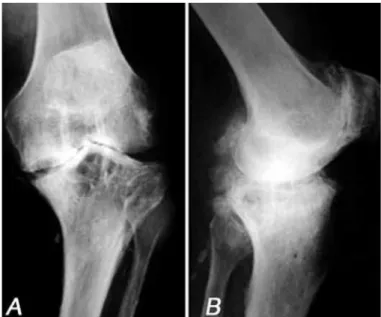
Figure 1 – Radiographic appearance of a patient with severe genu varus who underwent the proposed technique.
Rev Bras Ortop. 2011;46(1):27-30
was up to 25% with wedge use, 3.5 years after the operation. In the case of young patients and con-tained defects, Cuckler preferred to use autografts coming from the resected femoral condyles(4).
Rein-forcement with allografts is indicated in cases of large bone defects, severe trauma and, most commonly, knee arthroscopy revisions(6,7). When the defect is
not excessively large, another option is to use an autologous iliac graft, which is easy to obtain and presents little morbidity(1,7).
The options for treating defects show problems such as non-integration and/or migration of the graft, cement fatigue and aseptic loosening of the pros-thesis. So far, no ideal treatment for this relatively
common problem has been presented(8).
When the bone defect reaches the margin of the plateau, a situation known as an uncontained bone defect, there is a tendency for the graft to escape. In these cases, the use of a containment belt seems to be appropriate. This is the basis for our idea. This is therefore a new proposal for treating such defi-ciencies, through fixation of the graft using support osteosynthesis that acts as a containment belt, in or-der to avoid expulsion of the graft to the periphery of the defect.
PATIENTS AND METHODS
We reviewed the cases of six patients with bone deficiencies of type T2A according to the AORI classification. These patients had undergone total primary knee arthroplasty performed by the senior authors (CAJP and MKJ), between January 2007 and February 2009, and had presented significant bone loss, giving rise to an uncontained bone defect that reached a significant fraction of the margin of the tibial plateau. The patients were assessed using AP, lateral and oblique radiographs of the knee (Figure 1), along with a panoramic AP radiograph of the lower limb, to evaluate the mechanical axis.
The early postoperative stage was conducted rou-tinely, with the use of unfractionated heparin for 21 days and physical antithrombotic measures (elastic stocking and physiotherapy). Return visits were ar-ranged for the first, fourth and eighth weeks after the operation, and thereafter every two months, until graft integration was achieved. Weight bearing was
limited to touching the ankles, until there were indi-cations of bone integration.
Surgical technique
The transverse tibial incision for the defects of the medial plateau was done with the aim of resecting as much as possible of the bone of the medial plateau, while still allowing good bone support at the sides, so as to minimize the preexisting tibial defect that was to be filled.
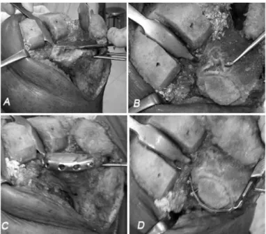
In the remaining defect, the eburnated bone was revived using curettage and perforated many times using a 2.5 mm drill bit. The receptor area was pre-pared and then the bone graft that fitted into the de-fect best was chosen. The graft chosen usually came from the condyle on the side of the deepening, since it would naturally be molded to the region. The graft was reduced and repeatedly tested until it fitted ap-propriately over the area of the defect. It was then fixed provisionally using 0.6 mm Kirschner wires. If any places were still not covered by the graft, these were completed with spongy bone obtained from the femoral and tibial incisions (Figures 2A and 2B).
29
Figure 2 – Sequence for preparation of the receptor area. A) Appearance of the defect before it was filled in; B) Defect viewed from above, after making the lateral edge of the defect vertical; C and D) After adaptation of the graft and placement of the plate.
USE OF AUTOLOGOUS BONE GRAFT ASSOCIATED WITH SUPPORT OSTEOSYNTHESIS FOR TIBIAL EDGE BONE LOSSES IN TOTAL KNEE PROSTHESES
Rev Bras Ortop. 2011;46(1):27-30 in the receptor area and avoid its shearing off to the
periphery (Figures 2C and 2D).
DISCUSSION
Biological solutions have obvious advantages in relation to solutions using special implants, parti-cularly for young patients who might subsequently require revision surgery(1,4,9-12). Reconstruction using
bone is a good option, regardless of whether it is au-tologous or homologous. However, the technique for making choices should take into account the patient’s activity, the extent of bone loss and the surgeon’s experience with the technique(13).
In our view, the technique proposed here has the advantage of providing mechanical support for the graft that is placed in the bone defect, thereby avoiding its escape through failure of the edge of the tibial cortical bone. The access is simple and only requires sufficient detachment of the soft tissue from the edge of the bone to accommodate the plate. In closing the operation site, care needs to be taken to do a good repair on the soft tissue above the plate, involving the gastrocnemius, posterior capsule and popliteus, with greater exploration of the posterior structures, without significant additional devitalization.
Associating the osteosynthesis with graft place-ment may also diminish the bone resection in the tibial incision, through making it viable to place the graft when the cortical edge is absent (Figure 3).
The stability provided only by the Kirschner wi-res and screws was not shown to be sufficient after a careful manual test. Moreover, the anchorages for the screws are rarely secure, which motivated us to add a support plate with a containment belt.
RESULTS
The mean follow-up among the cases was 27.1 months (minimum of nine and maximum of 35 mon-ths). Among the six cases operated, we observed complications in one case. This patient presented advanced arthrosis, secondary to rheumatoid dise-ase, and had not be able to walk over the past four years. After the operation, there was a problem of dehiscence of the surgical wound and necrosis of a large area of skin and subcutaneous tissue, which re-quired surgical cleaning, a local flap from the medial gastrocnemius and several sessions of hyperbaric oxygen therapy. Even after all the rescue measu-res, the patient presented chronic infection and had to undergo implant removal in order to control the infection. We judged that this complication had not come from the graft fixation technique but mainly from the comorbidities and complications inherent to the severity of the case.
As radiological signs of graft integration became visible, total weight-bearing was progressively allo-wed. Full weight-bearing was generally achieved
30
Rev Bras Ortop. 2011;46(1):27-30
CONCLUSION
We are aware of the limitations of our study, which was a limited series of cases. Except for a single poor result, which we ascribed to unfavora-ble patient-related preoperative factors, all the cases
had good results. This allows us to recommend the technique described here.
However, a study with a greater number of cases and long-term follow-up is necessary in order to re-ach conclusions that are more secure.
REFERENCES
1. Whittaker JP, Dharmarajan R, Toms AD. The management of bone loss in revision total knee replacement. J Bone Joint Surg Br. 2008;90(8):981-7.
2. Engh GA, Ammeen DJ. Use of structural allograft in revision total knee arthroplasty in knees with severe tibial bone loss. J Bone Joint Surg Am. 2007;89(12):2640-7.
3. Cuckler JM. Bone loss in total knee arthroplasty: graft augment and options. J Arthroplasty. 2004;19(Suppl 1):56-8.
4. Brand MG, Daley RJ, Ewald FC, Scott RD. Tibial tray augmentation with modular metal wedges for tibial bone stock deficiency. Clin Orthop Relat Res. 1989;(248):714.
5. Lyall HS, Sanghrajka A, Scott G. Severe tibial bone loss in revision total knee replacement managed with structural femoral head allograft: a prospective case series from the Royal London Hospital. Knee. 2009:16(5):326-31.
6. Backstein D, Safir O, Gross A. Management of bone loss: structural grafts in revision total knee arthroplasty. Clin Orthop Relat Res. 2006;446:104-12.
7. Dorr LD, Ranawat CS, Sculco TA, McKaskill B, Orisek BS. Bone graft for tibial defects in total knee arthroplasty. Clin Orthop Relat Res. 1986;(205):153–65.
8. Lotke PA, Carolan GF, Puri, N. Technique for impaction bone grafting of large bone defects in revision total knee arthroplasty. J Arthroplasty. 2006;21(Suppl 1):57-60.
9. Toms AD, Barker RL, McClelland D, Chua l, Spencer-Jones R, Kuiper JH. Repair of defects and containment in revision total knee replacement: a com-parative biomechanical analysis. J Bone Joint Surg Br. 2009;91(2): 271-7.
10. Ghazavi MT, Stockley I, Yee G, Davis A, Gross AE. Reconstruction of massive bone defects with allograft in revision total knee arthroplasty. J Bone Joint Surg Am. 1997;79(1):17-25.
11. Radnay CS, Scuderi GR. Management of bone loss: augments, cones, offset stems. Clin Orthop Rel Res. 2006;446:83-92.
12. Mabry T, Hanssen AD. The role of stems and augments for bone loss in revision knee arthroplasty. J Arthroplasty. 2007;22(Suppl 1):56-60.