Intrauterine growth restriction inluence on the nutritional evolution
and growth of preterm newborns from birth until discharge
Inluência do crescimento intrauterino restrito sobre a evolução nutricional e crescimento de recém-nascidos pré-termo até a alta hospitalar
Igor Tadeu da Costa1, Cléa Rodrigues Leone2
Institution: Departamento de Pediatria da Faculdade de Medicina da Uni-versidade de São Paulo (FMUSP), São Paulo, SP, Brasil
1Aluno do Programa de Iniciação Científica do Departamento de Pediatria
da FMUSP, São Paulo, SP, Brasil
2Professora-associada do Departamento de Pediatria da FMUSP,
coordena-dora da Área Técnica de Saúde da Criança e do Adolescente da Secretaria Municipal de Saúde de São Paulo, São Paulo, SP, Brasil
ABSTRACT
Objective: Analyze the growth of intrauterine growth restricted (IUGR) preterm newborns infants (PTNB) from birth until hospital discharge.
Methods: Cohort study of PTNB from single gesta-tions with gestational age of 30 to 34 weeks, Apgar score at ive minutes >6, without perinatal infectious risk and/ or malformations. Patients were divided into two groups. Group I: PTNB with IUGR (Kramer index: birth weight/ weight at 50th percentile <0,85); Group II: PTNB with-out IUGR. Weight (W), length (L), head circumference (HC) and body mass index (BMI) were evaluated at birth and at 40 weeks corrected GA or discharge. Statistical analysis included Student t test, paired t test, chi-square test, Pearson’s correlation and linear regression, being signiicant p<0.05.
Results: At birth, anthropometric signiicant differ-ences (p<0.0001) were seen between the 24 Group I PTNB (W=1192g, L=37.7cm, HC=26.9cm) and the 27 Group II infants (W=2081g, L=43.2cm, HC=30.9cm). At discharge, Group II PTNB were heavier (p=0.03), but L, HC and BMI were similar between groups. From birth until discharge, the W, L and HC increased in both groups. BMI increased from birth to discharge only in Group I (p<0.0001), with a negative correlation between BMI at birth and the BMI dif-ference between birth and discharge (r=-0.79; p<0.0001).
Conclusion: IUGR at birth was associated to signiicant BMI increase until discharge, which was inversely correlated to birth BMI, suggesting a higher risk of future obesity in
Corresponding author: Cléa Rodrigues Leone Alameda Itu, 433, apto. 42 CEP 01421-000 – São Paulo/SP E-mail: [email protected]
Submission: Aug 4, 2008 Approval: Oct 10,2008
these infants if this trend persists throughout infancy and childhood.
Key-words: infant nutrition; infant, newborn; fetal growth retardation.
RESUMO
Objetivo: Analisar o crescimento de recém-nascidos pré-termo (RNPT) com crescimento intrauterino restrito (CIUR) do nascimento até a alta hospitalar.
Métodos: Coorte de RNPT provenientes de gestação única, com idade gestacional (IG) de 30 a 34 semanas, Apgar de cinco minutos >6, sem risco infeccioso perinatal e sem malformações. Grupos de estudo: I: CIUR (índice de Kramer=peso ao nascer/peso P50<0,85); II: sem CIUR. Analisaram-se: peso (P), comprimento (C), perímetro cefálico (PC) e índice de massa corpórea (IMC) ao nascimento e à alta ou com 40 semanas de IG corrigida. Análise estatística: comparação de médias (teste t de Student e t pareado), teste do qui-quadrado, correlação de Pearson e regressão linear, sendo signiicante p<0,05.
inversa entre IMC ao nascimento e diferença do IMC (alta-nascimento): r=-0,79 (p<0,0001) no grupo com CIUR.
Conclusões: CIUR ao nascimento associou-se à elevação do IMC até a alta, que se correlacionou inversamente com o IMC ao nascer, o que sugere um risco maior de obesi-dade futura nos RNPT com maiores déicits de peso em relação ao comprimento ao nascimento, se essa tendência se mantiver.
Palavras-chave: nutrição do lactente; recém-nascido; retardo do crescimento fetal.
Introduction
Many studies have attempted to identify newborn in-fants (NB) with intrauterine growth restriction (IUGR) at birth, but no early indicator has been validated so far for this end. The need for this identiication is based on the observation of greater morbidity and mortality among these NB, in addition to an increased likelihood of development of chronic diseases in adulthood, such as type 2 diabetes, systemic arterial hypertension, obesity, and cardiovascular diseases(1-5).
IUGR is deined as the incomplete expression of an indi-vidual’s genetic potential for fetal growth. Among the many causes of IUGR, restricted nutrition and/or oxygenation in particular trigger adaptive mechanisms to guarantee the individual’s survival in this adverse intrauterine environ-ment. As part of these mechanisms, metabolic adaptations will “program” the fetus to save energy and deposit fat(6,7). On the other hand, during the neonatal period, increased nutrient supply and the occurrence of catch-up growth dur-ing the irst months after reachdur-ing term can increase the risk of developing obesity and other manifestations of metabolic syndrome over time(8,9).
Classic examples of this association can be found in epidemiological studies of people born after periods of extreme nutritional deprivation, as occurred in Holland in 1944-45(10), and also in research using experimental models(2). In the same way, the longitudinal cohort study carried out in Avon in the United Kingdom (The Avon Longitudinal Study of Parents and Children, ALSPAC) en-rolled full term NB and detected the occurrence of catch-up growth between birth and 2 years of age among neonates with smaller weight and length at birth. This accelerated period of growth was related to increased weight, height and body mass index (BMI) at ive years of age(11),
provid-ing evidence of the importance of the early growth pattern in this population.
Published data indicate that newborn infants with IUGR have increased susceptibility to the development of obesity during postneonatal life and that early catch-up growth is a factor that possibly accelerates this process. Therefore, it is important to analyze nutritional development and growth in NB with IUGR during the neonatal period, especially in preterm newborn infants (PTNB) with IUGR, since they show an accelerated pattern of postnatal growth in an at-tempt to attain growth paths that are closer to population medians. Within this group, the future risk may be even greater than for those born at full term.
The objective of this study was to analyze the charac-teristics of postnatal growth and nutritional development in PTNB with IUGR from birth up to discharge from the neonatal unit.
Methods
This was a cohort study based on a convenience sample of PTNB with gestational ages varying from 30 to 34 weeks admitted to the Nursery Annexed to the Maternity Unit run by the Neonatal and Intensive Service in the Pediatrics Department, Children’s Institute, School of Medicine, Uni-versidade de São Paulo (USP), São Paulo, Brazil, between January 1st, 2004, and May 31st, 2005. Data of the PTNB enrolled in the study were obtained retrospectively from the medical records. The study protocol was approved by the Research Ethics Committee.
Selection criteria were gestational age between 30 and 34 weeks, single fetuses, ifth minute Apgar >6 and no risk of perinatal infection. Children with congenital malforma-tions were excluded. Selected NB were then classiied into one of two study groups: Group I, with IUGR; Group II, without IUGR.
Each group was submitted to anthropometric assessment (weight, length, head circumference, Kramer index and BMI) at birth and at hospital discharge and/or 40 weeks’ cor-rected gestational age. Data on birth conditions, nutritional development and growth were also analyzed.
assessed by the New Ballard method. In all other cases, the gestational age calculated by the New Ballard method was considered deinitive.
IUGR diagnosis was based on the presence of a ratio <0.85 between actual birth weight and estimated weight at the 50th percentile for each gestational age on the Kramer index(12). Reference values for estimated weight at the 50th percentile were taken from Ramos’s intrauterine growth curve(13).
Anthropometric measurements were determined as fol-lows:
• weight: measured at birth on a balance accurate to 5g; • length: measured using a wooden anthropometer cali -brated in mm, with the baby’s head placed against the ixed part and the movable part adjusted to the baby’s feet;
• head circumference: measured using non-stretch measur -ing tape pass-ing the region above the supraorbital sulcus and over the most salient part of the occipital bone; • BMI: calculated as the ratio between weight in kg and
the square of length in meters(14).
Nutritional development was assessed based on the fol-lowing parameters:
• maximum weight loss (MaxWL): deined as the largest percentage of birth weight lost during the irst 10 days of life;
• age (AMax) at which MaxWL occurred;
• time taken to regain birth weight (TRec): measured in days;
• weight gain (WG in g/kg/day): mean weight gain per kg/day during the seven days following MaxWL; • type of feeding at discharge: breast milk or formula.
The means for each group were compared using Student t
test; when the test’s assumptions of normality were not met, the Mann-Whitney non-parametric test was employed. The
paired t test was used to compare the means at birth and at discharge. Proportions were compared using Fischer’s exact test or the chi-squared test, and Pearson’s correlation coeficient was used to analyze correlations. Linear regres-sion was also carried out for the birth variables against BMI at discharge. The level of statistical signiicance was set at p<0.05.
Results
A total of 158 PTNB with gestational ages varying between 30 and 34 weeks were enrolled in the study: 122 were born in 2004 and 36 were born between January and May of 2005. Of these, it was possible to retrieve data for 103 NB at the institution’s Medical Archives Department, due to localized dificulties experienced by that service. Of the 103 medical records, 50 cases met the inclusion criteria, while 53 were excluded due to risk of perinatal infection, inconsistencies regarding gestational age and/or because they were multiple births.
The average age of the mothers of children included in Group I was 26.8±6.8 years; all of them had attended prenatal consultations, just two were free from pathologies and 15 (62.5%) had suffered arterial hypertension during pregnancy. The average age of the mothers of children in-cluded in Group II was 27.9±7.5 years; four did not attend prenatal consultations, 13 (48%) had pathologies, and just one had suffered from arterial hypertension. Tables 1, 2 and 3 list the characteristics of the NB and the development of their weights and anthropometric variables. The frequency of breastfeeding (Table 2) was similar in both groups, which ruled out the possibility of analyzing the inluence of this factor on the neonates’ development.
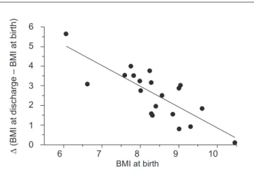
The correlation analysis revealed an inverse relationship between BMI at birth and change in BMI up to discharge (p<0.0001) in the IUGR group, with r=-0.79 (95%CI -0.91 to -0.54) and r2=0.63, which indicates that the correlation
Group I
(n=24)
Group II
(n=26) p
Birth weight (g) 1160±269 2081±321 <0.0001
Gestational age (weeks) 31.9±1.3 32.7±0.9 <0.0001
Sex (M:F) 1:2 2.2:1
Cesarean delivery 22/24(91.6%) 14/26(57.8%) 0.004
First minute Apgar ≤3 Fifth minute Apgar ≤5
3/24 (12.5%) 2/24 (8.3%)
2/26 (7.7%) –
Table 3 – Anthropometric measurements of preterm newborns at birth and at discharge, by study group
Group I (n=24)
Group II
(n=26) p
Birth
Weight (g) 1160.4±268.9 2081.5±321 <0.0001
Length (cm) 37.2±3.3 43.2±2 <0.0001
Head circumference (cm) 26.5±1.8 30.9±1.6 <0.0001
BMI 8.2±1 11.1±1 <0.0001
Discharge
Weight (g) 1956.8±399.3* 2301.8±519.4** 0.01
Length (cm) 43±2.1* 44.7±2.5*** 0.05
Head circumference (cm) 32.6±1.4* 32±1.2**** 0.16
BMI 10.9±0.7* 11.3±1.4 0.19
Discharge versus birth: Group I: *p<0.0001; Group II: **p=0.024; ***p=0.002; ****p=0.0002; BMI: body mass index.
6
6 7 8 9 10
5
4
3
2
1
0
∆
(BMI
a
t d
isch
arg
e
–
BMI
a
t b
irt
h)
BMI at birth
Figure 1 – Body mass index (BMI) increase from birth to dis-charge in preterm newborns with intrauterine growth restriction, by body mass index at birth
14 13
12 11
10 9
-15 -10 -5 0 5
∆
(BMI
a
t d
isch
arg
e
–
BMI
a
t b
irt
h)
BMI at birth
Figure 2 – Body mass index (BMI) increase from birth to discharge in preterm newborns without intrauterine growth restriction, by body mass index at birth
is strong and that for each variation of one unit (1kg/m2) of BMI, the change expected would be 63% (Figure 1). Linear regression results showed a slope of -1.06±0.19 (95%CI -1.46 to -0.66; p<0.0001) (Figure 1). Figure 2 shows
cor-relation analysis and linear regression results for Group II, with r=-0.06 (95%CI -0.42 to 0.33 and p=0.78), r2=0.003, and a slope of -0.17± 0.62 (95%CI -1.44 to 1.09; p=0.78). No correlations were observed with birth weight.
Group I (n=24)
Group II (n=26)
MaxWL (%) 11±4 12±7
AMax (days) 5.7±2.5 5.7±2.6
TRec (days) 10.6±6 12.2±6.5
Weight gain (g/Kg/d) 11.5±5.7 9.3±3.3
Breastfeeding 21/24 (87.5%) 23/26 (88.4%)
Table 2 – Ponderal development of the preterm newborns, by study group
MaxWL: largest percentage of birth weight lost during the irst days of life; AMax: age at maximum weight loss; TRec: time taken to regain
Discussion
The susceptibility of the fetal period to external fac-tors, with persistent effects on functional development, is a concept that is receiving more and more support from epidemiological and experimental research, with ever more speciic investigations on the cellular level being carried out in search of the mechanisms that probably cause these changes. In particular, the fetal and neonatal periods, in the presence of restrictions to fetal growth, have become the object of such studies(3).
In the present analysis, PTNB with IUGR progressed with a signiicant increase in BMI, in addition to growing in terms of weight, length and head circumference from birth until discharge from the neonatal unit; an inverse correlation was detected between BMI at birth and the change in BMI up to discharge. The nutritional pattern observed in our patients may be an early sign of the future development of overweight in this population, suggesting a possible relationship between fetal nutritional restriction and accelerated neonatal growth as risks factors for obesity in the future.
Although the inluence of the intrauterine period on “programming” metabolic behavior during the early post-natal period has been exhaustively investigated, it is still far from being known. In 2003, an international consensus meeting(15) deined as small for gestational age those new-borns whose weight and/or length are two or more standard deviations below the mean on a reference growth curve, which corresponds approximately to the third percentile. It should be considered that this is an arbitrary deinition made by convention and does not absolutely guarantee the presence of IUGR, since it still includes 3% of children whose dimensions may be appropriate for their genetic or family growth potential and not necessarily the result of factors restricting growth.
Other indicators, such as the Kramer index(12), have also been used to identify IUGR; however, to date, none has been accepted without restrictions. In the present study, the Kramer index was used for the selection of patients with IUGR; these patients presented signiicantly lower results for weight, length, head circumference and BMI at birth than the group without IUGR, which may further support the presence of IUGR. Additionally, the mothers of the children in this group had a greater frequency of pathologies; among these conditions, the most frequent was systemic arterial hy-pertension, which is known to restrict intrauterine growth,
depending on its intensity and on the period of gestation during which it occurs.
At the present moment, the relationships between birth weight, the future occurrence of obesity and centralized fat distribution in the adult phase are being widely investigated. Such studies are based on the premises that birth weight is an indicator of prenatal development and that the fetal period is critical to determining nutritional status in adulthood. Nevertheless, interpretation of the results found by these studies is made dificult by the variation in the deinitions of obesity used in the literature and also by the absence of con-trol for certain factors, such as gestational age, socioeconomic level, nutritional status of parents and nutrition during the neonatal period, among others. A systematic review of 56 studies(16) carried out with the objective of analyzing the as-sociation of these factors with birth weight concluded that there are elements supporting the association between birth weight and overweight/obesity later in life; the review also informed that this relationship was linear in some studies and had the shape of a “J” or a “U” in others(5).
In the results of this investigation, the IUGR group had a signiicantly lower weight than the group without IUGR both at birth and at discharge. Nevertheless, although the weight increased signiicantly in both groups during their stay in the neonatal unit, this increase was greater in the IUGR group. Despite this, there was no correlation between birth weight and BMI at discharge in the study group, which does not weaken the hypothesis that this may be an early indicator of the risk of overweight/obesity in the future; rather, it suggests that the period analyzed may have been insuficient to detect such an inluence.
Evidence obtained from a Dutch cohort from 1944-45 supports the importance of the fetal and neonatal periods to BMI programming. In that cohort, people assessed at 19 years of age who had been exposed to famine during the irst half of gestation had a greater prevalence of obesity, while those exposed during the third trimester and in the early neonatal period were less obese than the controls. These data suggest that changes affecting growth during earlier periods of gestation may affect differentiation of hypothalamic cen-ters regulating feeding and growth, whereas those affecting the last trimester may modify adipocyte regulation and rapid growth of body fat(5,17).
of the supply of nutrients, may be an important factor in, and perhaps a modulator of, the changes that have already been programmed during the intrauterine period in terms of nutritional development. With relation to this, the study carried out in Holland, which included PTNB below 32 weeks and assessed the patients at the age of 19 years, observed a positive relationship between birth weight and BMI at 19 years, and also between early weight gain and BMI in adulthood(5).
Therefore, the presence of catch-up growth, especially in preterms, may either be beneicial, aligning these neonates with the reference standards and guaranteeing them adequate development, or may be excessive for the “programming” of such development, as a result of the action of restrictive factors during critical periods of fetal development(18,19). The fact that in this sample signiicant BMI catch-up only oc-curred in the IUGR group may be an early indicator of the existence of an increased risk of future overweight/obesity were this tendency to be maintained. In agreement with this, data obtained in an English cohort of patients born in March 1958 in the United Kingdom indicated that the presence of rapid growth before seven years of age, especially
among low birth weight boys, increased the risk of obesity at 33 years of age(20).
Based on the indings that neonates with lower BMI at birth showed greater increases in BMI during the period analyzed, and that the repercussions of minimal variations in BMI at birth on the development of BMI were signiicant, it is possible to raise the question of whether the effect of nutritional restrictions during the third trimester of gesta-tion can be reversed by increased growth rates during the early postnatal period, increasing future nutritional risks. The indings of the longitudinal study carried out in Avon (United Kingdom) in 1991-92 corroborate the association between rapid weight catch-up before two years of age and lower weight and length at birth, although those children also had taller fathers and primaparous mothers with lower birth weight. At ive years of age, these children were heavier and taller and had higher BMI and body fat percentage(11).
The need for controlled studies to follow up the develop-ment of growth in newborn infants with IUGR, especially preterms, is becoming ever greater as evidence builds up on the contribution of the neonatal period to deining the future nutritional pattern of each individual.
Bibliography references
1. Morley R. Fetal origins of adult disease. Semin Fetal Neonatal Med 2006;11:73-8.
2. Ross MG, Beall MH. Adult sequelae of intrauterine growth restriction. Semin Perinatol 2008;32:213-8.
3. Waterland RA, Garza C. Potential mechanisms of metabolic imprinting that lead to chronic disease. Am J Clin Nutr 1999;69:179-97.
4. Regev RH, Lusky A, Dolin T, Litmanovitz I, Arnon S, Reichman B et al. Excess mortality and morbidity among small-for-gestational-age premature infants: a population-based study. J Pediatr 2003;143:186-91.
5. Euser AM, Finken MJ, Keijzer-Veen MG, Hille ET, Wit JM, Dekker FW; Dutch POPS-19 Collaborative Study Group. Associations between prenatal and infancy weight gain and BMI, fat mass, and fat distribution in young adulthood: a prospective cohort study in males and females born very preterm. Am J Clin Nutr 2005;81:480-7.
6. Barker DJ, Eriksson JG, Forsén T, Osmond C. Fetal origins of adult disease: strength of effects and biological basis. Int J Epidemiol 2002;31:1235-9. 7. Eriksson JG, Forsén T, Tuomilehto J, Osmond C, Barker DJ. Early adiposity
rebound in childhood and risk of type 2 diabetes in adult life. Diabetologia 2003;46:190-4.
8. Lucas A, Fewtrell MS, Cole TJ. Fetal origins of adult disease-the hypothesis revisited. BMJ 1999;319:245-9.
9. Singhal A, Wells J, Cole TJ, Fewtrell M, Lucas A. Programming of lean body mass: a link between birth weight, obesity, and cardiovascular disease? Am J Clin Nutr 2003;77:726-30.
10. Painter RC, de Rooij SR, Bossuyt PM, Simmers TA, Osmond C, Barker DJ et al. Early onset of coronary artery disease after prenatal exposure to the Dutch famine. Am J Clin Nutr 2006;84:322-7.
11. Ong KK, Ahmed ML, Emmett PM, Preece MA, Dunger DB. Association between postnatal catch-up growth and obesity in childhood: prospective cohort study. BMJ 2000;320:967-71.
12. Kramer MS, McLean FH, Olivier M, Willis DM, Usher RH. Body proportionality and head and length “sparing” in growth retarded neonates: a critical reappraisal. Pediatrics 1989;84:717-23.
13. Ramos JL. Avaliação do crescimento intra-uterino por medidas antropométricas do recém-nascido [tese de doutorado]. São Paulo (SP): FMUSP; 1983. 14. van’t Hof MA, Haschke F. Euro-Growth references for body mass index and
weight for length. J Pediatr Gastroenterol Nutr 2003;31(Suppl 1):S48-59. 15. Lee PA, Chernausek SD, Hokken-Koelega AC, Czernichow P; International
Small for Gestational Age Advisory Board. International small for gestational age advisory board consensus development conference statement: management of short children born small for gestational age, April 24-October 1, 2001. Pediatrics 2003;111:1253-61.
16. Rogers I; EURO-BLCS Study Group. The influence of birthweight and intrauterine environment on adiposity and fat distribution in later life. Int J Obes Relat Metab Disord 2003;27:755-77.
17. Ravelli GP, Stein ZA, Susser MW. Obesity in young men after famine exposure in utero and early infancy. N Engl J Med 1976;295:349-53.
18. Rosenberg A. The IUGR newborn. Semin Perinatol 2008;32:219-24. 19. Thureen PJ. The neonatologist’s dilemma: catch-up growth or beneicial
undernutrition in very low birth weight infants-What are optimal growth rates? J Pediatr Gastroenterol Nutr 2007;45 (Suppl 3):S152-4.