VIEIRA MAHB ETAL.
718 REV ASSOC MED BRAS 2016; 62(8):718-720
IMAGE IN MEDICINE
Development of exclusively cutaneous sarcoidosis in patient with
rheumatoid arthritis during treatment with etanercept
MARCELLA AMARAL HORTA BARBOSA VIEIRA1*, MARIA ISABEL RAMOS SARAIVA2, LARISSA KARINE LEITEDA SILVA2,
RAFAEL CAVANELLAS FRAGA3, PRISCILA KAKIZAKI4, NEUSA YURIKO SAKAI VALENTE5
1MD (Graduate degree in Dermatology), Resident in Dermatology, São Paulo, SP, Brazil 2Resident in Dermatology, Hospital do Servidor Público Estadual (HSPE), São Paulo, SP, Brazil 3Dermatologist, Specialist degree from the HSPE, São Paulo, SP, Brazil
4Dermatologist, Preceptor/Clinical Instructor, Instituto de Assistência Médica ao Servidor Público Estadual (IAMSPE), São Paulo, SP, Brazil
5PhD in Dermatology from Faculdade de Medicina da Universidade de São Paulo (FMUSP). Head Dermatologist at the HSPE. Assistant Physician, Dermatology Service, Hospital das Clínicas, FMUSP, São Paulo, SP, Brazil
S
UMMARYStudy conducted at Hospital do Servidor Público Estadual (HSPE), São Paulo, SP, Brazil
Article received: 6/16/2015
Accepted for publication: 11/8/2015
*Correspondence:
Address: Rua Borges Lagoa, 1755 São Paulo, SP – Brazil Postal code: 04038-004 [email protected]
http://dx.doi.org/10.1590/1806-9282.62.08.718
We report the case of a patient with rheumatoid arthritis who, after 2 months of treatment with etanercept, showed disseminated asymptomatic violaceous papules. Biopsy of the skin lesion showed chronic granulomatous dermatitis with negative staining for fungi and acid-fast bacilli (AFB). After discontinuation of etanercept, the patient’s condition improved. Although apparently para-doxical, cases of cutaneous and systemic sarcoidosis after anti-TNF medications have been reported in the literature, with very few cases presenting exclusive cutaneous involvement.
Keywords: sarcoidosis, tumor necrosis factor-alpha, chronic granulomatous disease, rheumatoid arthritis.
I
NTRODUCTIONSarcoidosis is a systemic granulomatous disease of un-known etiology.1 It commonly affects the lungs, lymphoid
system, and skin.2 Diagnosis is based on clinical
presenta-tion and histopathological indings.3
Tumor necrosis factor is produced by inlammatory cells such as macrophages, keratinocytes, and T helper 1 (Th1) lymphocytes.4 It is implicated in the
pathogen-esis of granulomatous inlammatory diseases. Granu-loma formation requires a Th1 pattern of response in-volving macrophages and CD4+ T lymphocytes activated with interferon (IFN) and IL-2 secretion, and macrophage activation causes increased production of tumor necro-sis factor-alpha (TNF-α). There is a positive correlation between TNF-α levels and sarcoidotic disease activity.3,4
Thus, sarcoidosis would be effectively treated with TNF-α blockers. However, cases of cutaneous, pulmonary, and ocular sarcoidosis due to an unexpected and para-doxical effect of these medications have been reported.3,4
One of the hypotheses that have been reported is that the increased incidence of infections treated with anti-TNF-α could lead to the development of granulomas.5
We report the case of a patient who developed sar-coidotic skin lesions during treatment with etanercept
(ETN), without associated systemic manifestations. Sim-ilar cases reported in the literature are rare.
C
ASE REPORTDEVELOPMENTOFEXCLUSIVELYCUTANEOUSSARCOIDOSISINPATIENTWITHRHEUMATOIDARTHRITISDURINGTREATMENTWITHETANERCEPT
REV ASSOC MED BRAS 2016; 62(8):718-720 719
etanercept discontinuation. The current treatment is done with prednisone and abatacept.
D
ISCUSSION AND CONCLUSIONThe observation of granulomatous disease during treat-ment with TNF-α blocking agents seems paradoxical. However, the temporal relationship between beginning of therapy/onset of disease and regression of lesions after discontinuation of treatment strongly suggests that the TNF-α blocker is involved with the development of a granuloma.6 Based on the clinical history,
histopathol-ogy, and chronological sequence of events, our case report suggests the diagnosis of ETN-induced sarcoidosis.
Cases of sarcoidosis have been reported in the litera-ture after use of TNF-α blockers. The use of anti-TNF
agents would result in increased T lymphocyte reactivity and imbalance of cytokines, altering the homeostasis of the immune system responsible for the formation and maintenance of granuloma.7,8 Diagnosis remains a
chal-lenge probably because the safety proile of TNF-α block-ers is mainly directed to infections. This should be taken into account in case of lung symptoms and negative cul-tures. Angiotensin converting enzyme has a good sensitiv-ity (90%) and may help in the diagnosis.9
In a systematic review, we observed that more cases of sarcoidosis have been reported as a consequence of therapy with ETN (soluble TNF-α fusion protein), com-pared with other TNF-α inhibitors such as inliximab (IFX) and adalimumab (ADA). This could be related to the different binding characteristics of each of the three
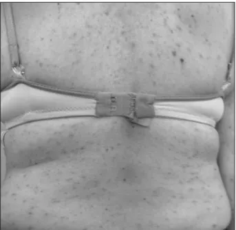
FIGURE 1 Numerous erythematous-violaceous papules on the back.
FIGURE 2 Epidermis slightely rectificated. Dermis with epithelioid and
VIEIRA MAHB ETAL.
720 REV ASSOC MED BRAS 2016; 62(8):718-720
TNF-α inhibitors (ETN, IFX, and ADA), since they present different clearances and binding avidities.4
ETN is a fusion protein that primarily neutralizes soluble TNF-α, with reduced binding activity to membrane TNF-α compared with IFX.6,10 Given that TNF-α is only
partially neutralized by ETN, there may be a redistribution of bioavailable cytokines that travel to low concentration sites such as the lungs. A new stimulus could thus result in the formation of granulomas.6
In the literature review, to date, we have found only nine reported cases of sarcoidosis induced by TNF-α blockers, whose clinical presentation was exclusively cu-taneous. The period between beginning of treatment and the onset of sarcoidosis ranged from 1 to 60 months. Cutaneous manifestations included papular eruptions, nodular eruptions (including facial nodules), scar lesions, and erythema nodosum. The non-cutaneous clinical manifestations reported included constitutional symp-toms, hilar lymphadenopathy, pulmonary interstitial iniltrate, pulmonary nodules and micronodules, pleural effusion, uveitis, and hepatic involvement. Most cases involved organs other than the skin.4 In most cases, there
was regression after discontinuation of anti-TNF-α ther-apy, with an average time of 4, 6 and 8 months for IFX, ADA, and ETN, respectively.11
Thus, ETN may trigger cutaneous and/or systemic sarcoidosis, so that cutaneous manifestation may be the irst or only manifestation of the disease. Physicians should be aware of this potential cutaneous and/or systemic adverse effect for early diagnosis and initiation of the appropriate therapy for each patient.4
R
ESUMODesenvolvimento de sarcoidose exclusivamente cutânea em paciente com artrite reumatoide, durante tratamento com etanercepte
Relata-se caso de uma paciente com artrite reumatoide que, após 2 meses de tratamento com o medicamento
imunobiológico anti-TNF-α etanercepte, apresentou quadro cutâneo compatível com sarcoidose. Notavam-se pápulas violáceas disseminadas e assintomáticas, cuja histopatologia revelou dermatite crônica granulomatosa, com pesquisa de fungos e bacilos álcool-ácido resistentes negativa. Após suspensão do etanercepte, houve regressão do quadro cutâneo. Apesar de paradoxal, têm sido re-latados na literatura casos de sarcoidose cutânea e sistêmi-ca após uso de medisistêmi-cações anti-TNF, sendo raríssimos os casos com acometimento cutâneo exclusivo.
Palavras-chave: sarcoidose, fator de necrose tumoral alfa, doença granulomatosa crônica, artrite reumatoide.
R
EFERENCES1. Santos G, Sousa LE, João AM. Exacerbation of recalcitrant cutaneous sarcoidosis with adalimumab – a paradoxical effect? A case report. An Bras Dermatol. 2013; 88(6 Suppl 1):26-8.
2. Buss G, Cattin V, Spring P, Malinverni R, Gilliet M. Two cases of interferon-alpha-induced sarcoidosis Koebnerized along venous drainage lines: new pathogenic insights and review of the literature of interferon-induced sarcoidosis. Dermatology. 2013; 226(4):289-97.
3. Daïen CI, Monnier A, Claudepierre P, Constantin A, Eschard JP, Houvenagel E, et al.; Club Rhumatismes et Inlammation (CRI). Sarcoid-like granulomatosis in patients treated with tumor necrosis factor blockers: 10 cases. Rheumatology (Oxford). 2009; 48(8):883-6.
4. Lamrock E, Brown P. Development of cutaneous sarcoidosis during treatment with tumour necrosis alpha factor antagonists. Australas J Dermatol. 2012; 53(4):e87-90.
5. Dhaille F, Viseux F, Caudron A, Dadban A, Tribout C, Boumier P, et al. Cutaneous sarcoidosis occurring during anti-TNF-alpha treatment: report of two cases. Dermatology. 2010; 220(3):234-7.
6. Toussirot E, Pertuiset E, Kantelip B, Wendling D. Sarcoidosis occuring during anti-TNF-α treatment for inlammatory rheumatic diseases: report of two cases. Clin Exp Rheumatol. 2008; 26(3):471-5.
7. Wallis RS, Ehlers S. Tumor necrosis factor and granuloma biology: explaining the differential infection risk of etanercept and inliximab. Semin Arthritis Rheum. 2005; 34(Suppl 1):34-8.
8. Gifre L, Ruiz-Esquide V, Xaubet A, Gómez-Puerta JA, Hernández MV, Sanmartí R. Lung sarcoidosis induced by TNF antagonists in rheumatoid arthritis: a case presentation and a literature review. Arch Bronconeumol. 2011; 47(4):208-12.
9. Vigne C, Tebib JG, Pacheco Y, Coury F. Sarcoidosis: an underestimated and potentially severe side effect of anti-TNF-alpha therapy. Joint Bone Spine. 2013; 80(1):104-7.
10. Tong D, Manolios N, Howe G, Spencer D. New onset sarcoid-like granulomatosis developing during anti-TNF therapy: an under-recognised complication. Intern Med J. 2012; 42(1):89-94.