SPONTANEOUSINTRACRANIALEPIDURALHEMATOMADURINGRIVAROXABANTREATMENT
REV ASSOC MED BRAS 2016; 62(8):721-724 721
IMAGE IN MEDICINE
Spontaneous intracranial epidural hematoma during
rivaroxaban treatment
LEONARDO GILMONE RUSCHEL1*, FELIPE MARQUES MONTEIRODO REGO1, JERÔNIMO BUZETTI MILANO2, GUSTAVO SIMIANO JUNG1,
LUIS FERNANDO SILVA JR3, RICARDO RAMINA4
1MD, Resident of the Neurosurgery Residency Program at Instituto de Neurologia de Curitiba (INC), Curitiba, PR, Brazil 2PhD, Neurosurgeon, INC, Curitiba, PR, Brazil
3MD, Neurosurgeon, INC, Curitiba, PR, Brazil
4PhD, Head of the Neurosurgery Department, INC, Curitiba, PR, Brazil
S
UMMARYStudy conducted at Instituto de Neurologia de Curitiba (INC),
Curitiba, PR, Brazil
Article received: 7/23/2015
Accepted for publication: 8/10/2015
*Correspondence:
Address: Rua Jeremias Maciel Perretto, 300 Curitiba, PR – Brazil Postal code: 81210-310 [email protected]
http://dx.doi.org/10.1590/1806-9282.62.08.721
According to our research, this is the irst case described in the literature of spontaneous intracranial epidural hematoma secondary to the use of Xarel-to®. Spontaneous intracranial epidural hematomas are rarely described in
the literature. They are associated with infectious diseases of the skull, co-agulation disorders, vascular malformations of the dura mater and metasta-sis to the skull. Long-term post-marketing monitoring and independent reports will probably detect the full spectrum of hemorrhagic complications of the use of rivaroxaban.
Keywords: cranial epidural hematoma, hemorrhage, cerebrovascular circulation.
I
NTRODUCTIONEpidural hematomas are fairly common in the literature. About 83 to 95% of the cases are associated with skull fractures1 with vascular injury and detachment of the
dura mater from the bone’s inner surface.2 Except for
traumatic causes, this type of hematoma is associated with four etiological categories: pericranial infections (sinusitis, otitis),3,4 dural arteriovenous malformations,3
metastases to the bony skull,5 and coagulation disorders.6
New anticoagulants are emerging, since warfarin requires frequent monitoring, and has multiple drug and food interactions. Rivaroxaban and other new anticoagu-lants were developed with the goal of predictable phar-macokinetics, which eliminates the need for monitoring the International Normalized Ratio (INR). Previous stud-ies have shown a statistically signiicant decrease in in-tracranial hemorrhage with rivaroxaban against warfarin, but the risk still exists.7
We describe the irst case of spontaneous intracra-nial epidural hematoma secondary to rivaroxaban use to be published in a medical journal.
C
ASE REPORTA 39 year-old male, with a previous hospitalization due to atrial ibrillation with rapid ventricular response and med-icated with rivaroxaban (Xarelto®, Janssen Pharmaceuticals
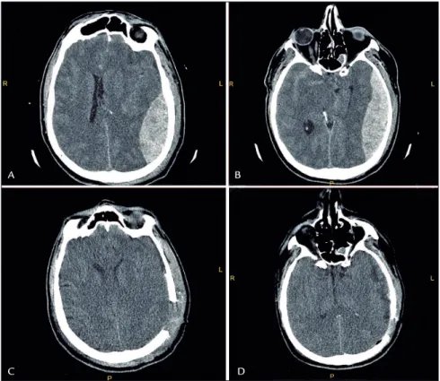
Inc.). Two months later, he was admitted into our emer-gency service. The family reported generalized tonic-clonic seizures 2 hours before. This was the irst event in his life and there was no reported trauma, not even minor. He temporarily recovered his consciousness but a few minutes later became progressively comatose (lucid interval). On arrival, his Glasgow Coma Score (GCS) was 4, the left pu-pil was mydriatic and nonreactive, and the Babinski relex was positive on the right. Emergency orotracheal intubation was performed; blood samples were taken, as well as a brain CT scan, which depicted epidural hematoma on the left with midline deviation (Figure 1 A and B).
Laboratory test results included white blood cells 0.02 x 10⁹/L, hemoglobin 17.29 g/dL, hematocrit 0.50, platelet 401.5 x 10⁹/L, sodium 142 mmol/L, potassium 3.17 mmol/L, creatinine 1.18 µmol/L, urea 2.5 mmol/L, prothrombin time 12.2 seconds, INR 1.07 IU, and partial thromboplas-tin time (PTT) 23.9 seconds.
RUSCHEL LG ETAL.
722 REV ASSOC MED BRAS 2016; 62(8):721-724
level was altered from 17.29 g/dL to 6.0 g/dL. An infusion of 900 mL of concentrated erythrocytes and 1,500 mL of fresh frozen plasma was used. Closure was performed anchoring the dura mater to the bone, and placing an intracranial pressure monitor and a subcutaneous drain. Forwarded to the intensive care unit, the brain CT scan was repeated in the second postoperative period depicting no evidence of hematoma in the surgical ield (Figure 1 C and D). Coma lasted for 4 days. He remained hospitalized in rehabilitation and for treatment of respiratory infection being discharged after 40 days, ambulating, speaking with a slight right-sided hemiparesis grade IV.
D
ISCUSSIONIntracranial epidural hematoma is an uncommon, but serious complication of head injury. While its exact inci-dence is still unknown, it is estimated at 1 to 4% of trau-matic head injury cases, and 5 to 15% of autopsy series. Spontaneous intracranial epidural hematoma is the most uncommon neurological emergency and requires urgent investigation and treatment.8
The dura mater is adhered to the bone with its most external layer along the base level and the sutures. There is a region in which the dura can be easily detached from bone surface called the “Marchand Zone”. This zone
ex-tends antero-posteriorly from the pterional region until 2 to 3 cm of the external occipital protuberance. In the cephalocaudal direction, it runs from the middle line to the base of the skull. Whichever the causal mechanism, this region can be easily detached from the skull.9
Schneider and Hegarty in 1951 wrote the irst report on spontaneous intracranial epidural hematoma. Until this date, only 20 cases were documented in the litera-ture.3-6,10 Hematologic abnormalities are associated with
up to 8% of all intracerebral hemorrhage cases, including anticoagulant-induced coagulopathy.11 Bleeding events
are the most frequent adverse reactions associated with oral anticoagulants. Warfarin anticoagulation increases the risk of intracerebral hemorrhage in two- to ive-fold. The risk is directly related to anticoagulation intensity. Nevertheless, most warfarin-associated intracerebral hem-orrhage cases occur during times when the drug is in the therapeutic range (INR 2.0-3.0).12
The Food and Drug Administration (FDA) approved rivaroxaban (Xarelto®) in 2011, a new orally administered direct factor Xa inhibitor with superior eficacy and a sim-ilar safety proile to warfarin, prescribed for stroke, sys-temic embolism prophylaxis in patients with non valvular atrial ibrillation, treatment, and prevention of pulmonary embolism and deep vein thrombosis. The ROCKET AF
FIGURE 1 A and B. Pre-op CT scan image. C and D. Post-op CT scan image.
A
C
B
SPONTANEOUSINTRACRANIALEPIDURALHEMATOMADURINGRIVAROXABANTREATMENT
REV ASSOC MED BRAS 2016; 62(8):721-724 723
study showed that rivaroxaban had 40% lower risk of in-tracranial and fatal bleeding compared to warfarin.13
A recent study about characteristics of intracerebral hemorrhage during rivaroxaban treatment reported its occurrence in patients at high risk for major bleeding. However, when compared with combined warfarin, the hematomas were smaller, had no expansion, and had favorable functional/survival outcomes.14
In this context, the once-a-day dosage, the need for monitoring the INR, no inferiority to warfarin in treating the atrial ibrillation, and the decreased risk of bleeding in comparison to warfarin have made rivaroxaban an attractive alternative in patients who plan to use this anticoagulant.
Despite the promise to be a medication with better com-pliance and safer for intracranial hemorrhage, we must be cautious and careful with its use. Traditional coagulation studies do not determine the degree of anticoagulation of rivaroxaban15 in patiens with decreased creatinine clearance
as drug exposure is increased, and the risk of bleeding is elevated:16 both thrombotic and bleeding event rates were
higher in patients over the age of 65.17 It is also
contraindi-cated in patients with hepatic disease associated with co-agulopathy,18 and associated with increased risk of
gastro-intestinal bleeding compare to warfarin.19 Moreover,
multiple cerebral micro bleeds were detected more frequent-ly in the rivaroxaban group than in the warfarin group.14
These are important data that we take into account in the selection of the patient for the use of the drug. Even in a young patient, as in the case presented and without other risk factors for bleeding, the use of rivaroxaban can be a triggering factor for the intracranial bleeding. As reported in the described case, the control of bleeding can be extreme-ly dificult. Currentextreme-ly, there are no reports of spontaneous subdural hematomas associated with rivaroxaban. Our patient developed spontaneous subdural hematoma, which is a serious complication, even in the absence of other factors (trauma, renal dysfunction, or uncontrolled blood pressure). Fresh frozen plasma is unlikely to be effective in pa-tients treated with these drugs who are acutely bleeding. Prothrombin complex concentrate can be considered in patients on rivaroxaban regimen.15 Assays are needed to
establish the degree of anticoagulation produced by riva-roxaban, and to evaluate reversal agents that could be ef-fective in the setting of acute bleeding.
C
ONCLUSIONRivaroxaban is a newer anticoagulant that has several advantages compared to traditional anticoagulants. Nev-ertheless, there are several factors to be considered before prescribing it. To our knowledge, this is the irst report
of spontaneous subdural hematoma associated with ri-varoxaban usage. Long-term post-marketing monitoring and independent reports will probably detect the full spectrum of hemorrhagic complications triggered by the use of this medication.
R
ESUMOHematoma epidural intracraniano espontâneo durante tratamento com rivaroxaban
Segundo nossa pesquisa, descrevemos o primeiro caso na literatura de hematoma epidural intracraniano espontâ-neo secundário ao uso de Xarelto®. Hematomas epidurais
intracranianos espontâneos raramente são descritos na literatura, sendo comumente associados a doenças infec-ciosas cranianas, distúrbios de coagulação, malformações vasculares da dura-máter e metástases cranianas. A ela-boração de relatórios de monitoramento em longo prazo de pós-comercialização e relatórios independentes pro-vavelmente irá detectar o espectro completo de compli-cações hemorrágicas do uso desse medicamento. Palavras-chave: hematoma epidural craniano, hemorra-gia, circulação cerebrovascular.
R
EFERENCES1. Cooper RP. Post-traumatic intracranial mass lesion. Head injury. 2. ed. Baltimore: Willians & Wilkins 1987; p. 238-84.
2. Ford LE, McLaurin R. Mechanism of extradural hematomas. J Neurosurg. 1963; 20(9):760-9.
3. Grifiths SJ, Jatavallabhula NS, Mitchell RD. Spontaneous extradural haematoma associated with craniofacial infections: case report and review of the literature. Br J Neurosurg. 2002; 16(2):188-91.
4. Chaiyasate S, Halewyck S, Van Rompaey K, Clement P. Spontaneous extradural hematoma as a presentation of sinusitis: case report and literature review. Int J Pediatr Otorhinolaryngol. 2007; 71(5):827-30.
5. Wani AA, Ramzan AU, Kirmani AR, Bhatt AR, Hamdani N, Zargar J. Intradiploic epidermoid causing spontaneous extradural hematoma: case report. Neurosurgery. 2008; 62(4):E971.
6. Zheng FX, Chao Y. Spontaneous intracranial extradural hematoma: case report and literature review. Neurol India. 2009; 57(3):324-6.
7. Patel MR, Mahaffey KW, Garg J, Pan G, Singer DE, Hacke W, et al.; ROCKET AF Investigators. Rivaroxaban versus warfarin in nonvalvular atrial ibrillation. N Engl J Med. 2011; 365(10):883-91.
8. Bullock MR, Chesnut R, Ghajar J, Gordon D, Hartl R, Newell DW, et al.; Surgical Management of Traumatic Brain Injury Author Group. Surgical management of acute epidural hematomas. Neurosurgery. 2006; 58(3 Suppl):S7-15. 9. Rebollo MA, Soria VR. Neuroanatomía. 2. ed. Buenos Aires: Inter-Médica;
1988. p. 422-35.
10. Shahlaie K, Fox A, Butani L, Boggan JE. Spontaneous epidural hemorrhage in chronic renal failure. A case report and review. Pediatr Nephrol. 2004; 19(10):1168-72.
11. del Zoppo GJ, Mori E. Hematologic causes of intracerebral hemorrhage and their treatment. Neurosurg Clin N Am. 1992; 3(3):637-58.
RUSCHEL LG ETAL.
724 REV ASSOC MED BRAS 2016; 62(8):721-724
13. ROCKET AF Study Investigators. Rivaroxaban-once daily, oral, direct factor Xa inhibition compared with vitamin K antagonism for prevention of stroke and Embolism Trial in Atrial Fibrillation: rationale and design of the ROCKET AF study. Am Heart J. 2010; 159(3):340-7.e1.
14. Hagii J, Tomita H, Metoki N, Saito S, Shiroto H, Hitomi H, et al. Characteristics of intracerebral hemorrhage during Rivaroxaban treatment: comparison with those during warfarin. Stroke. 2014; 45(9):2805-7. 15. Brem E, Koyfman A, Foran M. Review of recently approved alternatives to
anticoagulation with warfarin for emergency clinicians. J Emerg Med. 2013; 45(1):143-9.
16. EINSTEIN Investigators, Bauersachs R, Berkowitz SD, Brenner B, Buller HR, Decousus H, et al. Oral rivaroxaban for symptomatic venous thromboembolism. N Engl J Med. 2010; 363(26):2499-510.
17. Kubitza D, Becka M, Mueck W, Halabi A, Maatouk H, Klause N, et al. Effects of renal impairment on the pharmacokinetics, pharmacodynamics and safety of rivaroxaban, an oral, direct Factor Xa inhibitor. Br J Clin Pharmacol. 2010; 70(5):703-12.
18. Graff J, Harder S. Anticoagulant therapy with the oral direct factor Xa inhibitors rivaroxaban, apixaban and edoxaban and the thrombin inhibitor dabigatran etexilate in patients with hepatic impairment. Clin Pharmacokinet. 2013; 52(4):243-54.