Conduct protocol in emergency: Acute adrenal insufficiency
ADIL BACHIR FARES1*, RÔMULO AUGUSTODOS SANTOS2
1Medical Student, 6th year, Faculdade de Medicina de São José do Rio Preto (Famerp), São José do Rio Preto, SP, Brazil
2Degree in Endocrinology and Metabology from Sociedade Brasileira de Endocrinologia e Metabologia (SBEM). Assistant Physician at the Internal Medicine Service of Hospital de Base. Researcher at Centro Integrado de Pesquisa (CIP), Hospital de Base, São José do Rio Preto. Endocrinology Coordinator of the Specialties Outpatient Clinic (AME), São José do Rio Preto, SP, Brazil
S
UMMARYStudy conducted at Faculdade de Medicina de São José do Rio Preto (Famerp), São José do Rio Preto, SP, Brazil
Article received: 9/3/2015 Accepted for publication: 9/28/2015
*Correspondence: Address: Av. Deputado Emílio Carlos, 586
São Paulo, SP – Brazil Postal code: 02721-100 [email protected]
http://dx.doi.org/10.1590/1806-9282.62.08.728
Introduction: Acute adrenal insuficiency or addisonian crisis is a rare comor-bidity in emergency; however, if not properly diagnosed and treated, it may progress unfavorably.
Objective: To alert all health professionals about the diagnosis and correct treatment of this complication.
Method: We performed an extensive search of the medical literature using spe-ciic search tools, retrieving 20 articles on the topic.
Results: Addisonian crisis is a dificult diagnosis due to the unspeciicity of its signs and symptoms. Nevertheless, it can be suspected in patients who enter the emergency room with complaints of abdominal pain, hypotension unresponsive to volume or vasopressor agents, clouding, and torpor. This situation may be associated with symptoms suggestive of chronic adrenal insuficiency such as hyperpigmentation, salt craving, and association with autoimmune diseases such as vitiligo and Hashimoto’s thyroiditis. Hemodynamically stable patients may undergo more accurate diagnostic methods to conirm or rule out addiso-nian crisis. Delay to perform diagnostic tests should be avoided, in any circum-stances, and unstable patients should be immediately medicated with intravenous glucocorticoid, even before conirmatory tests.
Conclusion: Acute adrenal insuficiency is a severe disease that is dificult to di-agnose. It should be part of the differential diagnosis in cases of hypotensive patient who is unresponsive to vasoactive agents. Therefore, whenever this complication is considered, health professionals should aim speciically at this pathology.
Keywords: acute adrenal insuficiency, Addison’s disease, addisonian crisis, adrenal crisis.
I
NTRODUCTIONThe adrenal are glands divided in two histological areas: cortex and medulla. The adrenal cortex is composed of three layers: zona fasciculata, zona glomerulosa and zona reticularis, producing mainly cortisol, aldosterone, and estrogens and androgens, respectively.1
Aldosterone, a potent mineralocorticoid, is very im-portant for the control of blood pressure and volume, by increasing sodium reabsorption and potassium secretion in the distal tubules of the kidneys. Cortisol is a glucocor-ticoid of great relevance in body stress, acting on the in-crease of blood glucose, on plasma concentrations of proteins and lipids, and as an anti-inlammatory agent. At high concentrations, it has relevant lymphopenic and
eosinopenic effects. Among the many adrenal androgens, the main one is dehydroepiandrosterone (DHEA), which has weaker effect in humans, contributing to the develop-ment of secondary sexual characteristics.1,2,6
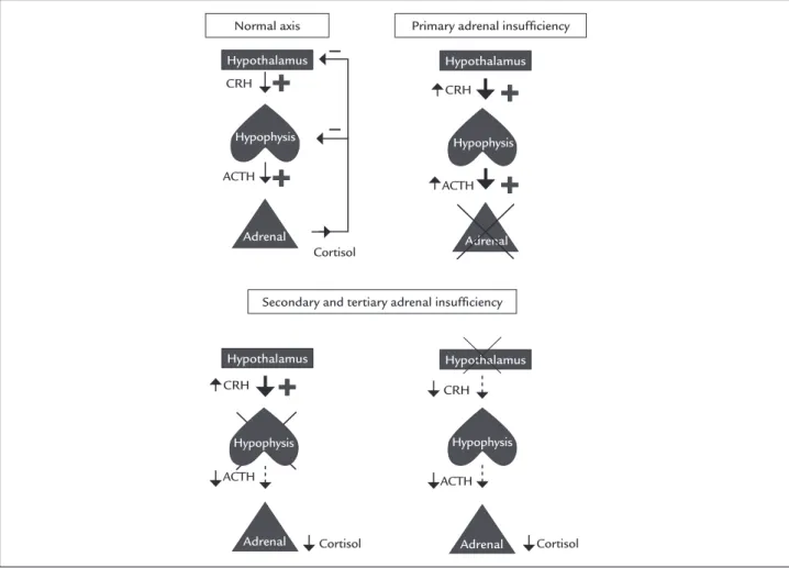
cor-ticotropin (ACTH) and corcor-ticotropin releasing hormone (CRH), respectively.2,17 ACTH is a hormone secreted by corticotropic cells of pars distalis in the hypophysis. It is upregulated by CRH, which is produced in the paraven-tricular nucleus of the hypothalamus (Figure 1).
M
ETHODBetween the months of April and June 2015, we performed extensive literature search and review to ind the most current articles on acute adrenal insuficiency (acute AI). Initially, we selected synonyms for acute AI, including: addisonian crisis and adrenal crisis, which were used as keywords in our search for articles and dissertations from databases such as Uptodate, Pubmed, and Scielo.
This study aimed to ind and review the most recent articles in English or Portuguese on the subject studied, but without being oblivious to very important articles that, though older, bring useful and still prevalent in-formation on the subject. Therefore, all relevant articles that contributed with solid knowledge regarding conduct
and the diagnosis of possible acute AI in humans (some articles refer to this pathology in animals) were includ-ed, while those that did not follow this logic were
ex-cluded from the analysis.175 articles were found
ini-tially, of which we selected 20.For better anatomical and pathophysiological understanding of the subject, important books in the academic circles were also used as a source of knowledge.
A
CUTE ADRENAL INSUFFICIENCY(
ADDISONIAN CRISIS OR ADRENAL CRISIS)
The addisonian crisis is a relatively uncommon endocrine emergency that translates to serious risk of death if not diagnosed and treated in time. It is usually seen in patients with chronic AI exposed to acute stress such as trauma, surgery, infections, and dehydration. Other causes are displayed in Chart 1. 3,7,8,11,16-18
The physician should always be alert to any changes in the vital signs of a patient with known chronic AI. Be-ing aware and knowBe-ing how to correlate certain signs and
FIGURE 1 Schematic comparison of the adrenal physiology and pathophysiology of the adrenal insufficiency.
CRH: corticotropin-releasing hormone; ACTH: adrenocorticotropic hormone.
Normal axis Primary adrenal insuficiency
Secondary and tertiary adrenal insuficiency
Hypothalamus Hypothalamus
Hypothalamus Hypothalamus
CRH
CRH CRH
CRH
ACTH
Cortisol
Cortisol Cortisol
ACTH
Hypophysis
Hypophysis Hypophysis
Adrenal
Adrenal Adrenal
Adrenal Hypophysis
ACTH
symptoms that suggest acute AI is also necessary, as over 25% of patients with Addison’s disease are diagnosed at the time of an adrenal crisis, probably triggered by a stress-ful event (surgery, infection or trauma).4,16,18
Adrenal crisis should be included in the differential diagnosis when a patient enters the emergency room with hypotension that does not respond to the administration of volume or vasopressors, worsening on standing posi-tion, sometimes leading to shock, associated with non-speciic symptoms such as anorexia, nausea, vomiting, pain unexplained abdominal bloating, constipation, fever secondary to an infectious process, apathy, confusion, and drowsiness, which may progress to coma.3,9,13,14,17,18,20
A suggestive sign of past Addison’s disease, which can help make the diagnosis, is hyperpigmentation of mucous membranes and skin by melanin due to the excessive production of alpha-MSH (melanocortin stim-ulating hormone) derived from the same precursor pro-tein of ACTH called POMC (pro-opiomelanocortin). This change in skin mainly affects areas exposed to the sun or pressure areas like ingers, knees, and ankles, in ad-dition to the lips and nipples. Other signs and symptoms that should draw attention to a previous primary AI are salt craving and vitiligo.4,5,20
The clinical picture that allows us to suspect a second-ary AI is: pallor, but with no changes in red blood cells that could suggest anemia, and symptoms of hypogonad-ism (loss of axillary and pubic hair, loss of libido, and amenorrhea in women). In secondary AI, usually there is no aldosterone deiciency because the renin-angiotensin system is intact; however, it may be associated with hy-ponatremia due to reduced water clearance resulting from
hypocortisolism.3,4 The most common manifestations
of acute AI can be seen in Chart 2.
CHART 2 Clinical manifestations of acute adrenal insuficiency.
High fever Abdominal pain Tachycardia or bradycardia Apathy and tiredness Dehydration Nausea and vomiting Hypotension and shock Anorexia
Cyanosis or paleness Malaise Petechiae and ecchymosis
(Waterhouse--Friderichsen syndrome)
Constipation
Faintness, numbness Myalgia and arthralgia
Coma Syncope
L
ABORATORY TESTSSome immediate laboratory changes may be observed with plain doses of Na+1, Ca+2 , K+, blood glucose, and urea. Sodium tends to fall (hyponatremia) and potassium tends to rise (hypercalcemia) due to decreased aldosterone. There is a drop in blood glucose due to serum decrease of an important hormone counter-regulator of insulin, cortisol. Decreased levels of this hormone also lead to an increase in lymphocytes and eosinophils, as a result of decreased immune-modulatory action of hydrocortisone. In some cases, mild hypercalcemia (rarely) and uremia (55% of cases) are observed.3,9,12,18
Diagnosis can generally be achieved by measuring baseline levels of cortisol between 8:00 and 9:00 AM. The diagnosis of AI is conirmed if the value is less than 5 μg/mL (some authors consider < 3 μg/mL). Values greater than 18 μg/mL should prompt physicians to seek alternative diagnoses.18
In case of diagnostic uncertainty, cortrosyn test can be used. It consists of the administration of a semi-syn-thetic ACTH (cosyntropin) aiming to stimulate the cortex of the adrenal glands. Then, a 250 μg dose is intramus-cular (IM) or intravenous (IV) administered and cortisol is measured 30 minutes after infusion (some authors recommend dosing at 0, 30 and 60 minutes). Values equal to or greater than 18 μg/dL indicate a normal response
CHART 1 Triggering factors of adrenal insuficiency.
Infections Surgeries Dehydration Lack of early diagnosis
Improper adjustment of glucocorticoid dose, or abrupt withdrawal in chronic users
Isolated and hasty replacement of thyroid hormones in patients with hypothyroidism and hypocortisolism
Use of drugs that inhibit adrenal steroidogenesis (ketoconazole, aminoglutethimide, metyrapone, mitotane, etc.)
Bilateral adrenal hemorrhage
Meningococcemia (Waterhouse-Friderichsen syndrome) Use of anticoagulants
Trauma or abdominal surgeries
Sepsis due to other bacteria (P. aeruginosa) Coagulopathies
Leukemia Metastasis
Sequel from bilateral venography Primary antiphospholipid syndrome Acute myocardial infarction
excluding primary and secondary AI with adrenal atrophy; however, this does not rule out a mild or recent deicien-cy of ACTH. A cortisol peak < 18 μg/dL conirms the diagnosis of AI, without, however, differing from pri-mary or secondary AI, which does not change in thera-peutic management table adrenal crisis.14
The normal value of plasma ACTH is typically 10 to 60 pg/mL in most of the laboratory methods used. At levels greater than 100 pg/mL, diagnosis of Addison’s syndrome is invariably established. In secondary AI, how-ever, values less than 10 pg/mL or values in the lower limits of normality are observable.3,14,20
Although the cortrosyn test is the most suitable for diagnosis, it should not be chosen for unstable patients, in which case the serum level of cortisol should be dosed when the central venous access is obtained. Values < 5 μg/dL and > 34 μg/dL, respectively, conirm and rule out the
diagno-sis of hypercortisolism.13
It is also important to point out that electrocardio-gram (ECG) changes are commonly observed. These changes are due to hyperkalemia, which occurs with peaked T wave (“tent-shaped”), P wave lattening, and widening of the QRS complex, and in some cases, atrial asystole, intraventricular block, and ventricular asystole.
T
REATMENTAs discussed so far, an adrenal crisis consists of a set of speciic signs and symptoms that require rapid action if there is clinical and hemodynamic instability. In this case, the consensus in all services is to treat hypotension or shock, and to correct hypoglycemia and electrolyte imbalance, even before adopting measures to reach a speciic diagnosis.
Therefore, adrenal crisis should always be in our di-agnostic spectrum when we encounter a patient who comes to the emergency room dehydrated, hypotensive, pale, presenting abdominal pain, hypoglycemia, weakness, anorexia, and hyperpigmentation.
After raising the hypothesis of acute AI, we must irst evaluate the hemodynamic status of the patient. In patients who are hypotensive or inadequately perfused, it is possible to take blood samples, after obtaining venous access, and dose plasma levels of ACTH, cortisol, and electrolytes, im-mediately correcting luid imbalance. Infusion of saline crystalloid solution at 0.9% should be carried out intrave-nously, 20 to 30 mL/kg in 1 hour, observing possible co-morbidities related to volume overload. After correction of hypovolemia, intravenous hydration should be maintained at a slow speed rate of 125 to 250 mL/h for 24 to 48 hours. Importantly, administration of hypotonic solutions is con-traindicated, as this would lead to dilution of natremia.5,17,18
In this patient, glucocorticoid administration should be started:
• Hydrocortisone, 100 mg, IV, every 6 to 8 hours during the irst 24 hours (in unstable patients, the dose of 100 mg every 6 to 8 hours can be maintained until their condition stabilizes). On the second day, when the patient is already stabilized, 50 mg are administe-red every 6h. After this, the dose can be gradually re-duced on the 4th or 5th day until reaching a mainte-nance dose.5,14,17-20 [In primary AI, ludrocortisone can be administered (0.05-0.2 mg at 8:00 AM, orally)].5,18,20
Maintenance treatment in this case is based on the treat-ment of chronic AI:
1. In Brazil, the drug of choice is prednisone at a dose of 5 to 10 mg in the morning.
2. Fludrocortisone, 0.05 to 0.2 mg in the morning. If the dose is administered in excess, the patient may present with hypertension and hypokalemia.5,18,20 3. Dehydroepiandrosterone (DHEA) 25 to 50 mg/day,
orally (especially for women).3,19,20
4. Clinical follow-up: maintaining blood pressure, body weight, electrolytes, and regression of symptoms. 5. Advise the patient to carry an ID card or bracelet. 6. Increase 2 to 3 fold glucocorticoid dose in periods of
stress (infections, sepsis, surgery). The injectable form should be offered in times of crisis.18,19
During the replacement of glucocorticoids in an outpa-tient setting, other forms can be chosen, taking into ac-count the equivalent dosages cited in Chart 3.
Nevertheless, dexamethasone or betamethasone should be avoided because of the increased likelihood of developing exogenous Cushing’s syndrome and espe-cially due to their low mineralocorticoid effect.2
CHART 3 Pharmacokinetics of glucocorticoids.
Drug Equivalent
dose (mg)
Plasmatic half-life (hours)
Hydrocortisone 20 8-12 Cortisone 25 8-12 Prednisone 5 12-36 Prednisolone 4 12-36 Methylprednisolone 4 12-36 Triamcinolone 4 12-36 Betamethasone 0.6 36-72 Dexamethasone 0.75 36-72
P
REVENTION OF A NEW ADDISONIAN CRISISAfter the end of an acute adrenal crisis, the patient must necessarily take some simple steps to prevent a new acute episode:
1. The patient should be informed of the existence of AI (if it has been discovered only during the crisis), the need to maintain a permanent treatment th-roughout life, and the severity of adrenal crises. 2. As mentioned, the patient must carry some form of
identiication of their underlying condition.15,17 3. The maintenance dose used in cases of mild stress
(infections) should be doubled or tripled in periods of more intense stress (surgeries), and may return to the prior dose after 24 to 48 hours.17
4. Follow-up required with a medical specialist. 5. If possible, the patient should take with them a kit with
parenteral hydrocortisone for self-administration.15,17,19
C
ONCLUSIONAs noted in our review, although rare, acute AI is a serious complication and requires rapid and speciic therapeutic action, which will be critical to the patient’s prognosis.
Therefore, it is essential that all health professionals are able to recognize the nonspeciic symptoms that in-clude orthostatic hypotension unresponsive to volume, anorexia, muscle weakness, paleness, nausea, and vomit-ing, and at least suspect a possible addisonian crisis.
There are some classical signs and many signs sug-gestive of AI that may be useful for diagnosis, such as hyperpigmentation or salt craving.
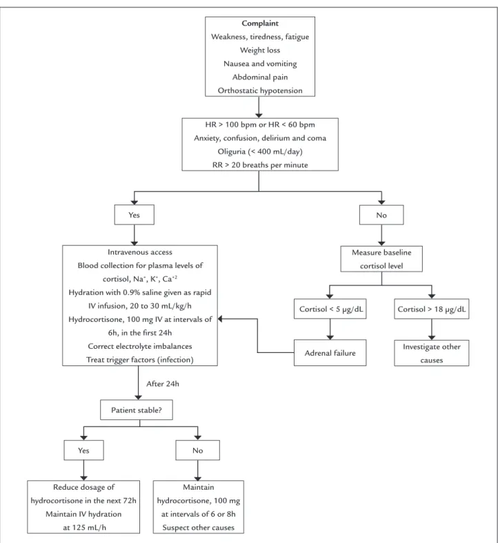
In case of clinical suspicion, it is very important that glucocorticoids (hydrocortisone additional intrinsic miner-alocorticoid effect) are promptly administrated, together with luid replacement to the patient, leaving the inal diag-nosis to be established when the patient’s condition is stable. So to make it easier to understand and increase the effectiveness in routine emergency services, we have cre-ated an algorithm displayed in Figure 2.
R
ESUMOProtocolo de condutas em emergência: insuiciência adre-nal aguda
Introdução: a insuiciência adrenal aguda ou crise addi-soniana é uma comorbidade rara na emergência; porém, se não diagnosticada e tratada de forma correta, pode evoluir de maneira desfavorável.
Objetivo: alertar a todos os proissionais da saúde sobre o diagnóstico e tratamento corretos dessa complicação.
Método: foi realizada uma ampla pesquisa na literatura médica, por meio de ferramentas especíicas, sendo sele-cionados 20 artigos sobre o tema.
Resultados: a crise addisoniana é de difícil diagnóstico pela inespeciicidade de seus sinais e sintomas. No en-tanto, pode ser suspeitada em pacientes que chegam à emergência com queixa de dor abdominal, hipotensão não responsiva a volume ou a agentes vasopressores, ob-nubilação e torpor. Esse quadro pode vir associado a sintomas sugestivos de insuiciência adrenal crônica, como hiperpigmentação e avidez por sal, bem como a doenças autoimunes, como vitiligo e tireoidite de Ha-shimoto. Pacientes estáveis hemodinamicamente podem passar por métodos diagnósticos mais apurados para se conirmar ou descartar a crise addisoniana. Os exames diagnósticos não podem retardar, em hipótese alguma, o tratamento de pacientes instáveis, que deve ser inicia-do imediatamente com glicocorticoide eninicia-dovenoso, in-clusive antes das provas conirmatórias.
Conclusão: a insuiciência adrenal aguda é uma patolo-gia grave e de difícil diagnóstico, que deve fazer parte do diagnóstico diferencial do médico ao atender um pacien-te hipopacien-tenso sem reposta à infusão de drogas vasoativas. Logo, na suspeita dessa complicação, o proissional não deve tardar a agir especiicamente nessa patologia.
FIGURE 2 Acute adrenal insufficiency algorithm.
HR: heart rate; bpm: beats per minute; RR: respiratory rate; IV: intravenous.
Complaint
Weakness, tiredness, fatigue Weight loss Nausea and vomiting
Abdominal pain Orthostatic hypotension
HR > 100 bpm or HR < 60 bpm Anxiety, confusion, delirium and coma
Oliguria (< 400 mL/day) RR > 20 breaths per minute
Intravenous access Blood collection for plasma levels of
cortisol, Na+, K+, Ca+2
Hydration with 0.9% saline given as rapid IV infusion, 20 to 30 mL/kg/h Hydrocortisone, 100 mg IV at intervals of
6h, in the irst 24h Correct electrolyte imbalances Treat trigger factors (infection)
Measure baseline cortisol level Yes
Yes
Cortisol < 5 μg/dL No
No
Cortisol > 18 μg/dL
After 24h
Patient stable?
Maintain hydrocortisone, 100 mg
at intervals of 6 or 8h Suspect other causes Reduce dosage of
hydrocortisone in the next 72h Maintain IV hydration
at 125 mL/h
Adrenal failure Investigate other causes
R
EFERENCES1. Junqueira LCU, Carneiro J. Adrenais. In: Histologia básica. 11. ed. Rio de Janeiro: Guanabara Koogan; 2008. p. 397-403.
2. Guyton AC, Hall JE, editors. Hormônios adrenocorticais. 12. ed. Rio de Janeiro: Elsevier; 2011. p. 944-60.
3. Kater CE, Silva RC, Vilar L. Doenças das adrenais. Insuiciência adrenal – Diagnóstico e tratamento. In: Vilar L, editor. Endocrinologia Clínica. 5. ed. Rio de Janeiro: Guanabara Koogan; 2013. p. 399-414.
4. Alves M, Souto SB, Neves C, Carvalho Braga D, Medina JL. Protocolo de diagnóstico e tratamento de insuiciência supra-renal aguda. Rev Port Endocrinol Diabetes Metab. 2008; 1:23-30.
5. Santos RA. Síndrome adrenal aguda. In: Santos RA, Marino EC, Campos RG. Manual de condutas práticas em endocrinologia e metabologia. São José do Rio Preto: THS Editora; 2013. p. 245-6.
7. Faiçal S, Silva RC, Moritmisu LK. Insuiciência adrenocortical aguda. In: Frisoli Jr AJ, Lopes AC, Amaral JLG, Blum VF, Ferraro JR. Emergências: manual de diagnóstico e tratamento. 2. ed. São Paulo: Savier; 2004. 8. Betterle C, Morlin L. Autoimmune Addison’s disease. Endocr Dev. 2011;
20:161-72.
9. Brandão Neto RA, de Carvalho JF. Autoimmune diagnosis and classiication of Addison’s disease (autoimmune adrenalitis). Autoimmun Rev. 2014; 13(4-5):408-11.
10. Hahner S, Allolio B. Therapeutic management of adrenal insuficiency. Best Pract Res Clin Endocrinol Metab. 2009; 23(2):167-79.
11. Ten S, New M, Maclaren N. Clinical review 130: Addison’s disease 2001. J Clin Endocrinol Metab. 2001; 86(7):2909-22.
12. Lau SY, Yong TY. Rhabdomyolysis in acute primary adrenal insuficiency complicated by severe hyponatraemia. Intern Med. 2012; 51(17):2371-4. 13. Cooper MS, Stewart PM. Corticosteroid insuficiency in acutely ill patients.
N Engl J Med. 2003; 348(8):727-34.
14. Charmandari E, Nicolaides NC, Chrousos GP. Adrenal insuficiency. Lancet. 2014; 383(9935):2152-67.
15. Hahner S, Hemmelmann N, Quinkler M, Beuschlein F, Spinnler C, Allolio B. Timelines in the management of adrenal crisis - targets, limits and reality. Clin Endocrinol (Oxf). 2015; 82(4):497-502.
16. Smans LC, Van der Valk ES, Hermus AR, Zelissen PM. Incidence of adrenal crisis in patients with adrenal insuficiency. Clin Endocrinol (Oxf). 2016; 84(1):17-22. 17. Allolio B. Extensive expertise in endocrinology. Adrenal crisis. Eur J
Endocrinol. 2015; 172(3):R115-24.
18. Tucci V, Sokari T. The clinical manifestations, diagnosis, and treatment of adrenal emergencies. Emerg Med Clin North Am. 2014; 32(2):465-84. 19. Grossman A, Johannsson G, Quinkler M, Zelissen P. Therapy of endocrine
disease: Perspectives on the management of adrenal insuficiency: clinical insights from across Europe. Eur J Endocrinol. 2013; 169(6):R165-75. 20. Michels A, Michels N. Addison disease: early detection and treatment