Alzheimer’s disease and periodontitis – an elusive link
ABHIJIT N. GURAV
Professor Department of Periodontics Tatyasaheb Kore Dental College & Research Centre
S
UMMARYArticle received: 01/16/13
Accepted for publication: 03/27/13
Correspondence:
Department of Periodontics Tatyasaheb Kore Dental College & Research Centre
New Pargaon Kolhapur-416137 Maharashtra State
INDIA Phone +91 230 2477081
Fax +91 230 2477082 [email protected]
http://dx.doi.org/10.1590/1806-9282.60.02.015
Conflict of interest: none
Alzheimer’s disease is the preeminent cause and commonest form of dementia. It is clinically characterized by a progressive descent in the cognitive function, which commences with deterioration in memory. The exact etiology and patho-physiologic mechanism of Alzheimer’s disease is still not fully understood. Ho-wever it is hypothesized that, neuroinlammation plays a critical role in the pa-thogenesis of Alzheimer’s disease. Alzheimer’s disease is marked by salient inlammatory features, characterized by microglial activation and escalation in the levels of pro-inlammatory cytokines in the affected regions. Studies have suggested a probable role of systemic infection conducing to inlammatory sta-tus of the central nervous system. Periodontitis is common oral infection afi-liated with gram negative, anaerobic bacteria, capable of orchestrating localized and systemic infections in the subject. Periodontitis is known to elicit a “low gra-de systemic inlammation” by release of pro-inlammatory cytokines into syste-mic circulation. This review elucidates the possible role of periodontitis in exa-cerbating Alzheimer’s disease. Periodontitis may bear the potential to affect the onset and progression of Alzheimer’s disease. Periodontitis shares the two im-portant features of Alzheimer’s disease namely oxidative damage and inlamma-tion, which are exhibited in the brain pathology of Alzheimer’s disease. Perio-dontitis can be treated and hence it is a modiiable risk factor for Alzheimer’s disease.
Key words: Alzheimer’s disease, periodontitis, cytokines, systemic
inlamma-tion, periodontal pathogen.
I
NTRODUCTIONAlzheimer’s disease (AD) is the most common cause of dementia in the elderly age group and a major health pro-blem in the geriatric subjects worldwide. The incidence of AD rises signiicantly with age, reaching almost 50% in subjects aged 85 years.1 AD is seen as an interaction between genetic and environmental factors. The hallmark of AD is progressive cognitive impairment with impaired judgment and decision making, followed by psycho-be-havioral disturbances and language disability.2 Periodon-titis is the most common oral infection aflicting the hu-man race. Prevalent worldwide, periodontitis is the major cause for tooth loss in adults worldwide. The present re-view elucidates the enigmatic link between AD and pe-riodontitis, showcasing the pathophysiology and possi-ble implications of the association. This review is prepared by screening the PubMed database, utilizing
keywords like “Alzheimer’s disease”, “periodontitis”, “cy-tokines”, “systemic inlammation” and “periodontal pa-thogen.” Systematic reviews, meta-analysis and original articles pertaining to the subject from 1994 to 2012, were referred. Human and animal studies published in english were considered.
P
ATHOGENESIS OFA
LZHEIMER’
S DISEASEhyper-phosphorylated tau protein, followed by consequent loss of neuronal synapses and neuronal degeneration. This
leads to diminution of essential neurotransmitters.3
Enhanced expression of the amyloid precursor pro-tein (APP) gene caused as a result of genetic aberration may be a risk factor for late-onset AD. Apolipoprotein ep-silon4 (APOEε4) allele is genetically linked to majority of the AD cases.4
AβP, the main component of amyloid plaques is
deri-ved from APP by proteolytic cleavage. Studies
corrobora-te the hypothesis that APP and AβP are instrumental in
the pathogenesis of AD.2 The NFTs are constituted of hy-perphosphorylated forms of the microtubule-associated protein tau. The microtubule-associated tau protein is res-ponsible for the stability of microtubules in neurons. Hy-perphosphorylated tau is insoluble with low afinity for microtubules, jeopardizing the microtubule stabilization, thus conducing to synaptic dysfunction and neurodege-neration. Hyperphosphorylation of tau takes place as a re-sult of inlammation, oxidative stress, up-regulation of tau
kinases and down-regulation of phosphatases.5 However
studies have revealed the interplay of other factors apart
from the characteristic AβP plaques and intraneuronal
NFTs for the complete evolution of AD.6 AβP exerts detri-mental effects on the neurovascular endothelial cells, ei-ther by direct action or causing local inlammation.
In-flammation leads to AβP formation in the cerebral
microvasculature and AβP, in turn, stimulates the release of pro-inlammatory mediators.7 Initially, AD was concei-ved as a disorder related to the augmentation in the syn-thesis and decline in the degradation of AβP. Now, impai-red clearance is also stated as a co-factor. This hypothesis has been proposed as the “amyloid cascade hypothesis” of AD, with APP playing a pivotal role.8 In AD, the neuroin-lammation is signiicantly exaggerated. It is hypothesized that neuroinlammation may be a result of pro-inlamma-tory cytokines, reactive oxygen and nitrogen species, ins-trumental in activation of microglia and abetting the for-mation of NFTs.9,10 AβP plaques in AD affected brains are closely afiliated with reactive astrocytes and activated mi-croglial cells, which exhibit exuberant expression of cyto-kines and acute-phase proteins.11 Microglia cells are mo-nonuclear phagocytes present in the brain, committed to thwart any noxious injury within the central nervous sys-tem and achieve brain homeostasis. In health, microglial cells maintain a neuroprotective function by clearing the AβP plaques.12
They also express several neuro trophic factors, such as insulin-like growth factor (IGF)-1, brain-derived
neu-rotrophic factor, transforming growth factor-β and
ner-ve growth factor. In states of peripheral or systemic in-lammation, the molecular and cellular components extend the inlammatory signals to the brain via different path-ways. Under normal conditions, the inlammatory res-ponse is suitably regulated to avoid uncontrolled
inlam-matory damage.13 However, the normal regulatory
mechanisms may become deicient with aging and
gene-tic predisposition.14,15 Thus, a sustained inlammatory
response persists. During these states, the microglial cells in the brain are programmed to switch their phenotypes to produce neurotoxic substances in event of exposure to the systemic inlammatory signals. Thus, instead of confronting with a protective response to these systemic inlammatory signals an exaggerated response is elicited by the diseased microglia, contributing to the pathoge-nesis of AD. The “ired up” microglia changes its mor-phology and releases a number of cell antigens. These are referred to as ‘activated microglia’. Activation of micro-glia results in expression of various pro-inlammatory factors. The uncontrolled release of these factors can in-duce neural damage. The microglial function may be li-kened to a “double-edged sword” being either damaging or protective depending on the context.13,16 Chronic in-lammation and cytokine up-regulation conduces to tau hyperphosphorylation in experimental mice model of AD.17 It has been observed that, chronic lipopolysaccha-ride (LPS)-induced neuroinlammation ensues in the ele-vated levels of intraneuronal AβP in transgenic mice. This may contribute to the deterioration of AD affected brain.18,19
P
ERIODONTITIS–
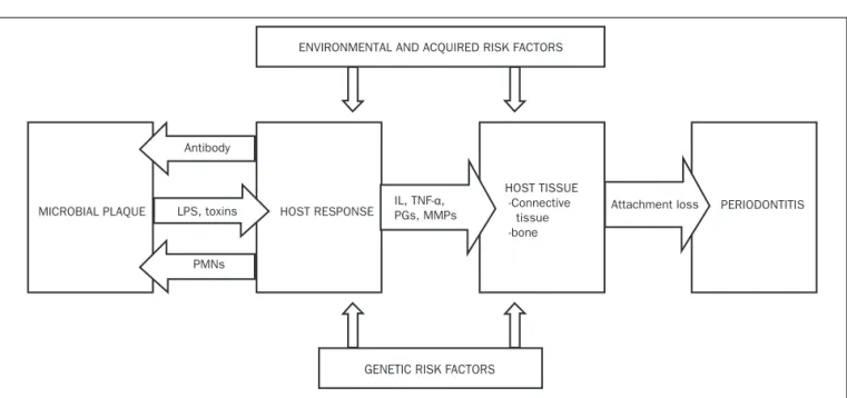
ALOWGRADESYSTEMICDISEASEPeriodontitis is a polymicrobial inlammatory disorder of the tooth investing tissues, resulting from microorga-nisms residing within the dental plaque. Periodontitis is characterized by bleeding and purulent discharge from the gums, progressive deepening of gingival sulcus (re-ferred as pocket formation), oral halitosis, spacing between the teeth and mobility of teeth in advanced stages.20 Den-tal plaque, the principal cause of periodontitis, exists in the form of bioilm. The gram negative and anaerobic species colonize in the periodontal pocket milieu. The predominant periodontal pathogens involved in perio-dontitis are Aggregatibacter actinomycetemcomitans (Aa), Por-phyromonas gingivalis (Pg), Prevotella intermedia (Pi), Fuso-bacterium nucleatum (Fn), Tannerella forsythensis (Tf),
Eikenella corrodens (Ec) and Treponema denticola (Td).21,22 The present model to explain the pathogenesis of
perio-dontitis was proposed by Page and Kornman (igure 1).23
pro-teolytic enzymes, which are implemental in destruction of soft and hard tissues supporting the teeth. The gram negative bacterial LPS also adds to the tissue destruction by amplifying the host response, resulting in the
expres-sion of pro-inlammatory factors like interleukin (IL)-1α
and -1β, IL-6, tumor necrosis factor (TNF) – α, prosta-noids, matrix metalloproteinases (MMP), by the host tis-sue cells.24 Host defense cells like neutrophils, monocy-tes secrete cytokines such as IL-1α and 1β, TNF - α in the diseased periodontal site. These cytokines act as crucial factors in host mediated bone resorption and periodon-tal tissue destruction.25 Host response in periodontal di-sease may act as the diabolical “double-edged sword” lea-ding to self destruction, due to the exaggerated expression of tissue proteolytic enzymes.26 The ulcerated periodon-tal pocket lining furnishes a porperiodon-tal access for the bacte-ria and their noxious products, into the systemic circula-tion. It is reported that the total surface area of the ulcerated periodontal pocket lining in patient with seve-re periodontitis is approximately 15-20 cm2.27 In perio-dontitis, the locally produced cytokines and pro-inlam-matory products are actually streamed through the ulcerated periodontal pocket lining, into systemic circu-lation. This alters the character of periodontitis from a local disease to that of a systemic disorder, capable of
sus-taining “low grade systemic inlammation.”28 This low
grade inlammation is conceived to perturb the general systemic health and exasperate other systemic disorders. Thus periodontitis can be marked as a “low grade
syste-mic disease”. Studies have isolated a number of systesyste-mic inlammatory biomarkers, reiterating a positive associa-tion of periodontitis with systemic inlammaassocia-tion. Surro-gate markers of host response against periodontal infec-tion like cytokines, chemokines, inlammainfec-tion markers, anti-phospholipid antibodies, antibodies to periodontal
pathogens can be demonstrated in serum.27,29
The concept of “periodontal medicine” associates pe-riodontitis as a risk factor with a large number of
syste-mic disorders.30 Periodontal inlammation and
atheros-clerotic cardiovascular diseases (ACD) display a concomitant increase in the levels of inlammatory sys-temic markers like acute phase reactants, interleukins
and TNF-α. Meta-analyses have concluded that subjects
with periodontitis are at a serious risk of ACD.31,32
AD
AND PERIODONTITIS–
A PLAUSIBLE LINKThe exact mechanism involved in the pathogenesis of AD is still unknown. Inlammation is known to play a pivo-tal role in this process. It is proposed that periodontitis can lead to progression of AD by two probable mecha-nisms (igure 2):
• Periodontitis preceding systemic
inlammation/in-fection
• Bacterial and viral inluence
According to the irst mechanism, periodontal patho-gens and the host response elevate the levels of lammatory cytokines. An array of cytokines and
pro-in-ENVIRONMENTAL AND ACQUIRED RISK FACTORS
MICROBIAL PLAQUE
Antibody
LPS, toxins
PMNs
HOST RESPONSE
HOST TISSUE -Connective tissue -bone IL, TNF-α,
PGs, MMPs Attachment loss PERIODONTITIS
GENETIC RISK FACTORS
LPS: Lipopolysaccharide; IL: Interleukin; TNF- α: Tumor necrosis factor- alfa; PGs: Prostaglandins; MMPs: Matrix metalloproteinases
Transforming Growth Factor- β, chemokines (Monocyte Chemotactic Protein, IL-8, Macrophage Migration Inhib-itory Factor, Monokine Induced by γ-Interferon, Fractal-kine) have been implicated as serum and plasma
biomark-ers for pathogenesis of AD.35 TNF- α expression is
up-regulated in AD and it is considered to be the crucial inlammatory cytokine, regulating cellular cascade of events
in neuroinlammatory response. TNF- α exacerbates
gli-osis, demyelination, inlammation, blood-brain-barrier de-terioration and cell death. Thus, TNF- α plays a pivotal role in the neurodegenerative disease process.36,37 Studies on mice models have revealed salutary effects of anti-in-lammatory agents in the amelioration of neuroinlamma-tion and amyloid plaque deposineuroinlamma-tion. A signiicant decrease in the levels of IL-1β and glial ibrillary acidic protein lev-els as well as diminished plaque load was observed in mice
treated with non steroidal anti-inlammatory agent.38,39
lammatory agents are spurted out in systemic circulation adding to the systemic inlammatory burden. Thus, peri-odontitis may produce a state of systemic/peripheral in-lammation. These pro-inlammatory molecules can com-promise the blood brain barrier (BBB) and gain access to the cerebral regions.33 This may result in priming/activa-tion of microglial cells and the adverse repercussions lead-ing to neuronal damage.
The second mechanism may involve invasion of the brain by bacteria and viruses residing in the dental plaque bioilm. This can occur directly through cerebral transport via blood stream or via peripheral nerves.34 There is appre-ciable evidence blaming the inlammatory mechanisms within the central nervous system for the cognitive impair-ment, as that presented in AD. This involves cytokine ar-bitrated interactions between neurons and glial cells. Var-ious cytokines consisting of interleukin family, TNF- α,
FIGURE 2 Possible pathways for the pathogenesis of Alzheimer’s disease.
Tau protein phosphorylation, Increased APP and AβP SYSTEMIC INFECTION/INFLAMMATION
Microglial activation
Neuro-inflammation
Platelet aggregation, atherogenesis
Cerebrovascular atherosclerosis NFTs
APP AβP ALZHEIMER’S DISEASE Apolipoprotein epsilon 4 PERIODONTITIS
PERIODONTOPATHOGENS (bacteria, viruses) LPS
INFLAMMATORY MEDIATORS (cytokines, acute phase reactants)
LPS-lipopolysaccharide; BBB-blood brain barrier; APP-amyloid precursor protein; A βP-amyloid beta protein; NFTs-neuroofibrillary tangles
The Alzheimer’s Disease Anti-inlammatory Prevention Trial (ADAPT), corroborates the hypothesis that the ben-eicial role of anti-inlammatory drugs is evident only in the early, asymptomatic, phases of the disease.40 Inlamma-tion could serve as a connecting link between periodonti-tis and AD. However there are no animal studies, specii-cally addressing the causal relationship of periodontal inlammation to AD, in the literature. Dementia may be designated as a complex disorder associated with an inter-action between genetics and diseases related to systemic inlammation, including diabetes mellitus and environ-mental factors like smoking. Cross-sectional and longitu-dinal studies have revealed dementia in subjects with poor oral health.41-43
Rai et al. observed statistically signiicant difference between patients and controls, concerned to the clinical periodontal parameters like gingival inlammation, den-tal plaque, bleeding on probing and probing pocket depth. Total counts of WBCs, neutrophils, thrombocytes and levels of pro-inlammatory markers like CRP, MMP-8,
MMP-9 and TNF-α were signiicantly elevated in subjects
with dementia and periodontitis in contrast to healthy individuals serving as controls. RBC counts, total IGF-1 and Hb levels were diminished in subjects with demen-tia and periodontitis, in comparison to healthy control subjects. However, these parameters scored signiicantly higher in dementia as compared to periodontitis patients.
An inverse relation was noted in the levels of TNF- α,
MMP-8, MMP-9 and CRP levels compared to free IGF-1 concentrations.45 There is a lack of direct clinical evidence for a causal relationship between periodontitis and AD. However, studies have observed that increased systemic/ peripheral inlammation can be a contributory risk fac-tor for AD.45-48
R
OLE OF PERIODONTAL PATHOGENS INAD
Periodontal pathogens in periodontitis like Aa, Pg, Pi, Tf, Fn are tissue invasive.49,50 This property enables the pa-thogens to escape from the extracellular host defense sys-tem and replicate in the host tissues. The spirochetal spe-cies in the periodontal plaque possess a wide range of virulence factors aiding in confronting with the host de-fense mechanisms and enhancing its ability to invade the periodontal host tissues.51 Spirochete plaques or masses in the brain resemble senile plaques of AD.52
Riviere et al. isolated spirochetal species like Td, Trepo-nema pectinovorum, Treponema vincentii, Treponema amylov-orum, Treponema maltophilum, Treponema medium and Trepo-nema socranskii from the brains of AD subjects, utilizing speciic PCR. Td was isolated in 14 of 16 AD subjects and
4 of 18 non AD subjects. Molecular and immunological techniques endorsed the existence of Palladium species in trigeminal ganglion specimen and cortex of AD affect-ed subjects. AD brain specimens depictaffect-ed more Trepone-ma species in comparison to control groups. It is specu-lated that Treponema from oral cavity must have gained access to the brain cortex via the trigeminal nerve.53 A sig-niicant association has been displayed between spiro-chetes and AD. Spirospiro-chetes were detected in the brain in 93.7% of AD cases and in 33.3% of controls. Borrelia burg-dorferi was isolated 13 times more frequently in AD cas-es than in controls. Considering all the studicas-es, involving spirochetal species detected in the brain, it can be rea-soned that the frequency of spirochetes exceeds more than eight times higher in AD cases (90/131; 68.7%) than in control groups (6/71; 8.41%). The spirochetes may nur-ture a perpetual infective and inlammatory process evok-ing neuronal damage and dysfunction.52,54 Study subjects with elevated levels of Pg antibodies in the serum had sig-niicantly greater odds of cognitive impairment. This ind-ing was constant even after adjustind-ing for the potential socio-demographic and vascular confounders. Neverthe-less, the association of cognitive impairment with anti-bodies to Aa was weak.55 In a longitudinal study, subjects with AD and moderate cognitive impairment (MCI) showed a signiicant increase in the levels of serum anti-bodies to Pi and Fn at the baseline, earlier to the diagno-sis of the neurological deicit. The subjects with AD dem-onstrated signiicantly higher level of antibody to Td and Pg observed at the baseline. The sera analysis of these sub-jects was carried out before the diagnosis of AD or MCI.56
Herpes simplex virus type 1(HSV-1) is a common neu-rotropic virus that infects elderly subjects. HSV-1 is shown to be present in the brain of AD subjects. A causal role was attributed to this virus for triggering AD. Studies
have noticed HSV-1 DNA in the brain of AD subjects.57,58
Polymerase chain reaction (PCR) technique has demon-strated HSV-1 DNA in the brains of large number of el-derly individuals, with or without AD. This was less
con-spicuous in younger subjects, serving as controls.59
Wozniak et al. utilized Enzyme-linked Immunosorbent
Assay (ELISA) to isolate antibodies to HSV-1 in the CSF of AD patients. Although the occurrence of anti-HSV-1 antibodies was signiicantly higher in AD patients than in younger controls, there was a lack of signiicant
differ-ence between the AD and age-matched control groups.60
Letenneur et al. observed an additional presence of IgM
Sub-jects nurturing IgM displayed a signiicantly higher risk of developing AD. The presence of IgM is indicative of active primary infection or reactivation of the viral infec-tion. Thus, the authors concluded a correlation between
reactivation of HSV-1 seropositivity and AD.61 Viruses
could be directly implicated in the pathogenesis of AD. HSV possesses glycoprotein structure that mimics the
amino acid sequence of AβP and tau protein and may
ac-cumulate in the brain like AβP.62 It was noted that HSV--1 is capable of interfering with APP metabolism and may
impart to AD development.63 HSV-1 infection is also a
predisposing factor for AD in subjects with the APOEε4
allele.64
D
ISCUSSIONCognitive disorders like AD have escalated steeply in the population of developed countries. This trend is
obser-ved at an alarming proportion in developing nations.65
The commonly accepted hypothesis for this disorder is the excessive accretion of AβP, including accumulation of abnormally phosphorylated tau proteins in the brain of the affected individuals. Neuroinlammation is a prin-cipal factor for the pathogenesis of AD. Systemic inlam-mation is instrumental in exacerbation of the neuronal degeneration, orchestrated by the activation of primed
microglia.66 Chronic periodontal inlammation, in
peri-odontitis serves as a perennial source for the up-regulat-ed levels of systemic pro-inlammatory factors. Periodon-titis is a polymicrobial infection, characterized by the presence of various bacteria and viruses in the periodon-tal pocket milieu.67,21 These agents, along with their prod-ucts are capable of compromising the BBB and entering the brain. In the brain, these agents can exert the adverse effects either directly or indirectly by affecting the vascu-lar integrity (igure 2). The brain invading spirochetal spe-cies can perpetuate a constant chronic inlammatory pro-cess operated by activation of the innate immune responses, involving the various signaling pathways, re-sulting in neuronal degeneration. Viruses, particularly HSV, can access the brain via blood stream or nerve ibers. Latent viruses may be reactivated by stress and inlamma-tion. Pg is known to express factors responsible for plate-let aggregation and induce atheromatous changes. This may contribute to the pathogenesis of atherosclerotic vas-cular diseases, conducting for cognitive impairment and AD.68-71 Recent literature has referred to the link between genetic polymorphisms, and progression of periodontal disease. Periodontitis susceptible subjects harbor a hy-per-inlammatory phenotype. In response to antigenic stimulus, these subjects exhibit a multifold expression of
pro-inlammatory mediators. Gene polymorphism in-volved in periodontal inlammation could be a conceiv-able nexus between periodontitis and AD.72,73 It is pro-posed that inlammation may act as an elusive link between periodontitis and pathogenesis of AD. Till date there is no evidence of a causal relationship between periodonti-tis and AD. Periodontiperiodonti-tis can intensify the systemic bio-burden and contribute to a “low grade systemic inlam-mation”. It may be accounted as one of the possible risk factors for perpetuating the neurodegenerative process in AD.
C
ONCLUSIONAD involves a complex pathophysiology; the exact etio-pathology of which is unknown. It is proposed that in-lammation could be operating as the central mecha-nism. Both, AD and periodontitis share the same characteristic features of chronicity with inlammation as the common link between them. Presently, studies addressing the role of periodontitis in cognitive func-tion are limited. Systematic, multicentric longitudinal studies, with large sample sizes, should be carried out to scrutinize the association between AD and periodon-titis. Periodontitis may lead to exacerbation and share risk factors with cognitive impairment related disorders. Interventional studies should be carried out to evalua-te a poevalua-tential beneit in periodontitis subjects with mild cognitive disorders. Levels of pro-inlammatory media-tors can be de-escalated with periodontal treatment, ab-breviating systemic inlammation. Presently, it may be stated that periodontitis may pose as a potential risk factor for the development of AD. An insuficient body of evidence based literature fails to endorse a causal re-lationship. Subjects, particularly in the geriatric cate-gory should be strongly motivated and frequent visits for periodontal maintenance should be duly emphasi-zed. The dental professional and neurologist need to co--ordinate consistently regarding the methodical
mana-gement of geriatric patients.
R
ESUMOA doença de Alzheimer e periodontite - um esquivo link
um papel crucial na patogênese da doença de Alzheimer. A doença de Alzheimer é caracterizada por importantes características inlamatórias, assinalada pela ativação mi-croglial e escalada dos níveis de citocinas pró-inlamató-rias nas regiões afetadas. Estudos têm sugerido um provável papel de infecção sistêmica imbuída de estado inlamatório do sistema nervoso central. Periodontite é uma infecção oral comum associada a germes Gram-ne-gativos, anaeróbios, capaz de orquestrar infecções locali-zadas e sistêmicas no paciente. É conhecida por suscitar um “baixo grau de inlamação sistêmica” pela liberação de citocinas pró-inlamatórias na circulação sistêmica. Esta revisão elucida o possível papel da periodontite no agravamento da doença de Alzheimer e pode ter o poten-cial de afetar o início e a progressão da doença de Alzhei-mer. Periodontite partilha as duas importantes caracte-rísticas da doença de Alzheimer: dano oxidativo e inlamação, que estão presentes na patologia do cérebro com doença de Alzheimer. Periodontite pode ser tratada e, portanto, é um fator de risco modiicável para a doen-ça de Alzheimer.
Unitermos: doença de Alzheimer, periodontite, citoci-nas, inlamação sistêmica, patógeno periodontal.
R
EFERENCES1. Ferri CP, Prince M, Brayne C, Brodaty H, Fratiglioni L, Ganguli M, et al.. Global prevalence of dementia: a Delphi consensus study. Lancet. 2005;366:2112-7.
2. Galimberti D, Scarpini E. Progress in Alzheimer’s disease. J Neurol. 2012;259:201-11.
3. Blennow K, de Leon MJ, Zetterberg H. Alzheimer’s disease. Lancet. 2006;368:387-403.
4. Bertram L, Lill CM, Tanzi RE. The genetics of Alzheimer disease: back to the future. Neuron. 2010;68:270-81.
5. Lee YJ, Han SB, Nam SY, Oh KW, Hong JT. Inlammation and Alzheimer’s disease. Arch Pharm Res. 2010;33:1539-56.
6. Holmes C, Boche D, Wilkinson D, Yadegarfar G, Hopkins V, Bayer A, et al.. Long-term effects of Aβ42 immunisation in Alzheimer’s disease: follow-up of a randomised, placebo-controlled phase I trial. Lancet. 2008;372:216-23. 7. Li M, Shang DS, Zhao WD, Tian L, Li B, Fang WG, et al.. Amyloid beta interaction with receptor for advanced glycation end products up-regulates brain endothelial CCR5 expression and promotes T cells crossing the blood-brain barrier. J Immunol. 2009;182:5778-88.
8. Claeysen S, Cochet M, Donneger R, Dumuis A, Bockaert J, Giannoni P. Alzheimer culprits: cellular crossroads and interplay. Cell Signal. 2012;24:1831-40.
9. Eikelenboom P, Veerhuis R, Scheper W, Rozemuller AJ, van Gool WA, Hoozemans JJ. The signiicance of neuroinlammation in understanding Alzheimer’s disease. J Neural Transm. 2006;113:1685-95.
10. Arnaud L, Robakis NK, Figueiredo-Pereira ME. It may take inlammation, phosphorylation and ubiquitination to ’tangle’ in Alzheimer’s disease. Neurodegener Dis. 2006;3:313-9.
11. Licastro F, Candore G, Lio D, Porcellini E, Colonna- Romano G, Franceschi G, et al.. Innate immunity and inflammation in ageing: a key for understanding age related diseases. Immun Ageing. 2005;2:8.
12. Fetler L, Amigorena S. Neuroscience. Brain under surveillance: the microglia patrol. Science. 2005;309:392-3.
13. Weitz TM, Town T. Microglia in Alzheimer’s disease: it’s all about context. Int J Alzheimers Dis. 2012; 2012:314185.
14. Schram MT, Euser SM, de Craen AJ, Witteman JC, Frolich M, Hofman A, et al.. Systemic markers of inlammation and cognitive decline in old age. J Am Geriatr Soc. 2007;55:708-16.
15. Arosio B, Trabattoni D, Galimberti L, Bucciarelli P, Fasano F, Calabresi C, et al.. Interleukin-10 and interleukin-6 gene polymorphisms as risk factors for Alzheimer’s disease. Neurobiol Aging. 2004;25:1009-15.
16. Perry VH, Cunningham C, Holmes C. Systemic infections and inlammation affect chronic neurodegeneration. Nat Rev Immunol. 2007;7:161-7. 17. Kitazawa M, Oddo S, Yamasaki TR, Green KN, LaFerla FM.
Lipopolysaccharide-induced inlammation exacerbates tau pathology by a cyclin-dependent kinase 5-mediated pathway in a transgenic model of Alzheimer’s disease. J Neurosci. 2005;25:8843-53.
18. Lee JW, Lee YK, Yuk DY, Choi DY, Ban SB, Oh KW, et al.. Neuro-inlammation induced by lipopolysaccharide causes cognitive impairment through enhancement of beta-amyloid generation. J Neuroinlammation. 2008;5:37. 19. Tan ZS, Seshadri S. Inlammation in the Alzheimer’s disease cascade: culprit
or innocent bystander? Alzheimers Res Ther. 2010;2:6.
20. Pihlstrom BL, Michalowicz BS, Johnson NW. Periodontal diseases. Lancet. 2005;366:1809-20.
21. Socransky SS, Haffajee AD. Periodontal microbial ecology. Periodontol 2000. 2005;38:135-87.
22. Filoche S, Wong L, Sissons CH. Oral bioilms: emerging concepts in microbial ecology. J Dent Res. 2010;89:8-18.
23. Page RC, Kornman KS. The pathogenesis of human periodontitis: An introduction. Periodontol 2000. 1997;14:9-11.
24. Ren L, Jiang, ZQ, Fu Y, Leung WK, Jin LJ. The interplay of lipopolysaccharide-binding protein and cytokines in periodontal health and disease. J Clin Periodontol. 2009;36:619-26.
25. Graves D. Cytokines that promote periodontal tissue destruction. J Periodontol. 2008;79:1585-91.
26. Preshaw PM, Taylor JJ. How has research into cytokine interactions and their role in driving immune responses impacted our understanding of periodontitis? J Clin Periodontol. 2011;38:60-84.
27. Loos BG. Systemic markers of inlammation in periodontitis. J Periodontol. 2005;76:2106-15.
28. D’Aiuto F, Graziani F, Tetè S, Gabriele M, Tonetti MS. Periodontitis: from local infection to systemic diseases. Int J Immunopathol Pharmacol. 2005;18:1-11.
29. Pussinen PJ, Paju S, Mäntylä P, Sorsa T. Serum microbial- and host-derived markers of periodontal diseases: a review. Curr Med Chem. 2007;14:2402-12.
30. Pizzo G, Guiglia R, Lo Russo L, Campisi G. Dentistry and internal medicine: from the focal infection theory to the periodontal medicine concept. Eur J Intern Med. 2010;21:496-502.
31. Humphrey LL, Fu R, Buckley DI, Freeman M, Helfand M. Periodontal disease and coronary heart disease incidence: a systematic review and meta-analysis. J Gen Intern Med. 2008;23:2079-8.
32. Azarpazhooh A, Tenenbaum HC. Separating fact from iction: use of high-level evidence from research syntheses to identify diseases and disorders associated with periodontal disease. J Can Dent Assoc. 2012;78:c25. 33. Lossinsky AS, Shivers RR. Structural pathways for macromolecular and
cellular transport across the blood brain barrier during inlammatory conditions. Review. Histol Histopathol. 2004;19:535-64.
34. Kamer AR, Dasanayake AP, Craiga RG, Glodzik-Sobanskac L, Bryc M, de Leon MJ. Alzheimer’s disease and peripheral infections: the possible contribution from periodontal infections, model and hypothesis. J Alzheimers Dis. 2008;13:437-49.
35. Lee KS, Chung JH, Choi TK, Suh SY, Oh BH, Hong CH. Peripheral cytokines and chemokines in Alzheimer’s disease. Dement Geriatr Cogn Disord. 2009; 28:281-7.
36. Park KM, Bowers WJ. Tumor necrosis factor-alpha mediated signaling in neuronal homeostasis and dysfunction. Cell Signal. 2010;22:977-83. 37. Montgomery SL, Bowers WJ. Tumor necrosis factor-alpha and the roles it
plays in homeostatic and degenerative processes within the central nervous system. J Neuroimmune Pharmacol. 2012;7:42-59.
38. Yan Q, Zhang J, Liu H, Babu-Khan S, Vassar R, Biere AL, et al.. Anti-inlammatory drug therapy alters β-amyloid processing and deposition in an animal model of Alzheimer’s disease. J Neurosci. 2003;23:7504-9. 39. Heneka MT, Sastre M, Dumitrescu-OzimekL, Hanke A, Dewachter I, Kuiperi
reduces glial inlammation and Aβ1-42 levels in APPV717I transgenic mice. Brain. 2005;128:1442-53.
40. Breitner JC, Baker LD, Montine TJ, Meinert CL, Lyketsos CG, Ashe KH, et al.. ADAPT Research Group. Extended results of the Alzheimer’s disease anti-inlammatory prevention trial. Alzheimers Dement. 2011;7:402-11. 41. Ship JA, Puckett SA. Longitudinal study on oral health in subjects with
Alzheimer’s disease. J Am Geriatr Soc. 1994;42:57-63.
42. Weyant RJ, Pandav RS, Plowman JL, Ganguli M. Medical and cognitive correlates of denture wearing in older community-dwelling adults. J Am Geriatr Soc. 2004;52:596-600.
43. Arrivé E, Letenneur L, Matharan F, Laporte C, Helmer C, Barberger-Gateau P, et al.. Oral health condition of French elderly and risk of dementia: a longitudinal cohort study. Community Dent Oral Epidemiol. 2012;40:230-8.
44. Rai B, Kaur J, Anand SC. Possible relationship between periodontitis and dementia in a North Indian old age population: a pilot study. Gerodontology. 2012;29:e200-5.
45. Engelhart MJ, Geerlings MI, Meijer J, Kiliaan A, Ruitenberg A, Van Swieten JC, et al.. Inlammatory proteins in plasma and the risk of dementia: the Rotterdam study. Arch Neurol. 2004;61:668-72.
46. Tan ZS, Beiser AS, Vasan RS, Roubenoff R, Dinarello CA, Harris TB, et al.. Inlammatory markers and the risk of Alzheimer disease: the Framingham Study. Neurology. 2007;68:1902-8.
47. Bermejo P, Martín-Aragón S, Benedí J, Susín C, Felici E, Gil P, et al.. Differences of peripheral inlammatory markers between mild cognitive impairment and Alzheimer’s disease. Immunol Lett. 2008;117:198-202. 48. Bonotis K, Krikki E, Holeva V, Aggouridaki C, Costa V, Baloyannis S. J
Neuroimmunol. 2008;193:183-7.
49. Dogan S, Gunzer F, Guenay H, Hillmann G, Geurtsen W. Infection of primary human gingival ibroblasts by porphyromonas gingivalis and Prevotella intermedia. Clin Oral Investig. 2000;4:35-41.
50. Gena D, Tribble, Richard J, Lamont. Bacterial invasion of epithelial cells and spreading in periodontal tissue. Periodontol 2000. 2010;52:68-83. 51. Visser MB, Ellen RP. New insights into the emerging role of oral spirochaetes
in periodontal disease. Clin Microbiol Infect. 2011;17:502-12.
52. Miklossy J. Emerging roles of pathogens in Alzheimer disease. Expert Rev Mol Med. 201;13:e30.
53. Riviere GR, Riviere KH, Smith KS. Molecular and immunological evidence of oral Treponema in the human brain and their association with Alzheimer’s disease. Oral Microbiol Immunol. 2002;17:113-8.
54. Miklossy J. Alzheimer’s disease - a neurospirochetosis. Analysis of the evidence following Koch’s and Hill’s criteria. J Neuroinlammation. 2011;8:90. 55. Noble JM, Borrell LN, Papapanou PN, Elkind MSV, Scarmeas Wright CB.
Periodontitis is associated with cognitive impairment among older adults: analysis of NHANES –III. J Neurol Neurosurg Psychiatry. 2009;80:1206-11. 56. Sparks Stein P, Steffen MJ, Smith C, Jicha G, Ebersole JL, Abner E, et al..
Serum antibodies to periodontal pathogens are risk factor for Alzheimer’s disease. Alzheimers Dement. 2012;8:196-203.
57. Itzhaki RF, Wozniak MA. Herpes simplex virus type 1 in Alzheimer’s disease: the enemy within. J Alzheimer’s Dis. 2008;13:393-405.
58. Itzhaki R, Lin WR, Shang D, Wilcock GK, Faragher B, Jamieson GA. Herpes simplex virus type 1 in brain and risk of Alzheimer’s disease. Lancet. 1997;349: 41-4.
59. Jamieson GA, Maitland NJ, Wilcock GK, Craske J, Itzhaki RF. Latent herpes simplex virus type 1 in normal and Alzheimer’s disease brains. J Med Virol. 1991;33:224-7.
60. Wozniak MA, Shipley SJ, Combrinck M, Wilcock GK, Itzhaki RF. Productive herpes simplex virus in brain of elderly normal subjects and Alzheimer’s disease patients. J Med Virol. 2005;75:300-6.
61. Letenneur L, Pérès K, Fleury H, Garrigue I, Barberger-Gateau P, Helmer C, et al.. Seropositivity to herpes simplex virus antibodies and risk of Alzheimer’s disease: a population-based cohort study. PLoS One. 2008;3:e3637. 62. Cribbs DH, Azizeh BY, Cotman CW, LaFerla FM. Fibril formation and
neurotoxicity by a herpes simplex virus glycoprotein B fragment with homology to the Alzheimer’s A beta peptide. Biochemistry. 2000;39:5988– 94.
63. Shipley SJ, Parkin ET, Itzhaki RF, Dobson CB. Herpes simplex virus interferes with amyloid precursor protein processing. BMC Microbiol. 2005;5:48-55. 64. Lin WR, Shang D, Wilcock GK, Itzhaki RF. Alzheimer’s disease, herpes simplex virus type 1, cold sores and apolipoprotein E4. Biochem Soc Trans. 1995;23:594S.
65. Nitrini R, Bottino CM, Albala C, Custodio Capuñay NS, Ketzoian C, Llibre Rodriguez JJ, et al.. Prevalence of dementia in Latin America: a collaborative study of population-based cohorts. Int Psychogeriatr. 2009;21:622-30. 66. Holmes C, Cunningham C, Zotova E, Woolford J, Dean C, Kerr S, et al..
Systemic inlammation and disease progression in Alzheimer disease. Neurology. 2009;73:768-74.
67. Parra B, Slots J. Detection of human viruses in periodontal pockets using polymerase chain reaction. Oral Microbiol Immunol. 1996;11:289-93. 68. Iwai T. Periodontal bacteremia and various vascular diseases. J Periodontal
Res. 2009;44:689-94.
69. Nakayama K. Porphyromonas gingivalis cell-induced hemagglutination and platelet aggregation. Periodontol. 2000 2010; 54:45-52.
70. Watts A, Crimmins EM, Gatz M. Inlammation as a potential mediator for the association between periodontal disease and Alzheimer’s disease. Neuropsychiatr Dis Treat. 2008;4:865-76.
71. Haan MN, Shemanski L, Jagust WJ, Manolio TA, Kuller L. The role of APOE
ε4 in modulating effects of other risk factors for cognitive decline in elderly persons. JAMA. 1999;282:40-6.
72. Rainero I, Bo M, Ferrero M, Valfrè W, Vaula G, Pinessi L. Association between the interleukin-1alpha gene and Alzheimer’s disease: a meta-analysis. Neurobiol Aging. 2004;25:1293-8.