Dynamic compared to rigid fixation in lumbar spine: a systematic review
RICARDO VIEIRA BOTELHO1, RAFAEL BASTIANELLO JUNIOR2, LUCIANA DINIGIANINIDE ALBUQUERQUE2, WANDERLEY MARQUES BERNARDO3
1Post Graduation in Health Sciences at IAMSPE-São Paulo, SP, Brazil 2Medicine School of the City University of São Paulo (UNICID), São Paulo, SP, Brazil 3University of São Paulo (USP) and Lusíadas University, São Paulo, SP, Brazil
S
UMMARYStudy conducted at the post graduation
program in health sciences- IAMSPE-São Paulo, SP, Brazil
Conflict of interest: none
Article received: 10/04/12
Accepted for publication: 07/01/13
Correspondence:
Address: Rua Tuim 585, (122A), Moema
São Paulo, SP - Brazil ZIP Code: 04514-102
http://dx.doi.org/10.1590/1806-9282.60.02.013
Conflict of interest: none
Objective: The objective of this review is to reveal the quality of published data and the effect size of DPFs compared to rigid fixation in lumbar spine. Sum-mary of background data: since 2002, several dynamic pedicle fixation (DPF) systems have been developed with the aim to stabilize the spine without the un-desirable effects of rigid lumbar spine fixation. Nearly ten years later, there are several studies on these dynamic systems.
Methods: A systematic review was done in MEDLINE/PubMED, Embase, Co-chrane Central Register of Randomized Trials and Google Scholar to assess the quality of published literature and the available studied outcomes in randomi-zed controlled trials of DPF.
Results: Only three papers described randomized trials studying DPF. One of them focused on protection of adjacent level disease provided by DPF.
Conclusion: It was not possible to reveal any evidence for benefits using DPF compared to rigid fixation in surgery for lumbar spine.
Key words: lumbar vertebrae, intervertebral disc degeneration, surgical fixation devices, internal fixators, bone screws, spinal fusion.
I
NTRODUCTIONFusion using spine fixation is a surgical intervention used for the treatment of degenerating lumbar spine.1,2 For pa-tients with vertebral instability and those with painful conditions secondary to disc degeneration, the elimina-tion of the vertebral joint movement could produce cli-nical improvement from the stabilization of the spine.3 Although there can be favorable clinical outcomes for cer-tain conditions, complications of fusion with rigid fixa-tion have appeared, such as intervertebral disc degenera-tion adjacent to fusion.4,5
To reduce or eliminate the undesirable effects of rigid fixation, dynamic systems have been developed based on flexible screws and rods (DPF). These implements could protect the spine from mechanical overload imposed by the rigid fixation of the spine while retaining spine move-ment and preventing adjacent disc degenerative disease.6,7,8
A decade after the first description of the dynamic fi-xation devices, some authors claim that indications for these devices are not yet defined, although there are po-tential diseases that could benefit from their use.9,10 The objective of this study is to evaluate the clinical
outco-mes of these procedures compared with rigid pedicle fi-xation systems.
M
ETHODSA systematic literature review was performed for publica-tions between January 2002 (the first DPF publication date 6) and August 18, 2011, in the following electronic primary literature databases: MEDLINE/PubMed, the Cochrane Central Register of Controlled Trials (CEN-TRAL) and EMBASE. Google Scholar and electronic da-tabase search engines were used, and publications in all languages were included.
The research protocol was based on the method of questions structured by types of participants (study popu-lation), comparing the two types of intervention (rigid, and semirigid or dynamic fixation) and selecting the type of study as a prospective randomized trial (appendix 1).11
Study population
sur-gery. Patients with infection, trauma, surgical treatment of osteoporosis, and comorbidities that interfere with walking were excluded.
Intervention
Patients who underwent DPF.
Control
Patients who underwent rigid fixation.
Outcome
To reveal the effect on all outcomes studied, all reported outcomes were compared.
Type of selected studies
Randomized trials.
Study selection
Searches were performed by three independent reviewers. The papers were initially assessed by title, and those selected were subsequently re-analyzed by abstract. The references of selected articles were studied to find other relevant citations. Disagreements were solved among the authors through dis-cussion. The search strategy is detailed in appendix 1.
Quality of papers selected for study
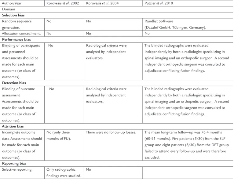
Because of the importance and prevalence of degenerative disease of the lumbar spine, only randomized controlled trials (RCTs) were included due to the more compelling evidence they provide. Assessments of methodological qua-lity were made according to Cochrane collaboration’s tool for assessing risk of bias (table 1).12
R
ESULTSThe electronic search revealed a total of 321 articles. Of these, 302 studies were categorized as human clinical trials and published as journal articles or conference pa-pers. Selection by title identified 27 articles that were sub-sequently reviewed by abstract, resulting in four articles. However, one of the papers (Putzier et al.) was not rando-mized and was excluded. 13
Description of included studies and assessment of methodolo-gical quality
Evaluation of inal three papers:
- Korovesis et al.14 The study population was composed of patients with lumbar canal stenosis. Patients were treated with rigid fixation and dynamic system (Twinflex). All pa-tients were fused with autograft. Thirty papa-tients were exa-mined 3 months after surgery. There was no description
of the randomization method and no blinded allocation attempt. This study did not detail clinical outcomes. Lum-bar lordosis, sacral inclination, intervertebral angle, and intervertebral disc index were evaluated. Comparisons were not made between the randomized groups, only within groups regarding pre- and postoperative outcomes.
- Korovesis et al.15 As in the first work by the same author, the study population was composed of patients with lum-bar canal stenosis. Patients were treated with dynamic, se-mirigid and rigid fixation. All patients underwent bone fu-sion with bone grafts from the iliac crest. Forty-five patients were randomized, but the randomization method was not described and there was no attempt to conceal allocation. Radiological evaluation was done by independent evalua-tors. Clinical outcomes assessed were the visual analogic scale VAS for back pain and lower extremity. The quality of life questionnaire F-36 was also used. Degeneration and complication rate adjacent to the surgery site and failures of the systems were evaluated. Apparently there were no differences in VAS between the groups, and authors did not provide statistical analyses between groups. There were no differences in the final clinical or radiological evalua-tions. The complication rate evaluation revealed that it was too low to allow for comparisons. There was no degenera-tion in any of the adjacent groups. The analysis of provi-ded radiological findings suggest that results were compa-red within each group, pre- and postoperatively, but not among the three randomized groups.
- Putzier et al.16 The authors studied patients with low back pain, presence of Modic signs, lumbar spine facet arthritis and spondylolisthesis. Sixty patients were stu-died comparing dynamic and rigid fixation. Randomiza-tion was performed by a specific software program, and there was no attempt to conceal allocation. The sample size of patients to be studied had been calculated previou-sly and there was an adequate description of follow-up loss of patients. The final follow-up period was 6 years. Clinical outcomes were the rate of satisfaction with treat-ment, the ODI, and the VAS. Radiological evaluation con-sisted of changes in vertebral plateau sign (Modic sign) and the Fujiwara facet arthritis degeneration index, in-tervertebral disc height and presence of dynamic instabi-lity in plain radiographs. There were no differences between groups in any of these outcomes.
Risk of bias in included studies (Table 1)
some kind of methodological weakness. Due to the na-ture of surgical intervention, blinding of treatment exe-cution and evaluation was not possible (double-blind). Only one of the three papers described the randomiza-tion method. In one study, there was no assessment of clinical outcome (Korovesis et al.) and in the others, only the VAS was used. Outcomes were determined by the staff that performed the procedures. Korovesis et al., followed the outcomes for just three months, and Korovesis et al., followed the outcomes for two years. The final pooled analysis consisted of only 135 patients, and the low me-thodological quality of the papers introduces considera-ble potential for bias.
D
ISCUSSIONLumbar spine Degenerative Disc Disease (DDD) causes social, economic and financial burden to patients affec-ted. It is estimated that annual expenditures for treat-ment cost are higher than those for all types of cancer
combined. Current studies suggest that DDD has a strong genetic component and progressive evolution.17 Due to complications of spinal fusion, semi-rigid and dynamic systems have been developed to protect the spine against adjacent disk degeneration disease and to eventually ame-liorate the mechanical effects superimposed on normal spine by rigid spine fusion.9 It has been suggested that these systems can return the degenerated intervertebral disc to a normal condition and eventually slow or rever-se the degenerative process.7 Here, randomized studies that compared the two types of systems were evaluated. Due to the economic and financial importance of these systems, only randomized trials were analyzed. Studies comparing effect sizes based on nonrandomized trials il-lustrate that lower quality manuscripts tend to increase effect size. Even among RCTs, distortion of the actual ef-fects can occur when randomization and allocation con-cealment are not performed properly, or when there is a high degree of follow-up loss.18,19,20
TABLE 1. The Cochrane Collaboration’s tool for assessing risk of bias
Author/Year Korovesis et al. 2002 Korovesis et al. 2004 Putzier et al. 2010
Domain
Selection bias
Random sequence generation.
No No Randlist Software
(DataInf GmbH, Tübingen, Germany).
Allocation concealment. No No No
Performance bias
Blinding of participants and personnel Assessments should be made for each main outcome (or class of outcomes).
No Radiological criteria were
analyzed by independent evaluators.
The blinded radiographs were evaluated independently by both a radiologist specializing in spinal imaging and an orthopedic surgeon. A second independent orthopedic surgeon was consulted to adjudicate conlicting fusion indings.
Detection bias
Blinding of outcome assessment
Assessments should be made for each main outcome (or class of outcomes).
No Radiological criteria were analyzed by independent evaluators.
The blinded radiographs were evaluated independently by both a radiologist specializing in spinal imaging and an orthopedic surgeon. A second independent orthopedic surgeon was consulted to adjudicate conlicting fusion indings.
Attrition bias
Incomplete outcome data Assessments should be made for each main outcome (or class of outcomes).
No (only three months of FU).
There were no follow-up losses. The mean long-term follow-up was 76.4 months (60-91 months). Five patients (5/30) from the SLF group and eight patients (8/30) from the DFT group failed to attend every follow-up and were therefore excluded.
Reporting bias
Selective reporting. Only radiographic indings were studied.
mais tarde, existe uma série de estudos sobre os sistemas dinâmicos. Revelar a qualidade dos dados publicados e o tamanho do efeito da FDP em comparação com a fixação rígida na coluna lombar.
Métodos: Uma revisão sistemática foi feita utilizando MEDLINE/ PubMed, Embase, a CENTRAL Cochrane de ensaios randomizados e Google Scholar para avaliar a qualidade da literatura publicada e os desfechos estuda-dos disponíveis em ensaios clínicos randomizaestuda-dos.
Resultados: Apenas três estudos randomizados foram en-contrados. Um deles estudou a proteção de degeneração no nível adjacente à fixação rígida proporcionada pela FDP.
Conclusão: Não foi possível revelar qualquer evidência de benefícios da FPD, em comparação com a fixação rí-gida em cirurgia para a coluna lombar.
Unitermos: doenças da coluna vertebral, dor lombar, ins-trumentação, fusão vertebral, degeneração do disco in-tervertebral.
R
EFERENCES1. Gelalis ID, Arnaoutoglou C, Christoforou G, Lykissas MG, Batsilas I, Xenakis T. Prospective analysis of surgical outcomes in Patients Undergoing decompressive laminectomy and posterior instrumentation for degenerative lumbar spinal stenosis. Acta Orthop Traumatol Turc. 2010;44:235-40. 2. Bono CM, Lee CK. Critical analysis of trends in fusion for degenerative disc
disease over the past 20 years: Influence of technique on fusion rate and clinical outcome. Spine. 2004;29:455-63.
3. Nockels RP. Dynamic stabilization in the surgical management of painful lumbar spinal disorders. Spine. 2005;30(16 Suppl):S68-S72.
4. Anandjiwala J, Seo JY, Ha KY, Oh IS, Shin DC. Adjacent segment degeneration after lumbar spinal posterolateral fusion with instrumentation in elderly patients. Arch Orthop Trauma Surg. 2002;122:39-43.
5. Cheh G, Bridwell KH, Lenke LG, Buchowski JM, Daubs MD, Kim Y, Baldus C. Adjacent segment disease following lumbar / thoracolumbar fusion with pedicle screw instrumentation: a minimum 5-year follow-up. Spine. 2007;32:2253-7.
6. Rajaratnam SS, Mueller M, Shepperd JAN, Mulholland RC. Dynesis stabilization of the lumbo-sacral presentation spine. Poster Britspine. In: The Second Combined Meeting of the BSS BASS BCSS SBPR, Birmingham; 2002.
7. Schmoelz W, Huber JF, Nydegger T, Dipl-Ing, Claes L, Wilke HJ. Dynamic stabilization of the lumbar spine and its effects on adjacent segments: an in vitro experiment. J Spinal Disord Tech. 2003;16:418-23.
8. Khoueir P, Kim KA, Wang MY.Classification of posterior dynamic stabilization devices. Neurosurg Focus. 2007;22(1):E3.
9. Mulholland RC, Sengupta DK. Rationale, principles and experimental evaluation of the concept of soft stabilization. Eur Spine J. 2002;11(Suppl 2):S198-205.
10. Schnake KJ, Schaeren S, Jeanneret B. Dynamic stabilization in addition to decompression for lumbar spinal stenosis with degenerative spondylolisthesis. Spine. 2006;31:442-9.
11. Van Tulder M, Furlan A, Bombardier C, Bouter L. Updated method guidelines for systematic reviews in the Cochrane Collaboration Back Review Group. Spine. 2003;28:1290-9.
12. Higgins JPT, Green S Altman DG. Assessing risk of bias in studies included. Cochrane Handbook for Systematic Reviews of Interventions: Chapter 8. Cochrane Book Series.
13. Putzier M, Schneider SV, Funk JF, Tohtz SW, Perka C. The surgical treatment of the lumbar disc prolapse: nucleotomy with additional transpedicular dynamic stabilization versus nucleotomy alone. Spine. 2005;30(5):E109-14.
The first description of dynamic screw fixation was published in 2002. Although there is extensive literature related to biomechanical and case series studies, 3,21,22,23,24,25 only three randomized trials compared dynamic pedicle fixation with rigid fixation.14,15,16 It is remarkable that the three RCTs used bone fusion in both rigid and dynamic fixation. Two of the three studies failed to make direct comparisons between rigid and dynamic techniques. Ra-ther, they compared the outcomes within each group, between pre- and postoperative status.14,15
Among all lumbar degenerative diseases, lumbar spi-nal stenoses were the focus in two studies,14,15 and the third study assessed a group of patients with heteroge-neous degenerative disc disease, characterized by Modic type changes in the vertebral plateau, facet arthritis and spondylolisthesis.16 In this study, which had the longest follow-up time and best design, the incidence of degene-ration adjacent to the fusion site was directly and objec-tively appraised. Six years after surgery, there was no evi-dence of protection against adjacent degeneration with dynamic systems.
C
ONCLUSIONImplications for practice
Although dynamic systems have been designed to protect the spine from undesirable rigid fixation effects, ten years of clinical use have failed to show superiority of these sys-tems in clinical or radiographic outcomes. There were no differences in the rate of degeneration in adjacent dyna-mical systems compared with rigid systems.
Implications for research
There are few randomized trials comparing both systems, and the methodological quality still needs to be improved. Specific areas to address include the categories of indepen-dent randomization, blinding and follow-up duration.
Disclosure of funding received for this work: Rafael Bastianello Junior, Luciana DiniGianini de Albuquerque received grants from the Institutional program of scien-tific initiation scholarships (PIBIC/CNPq).
R
ESUMOFixação pedicular dinâmica comparada com a fixação rí-gida na coluna lombar: uma revisão sistemática
14. Korovessis P Papazisis Z Lambiris E. The role of rigid vs..dynamic instrumentation for stabilization of the degenerative lumbosacral spine. Stud Health Technol Inform. 2002;91:457-61.
15. Korovessis P, Papazisis Z, Koureas G, Lambiris E. Rigid, semirigid versus dynamic instrumentation for degenerative lumbar spinal stenosis: a correlative radiological and clinical analysis of short-term results. Spine. 2004;29:735-42.
16. Putzier M, Hoff E, Tohtz S, Gross C, Perka C, Strube P. Dynamic stabilization adjacent to single-level fusion: part II. In the clinical benefit is asymptomatic, initially degenerated adjacent segments after 6 years follow-up. Eur Spine J. 2010;19:2181-9.
17. Chan D, Song Y, Sham P, Cheung KM. Genetics of disc degeneration. Eur Spine J. 2006;15(Suppl 3):S317-25.
18. Chalmers TC, Celano P, Sacks HS, Smith H Jr.Bias in treatment assignment in controlled clinical trials.N Engl J Med.1983;309:1358-61.
19. Moher D, Pham B, Jones A, Cook DJ, Jadad AR, Moher M, et al. Does quality of reports of randomized trials Affect Estimates of Intervention Efficacy Reported in meta-analysis? Lancet. 1998;325:609-13.
20. Schülke KF, Chalmers I, Hayes RJ, Altman DG. Empirical evidence of bias. JAMA.1995;273:408-12.
21. Scharer N., Dubois G, Braunsweiler R. Static and dynamic test of a dynamic biomechanical neutralization system for the spine. In: White AA, Panjabi MM, editors. Clinical biomechanics of the spine. 2nd ed. Philadelphia: JB Lippincott; 1990. p.19-24.
22. Stoll TM, Dubois G, Schwrzenbach O. The dynamic neutralization system for the spine: a multicenter study of a novel non-fusion system. Eur Spine J. 2002;11(Suppl 2):S170-S8.
23. Bono CM, Kadaba M, Vaccaro AR. Posterior fixation pedicle-based dynamic stabilization devices for the Treatment of degenerative diseases of the lumbar spine. J Spinal Disord Tech. 2009;22:376-83.
24. Markwald TM, Wenger M. Dynamic stabilization of lumbar motion segments by use of Graf ’s ligaments: results with an average follow-up of 7.4 years in 39 highly selected, consecutive patients. Neurochem Acta (Wien). 2003;145:209-14.