1
Echocardiographic Assessm ent of Cardiac
Resynchronizat ion Therapy
Carlos Eduardo Suaide Silva, Ant onio Carlos Pereira Barret t o
OM NI-CCNI M edicina Diagnóst ica/Diagnóst icos da América and Inst it ut o do Coração do Hospit al das
Clínicas - FM USP - São Paulo, SP - Brazil
Mailing address: Carlos Eduardo Suaide Silva - OMNI-CCNI - Rua Cubatão, 7 2 6 - 0 4 0 1 3 -0 0 2 - São Paulo, SP - Brazil
E-m a il: csua ide@ ca rdiol.br Received for publication: 1 0 /2 0 /0 4 Accepted for publication: 3 /4 /0 5 English version by Stela Maris Costalonga
Heart failure is one of the major causes of mortality, morbidity, and hospitalization in patients older than 6 0 years, accounting for 1 to 2 % of the total expenditure in the health sector in the United
States (approximately 2 0 billion dollars per year!)1 ,2. Despite the
great advances in therapy, the morbidity and mortality rates still
remain high3. Cardiac resynchronization therapy (CRT) was
intro-duced in the beginning of the 1 9 9 0 ’s and rapidly developed until its
approval in 2 0 0 1 by the FDA (Food and Drugs Administration)4.
In the American Heart Association guidelines, CRT has been
considered to be IIA evidence level5. Those guidelines were based
on 2 large trials: the MUSTIC6 and the MIRACLE7. In both, the
inclusion criteria were similar: a) significant heart failure despite appropriate therapy; b) low ejection fraction; and c) broad QRS with left bundle-branch block pattern (duration> 1 2 0 ms). Both studies have confirmed that CRT significantly improves symptoms, tolerance to exercise, and quality of life. Nevertheless, 2 0 % to
3 0 % of the patients do not improve with CRT8, emphasizing the
need for new criteria of patients’ selection.
Recent studies have reported that mechanical dyssynchrony
is not always related to electrical dyssynchrony9 ,1 0, and that the
presence of ventricular dyssynchrony is the best predictor of a good response to resynchronization therapy. In reality, some patients with broad QRS may not have mechanical dyssynchrony while
others with narrow QRS may1 1 -1 3.
Although QRS duration is a good prognostic marker for mortality in patients with heart failure and is present in more than 8 0 % of
the individuals in the 2 months preceding death1 4 ,1 5, studies
com-paring QRS alterations with the clinical outcome of patients seem
to show little or no relation to prognosis1 6.
These data suggest that electrocardiography may not be the best complementary diagnostic method for selecting candidates to CRT. Other imaging techniques, particularly the new methods for assessing ventricular function on echocardiography, seem to be better for selecting patients who will best respond to resynchroni-zation therapy.
Both the presence of broad QRS and the signs of interventricular
dyssynchrony are predictors of hospitalization and severe cardiac
events in patients with heart failure1 7 ,1 8.
Ventricular dyssynchrony
The mechanism of dyssynchrony includes regional delays of both ventricular contraction and relaxation. The right ventricle contracts during left ventricular telediastole leading to a septal bulging towards the left ventricle. In addition, the delay in the activation of the papillary m uscles causes, or worsens, m itral
incompetence1 9. All such factors contribute to a reduction in the
ejection fraction and a worsening of the clinical symptoms. Dyssynchrony may be inter- or intraventricular, and echocar-diography may evaluate both types through several techniques.
One way of evaluating interventricular dyssynchrony is by mea-suring the time between ventricular ejections. Conventional Doppler measures the time interval between the R wave in the electro-cardiogram and the beginning of the systolic waves of pulmonary and aortic ejection (fig. 1 ). A delay greater than 6 0 ms between those measurements indicates interventricular dyssynchrony. The limitation of such method is that those measurements are not
taken simultaneously2 0 -2 2.
Another way to assess delay is through the M mode, in which the time between septal contraction and posterior wall contraction is measured (fig. 2 ). This time is considered normal when under
1 3 0 ms2 3. In this case, the limitation is the fact that only the
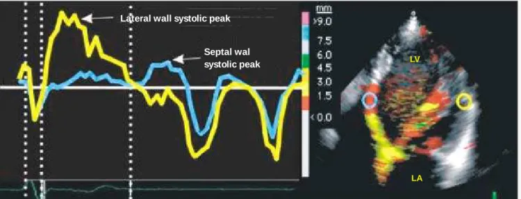
middle regions of the septal and posterior walls are assessed. Intraventricular dyssynchrony is considered one of the most important aspects of electromechanical delay and may be assessed through several echocardiographic techniques. The electrom e-chanical delay has been defined as the time between the beginning of the QRS complex and the peak systolic wave of tissue Doppler (echocardiographic technique that measures the velocity of myo-cardial motion) in the corresponding myomyo-cardial segment (fig. 3 ). Guidelines about this subject still lack, and several authors have published studies proposing several indices for diagnosing inter-and intraventricular dyssynchrony based on tissue Doppler
techni-ques. Yu et al1 1 studied 8 8 healthy individuals, 6 7 patients with
2
of intraventricular dyssynchrony: the maximum time difference between 2 distinct segments greater than 1 0 0 ms; and a 3 3 stan-dard deviation in the measurement of the 1 2 segments (named dyssynchrony index). The authors showed lack of dyssynchrony in the healthy group, 7 3 % of dyssynchrony in patients with CHF and broad QRS, and, the most interesting, 5 1 % of dyssynchrony in those with CHF and narrow QRS.
In other studies, dyssynchrony was assessed by using the measu-re of electromechanical delay in the basal measu-regions of the septal and lateral walls. A difference greater than 6 0 ms (septolateral delay)
was used as a substantial indicator of dyssynchrony2 4 -2 5 (fig. 4 ).
The intraventricular electromechanical delay may also be
de-monstrated by using the Tissue Trackingechocardiographic
tech-nique, which represents the integral of the velocity acquired by the tissue Doppler. This technique depicts in color the myocardial motion, from the basis to the ventricular apex. When there is no motion, there is no color. If the systolic and diastolic phases of the cardiac cycle are separately selected, the regions that are contracting may be identified in the respective phases. The regions that are colored in the diastolic phase represent a late myocardial contraction (or postsystolic contraction) and may be easily evi-denced by using that technique (fig. 5 ).
The postsystolic motion may be passive or active, in which case it should be called postsystolic contraction or shortening. However, care should be taken with patients with ischemic cardiomyopathy, because that phenomenon is not only a signal of dyssynchrony, but also a marker of ischemia or viability, or both, in akinetic or severely hypokinetic segments. In such cases, it should not be used as a
useful criterion to assess a positive response to CRT2 6.
Automatic detection of dyssynchrony
Tissue synchronization imaging (TSI) is a new echocardiogra-phic technique that encodes with colors the time intervals between the beginning of the QRS complex and the peak-systolic of tissue Doppler in each myocardial point. It allows the real time visuali-zation of dyssynchronous segments by superimposing the images of such time data over those of 2 -dimensional echocardiography. This analysis may be performed in all myocardial segments, but it should be carefully used in the apical segments. The principle is very simple: when the interval up to the peak-systolic (time to peak) is normal, the myocardium is represented in green; when the interval is between 1 5 0 and 3 0 0 ms, it is represented in
yellow; and when it is greater than 3 0 0 ms, in red2 7. The result
is very interesting and is shown in figure 6 .
Selection of patients for implantation of
biventricular pacemaker
Few published studies have used echocardiography as a tool for choosing patients for biventricular pacemaker implantation. One of
them2 8 has assessed 4 2 individuals with a pacemaker in the RV, 2 6
of whom had normal ejection fraction, and 1 6 had impairment of the systolic function and clinical findings of CHF. An electromechanical delay greater than 5 0 ms identified patients with significant dyssyn-chrony. No correlation was observed between dyssynchrony and broad QRS in patients with CHF. If only the electrocardiographic criterion had been used for biventricular pacemaker implantation, 4 4 % of the patients with dyssynchrony would have been excluded, showing the importance of performing echocardiography.
More studies in this area are required to definitively validate that
Fig. 1 - Mechanical interventricular delay measured in a patient with heart failure and left bundle-branch block. A) Time between QRS and aortic ejection of 2 3 0 ms. B) time between QRS and pulmonary ejection of 6 6 ms. The difference between those 2 measurements is the mechanical delay between the ventricles (1 6 4 ms, in this case).
AORTIC FLOW PULMONARY FLOW
RV
LV
3
methodology. This review, however, suggests that the criteria forbiventricular pacemaker implantation should be revised and tissue Doppler echocardiography should be used for selecting such patients.
Where should the electrode be implanted?
Tissue Doppler may help in determining the ideal site for im-planting the electrode within the coronary sinus.
The best site for im plantation, ie, the site where the best ventricular response is obtained (gain in ejection fraction), has already been documented as that with the greatest electromechanical delay. This site is in the lateral wall in 3 5 % of the cases, in the anterior and posterior walls in 2 6 % and 2 3 %, respectively, and rarely in the
inferior and septal walls (1 6 %)2 9.
The objective of the resynchronization therapy is to activate the site with the greatest electromechanical delay; therefore, the
Fig. 3 - Quantification of the intraventricular electromechanical delay on tissue Doppler. In this example, the blue and yellow curves represent the velocities of myocardial motion in the middle regions of the septal and lateral walls, respectively (LA - left atrium; LV - left ventricle).
Lateral wall systolic peak
Septal wal
systolic peak LV
LA
Fig. 4 - Measure of the electromechanical delay in the basal regions of the septal (green curve - 5 0 0 ms) and lateral (yellow curve - 2 3 5 ms) walls. A difference greater than 6 0 ms is highly indicative of intraventricular dyssynchrony. The red dotted lines delimitate the ventricular ejection period.
Lateral wall systolic peak
4
determination of such site is directly linked to a successful
proce-dure. And, in fact, Ansalone et al2 9 have demonstrated that the best
result was obtained in patients whose electrodes were implanted according to the site with the greatest electromechanical delay, determined on echocardiography.
Echocardiographic markers indicating
improvement w ith CRT
The most evident signs of improvement after biventricular pa-cemaker implantation are the increase in ejection fraction, the decrease in the degree of mitral incompetence, and the regression in ventricular remodeling. However, echocardiography may provide the following less evident markers: a) an improvement in the atrioven-tricular activation assessed through the increase in the time velocity integral of the aortic flow and extension of the time of diastolic filling (assessed through mitral flow) in 1 0 to 2 0 %; b) reversion of the interventricular electromechanical delay assessed on tissue
Dop-pler. The MIRACLE study reported a 1 9 % reduction in that marker3 0.
Yu et al3 1 reported a complete regression in the great interventricular
delay between the free wall of the RV and the lateral wall of the LV
after CRT; and c) intraventricular resynchronization. Several studies have confirmed the normalization of the intraventricular delay by using the different techniques above cited (M mode, pulsed Doppler
of the outflow tracts, and tissue Doppler)3 2 -3 5.
Conclusion
Cardiac resynchronization therapy has been defined by studies involving a reduced number of patients as an excellent therapeutic option for patients with heart failure. However, approxim ately 3 0 % of the cases have not responded adequately when the current electrocardiographic criteria of indication were used. The evidence above cited confirms that echocardiography seems to be the ide-al complementary method to identify those patients who will be effectively beneficiated by CRT. However, great studies definitively validating the method are yet to be carried out.
In addition to the quantitative diagnosis of resynchronization, the new echocardiographic techniques based on tissue Doppler (Tissue Tracking, Strain Rate, and TSI) may aid in choosing the best site for pacemaker electrode implantation with important benefits for the procedure and in following up the patients in a noninvasive form.
Fig. 5 - Tissue Trackingtechnique showing the area of late contraction (arrow) of the interventricular septum.
Systole
Diastole
LV
LV
LA
LA
Fig. 6 - Left: Apical 4 - chamber view showing electromechanical delay in the left ventricular lateral wall (red). Right: The same patient after resynchronization showing an improvement in the color pattern (green), representing the reestablishment of contractile synchrony. LA - left atrium; PM - pacemaker electrode; RV - right ventricle; LV - left ventricle.
LV
LV
RV
P M
5
1. American Heart Association. New Medicine Reports 1 9 9 7 ; 1 9 9 9 Heart and Stroke Statistical Update. Dallas, TX: Am erican Heart Association.
2. Cleland JGF. The heart failure epidem ic: exactly how big is it? Eur Heart J 2 0 0 1 ; 2 2 : 6 2 3 -6 .
3. Zannad F, Briancon S, Juillière Y et al. Incidence, clinical and etiologic features, and outcomes of advanced chronic heart failure: the EPICAL study. J Am Coll Car-diol 1 9 9 9 ; 3 3 : 7 3 4 -4 2 .
4. Abraham WT, Hayes DL. Cardiac resynchronization therapy for heart failure. Cir-culation 2 0 0 3 ; 1 0 8 : 2 5 9 6 -6 0 3 .
5. Gregoratos G, Abrams J, Epstein AE et al. ACC/AHA/NASPE 2 0 0 2 guideline update for im pla nta tion of ca rdia c pa cem a kers a nd a ntia rrhythm ia devices. J Am Coll Cardiol 2 0 0 2 ; 4 0 : 1 7 0 3 -1 9 .
6. Cazeau S, Leclercq C, Lavergne T et al. Effects of multisite biventricular pacing in pa tients with hea rt fa ilure a nd intra ventricula r conduction dela y. N Engl J Med 2 0 0 1 ; 3 4 4 : 8 7 3 -8 0 .
7. Abra ha m WT, Fisher WG, Sm ith AL et a l. Ca rdia c resynchroniza tion in chronic heart failure. N Engl J Med 2 0 0 2 ; 3 4 6 : 1 8 4 5 -5 3 .
8. Leclercq C, Kass DA. Retiming the failing heart: principles and current clinical sta-tus of cardiac resynchronization. J Am Coll Cardiol 2 0 0 2 ; 3 9 : 1 9 4 -2 0 1 . 9. Leclercq C, Faris O, Tunin R et al. Systolic improvement and mechanical
resynchro-nization does not require electrical synchrony in the dilated failing heart with left bundle-branch block. Circulation 2 0 0 2 ; 1 0 6 : 1 7 6 0 -3 .
10. Kass DA. Predicting cardiac resynchronization response by QRS duration: the long and short of it. J Am Coll Cardiol 2 0 0 3 ; 4 2 : 2 1 2 5 -7 .
11. Yu CM, Lin H, Zhang Q. High prevalence of left ventricular systolic and diastolic a synchrony in pa tients with congestive hea rt fa ilure a nd norm a l QRS dura tion. Heart 2 0 0 3 ; 8 9 : 5 4 -6 0 .
12. Bleeker GB, Schalij MJ, Molhoek SG et al. Relationship between QRS duration and left ventricular dyssynchrony in patients with end-stage heart failure. J Cardiovasc Electrophysiol 2 0 0 4 ; 1 5 : 5 4 4 -9 .
13. Achilli A, Sassara M, Ficili S et al. Long-term effectiveness of cardiac resynchroni-zation therapy in patients with refractory heart failure and “narrow” QRS. J Am Coll Cardiol 2 0 0 3 ; 4 2 : 2 1 1 7 -2 4 .
14. Iuliano S, Fisher SG, Karasik PE, Fletcher RD, Singh SN. QRS duration and m or-tality in patients with congestive heart failure. Am Heart J 2 0 0 2 ; 1 4 3 : 1 0 8 5 -9 1 . 15. Wilensky RL, Yudelman P, Cohen AI et al. Serial electrocardiographic changes in
idiopa-thic dilated cardiomyopathy confirmed at necropsy. Am J Cardiol 1 9 8 8 ; 6 2 : 2 7 6 -8 3 . 16. Sun JP, Chinchoy E, Donal E et al. Evaluation of ventricular synchrony using novel Doppler echocardiographic indices in patients with heart failure receiving cardiac resynchronization therapy. J Am Soc Echocardiogr 2 0 0 4 ;1 7 : 8 4 5 -5 0 . 17. Venkateshwar K, Gottipaty VK, Krelis SP, Lu F, Spencer EP, Shusterm an V, Weiss
R, Brode S, White A, Anderson KP, White BG, Feldman AM, For the VEST investi-gators: The resting electrocardiogram provides a sensitive and inexpensive marker of prognosis in patients with chronic congestive heart failure. J Am Coll Cardiol 1 9 9 9 , 3 3 : 1 4 5 (Abstract).
1 8 . Bode-Schnurbus L, Bocker D, Block M et a l. QRS dura tion: a sim ple m a rker for predicting ca rdia c m orta lity in ICD pa tients with hea rt fa ilure. Hea rt2 0 0 3 ; 8 9 : 1 1 5 7 -6 2 .
19. Bax JJ, Ansalone G, Breithardt OA et al. Echocardiographic evaluation of cardiac resynchronization therapy: ready for routine clinical use? A critical appraisal. J Am Coll Cardiol2 0 0 4 ; 4 4 : 1 -9 .
20. Kanzaki H, Jacques D, Sade LE, Severyn DA, Schwartzman D, Gorcsan J 3 rd. Re-gional correlation by color-coded tissue Doppler to quantify improvements in me-chanical left ventricular synchrony after biventricular pacing therapy. Am J Cardiol 2 0 0 3 ; 9 2 : 7 5 2 -7 5 .
21. Porciani MC, Puglisi A, Colella A et al., on behalf of the InSync Italian Registry In-vestigators. Echocardiographic evaluation of the effect of biventricular pacing: the InSync Italian Registry. Eur Heart J Suppl 2 0 0 0 ; 2 (Suppl J): J2 3 -3 0 .
22. Rouleau F, Merheb M, Geffroy S et al. Echocardiographic assessment of the inter-ventricular delay of activation and correlation to the QRS width in dilated cardio-myopathy. Pacing Clin Electrophysiol 2 0 0 1 ; 2 4 : 1 5 0 0 -6 .
23. Pitzalis MV, Iacoviello M, Romito R et al. Cardiac resynchronization therapy tailo-red by echocardiographic evaluation of ventricular asynchrony. J Am Coll Cardiol 2 0 0 2 ; 4 0 : 1 6 1 5 -2 2 .
24. Bax JJ, Molhoek SG, van Erven L et al. Usefulness of m yocardial tissue Doppler echocardiography to evaluate left ventricular dyssynchrony before and after bi-ventricular pacing in patients with idiopathic dilated cardiomyopathy. Am J Cardiol 2 0 0 3 ; 9 1 : 9 4 -7 .
25. Bax JJ, Marwick TH, Molhoek SG et al. Left ventricular dyssynchrony predicts be-nefit of cardiac resynchronization therapy in patients with end-stage heart failure before pacemaker implantation. Am J Cardiol 2 0 0 3 ; 9 2 : 1 2 3 8 -4 0 .
26. Mele D, Pasanisi G, Heimdal A et al. Improved recognition of dysfunctioning myo-cardial segments by longitudinal strain rate versus velocity in patients with myocar-dial infarction. J Am Soc Echocardiogr2 0 0 4 : 3 1 3 -2 1 .
27. Penicka M, Bartunek J, De Bruyne B et al. Improvement of left ventricular function after cardiac resynchronization therapy is predicted by tissue Doppler imaging echo-cardiography. Circulation2 0 0 4 ; 1 0 9 : 9 7 8 -8 3 .
28. Bordachar P, Garrigue S, Lafitte S et al. Interventricular and intra-left ventricular electromechanical delays in right ventricular paced patients with heart failure: im-plications for upgrading to biventricular stimulation. Heart2 0 0 3 ; 8 9 : 1 4 0 1 -0 5 . 29. Ansalone G, Giannantoni P, Ricci R, Tram baiolo P, Fedele F, Santini M. Doppler
myocardial imaging to evaluate the effectiveness of pacing sites in patients receiving biventricular pacing. J Am Coll Cardiol2 0 0 2 ; 3 9 : 4 8 9 -4 9 9 .
30. Kindermann M, Frolhig G, Doerr T, Schieffer H. Optimizing the AV delay in DDD pa-cemaker patients with high degree AV block: mitral valve Doppler versus impedance cardiography. Pacing Clin Electrophysiol 1 9 9 7 ; 2 0 : 2 4 5 3 -6 2 .
3 1 . Yu CM, Cha u E, Sa nderson JE et a l. Tissue Doppler echoca rdiogra phic evidence of reverse rem odeling a nd im proved synch ronicity by sim ulta neously dela ying regiona l contra ction a fter biventricula r p a cing th era p y in h ea rt fa ilure. Circu-lation 2 0 0 2 ; 1 0 5 : 4 3 8 -4 5 .
32. Breithardt OA, Stellbrink C, Kramer AP et al. Echocardiographic quantification of left ventricula r a synchrony predicts a n a cute hem odyna m ic benefit of ca rdia c resynchronization therapy. J Am Coll Cardiol 2 0 0 2 ; 4 0 : 5 3 6 -4 5 .
33. Kawaguchi M, Murabayashi T, Fetics BJ et al. Quantitation of basal dyssynchrony and acute resynchronization from left or biventricular pacing by novel echo-con-trast variability imaging. J Am Coll Cardiol 2 0 0 2 ; 3 9 : 2 0 5 2 -8 .
34. Yu CM, Fung WH, Lin H et al. Predictors of left ventricular reverse remodeling after cardiac resynchronization therapy for heart failure secondary to idiopathic dilated or ischemic cardiomyopathy. Am J Cardiol 2 0 0 3 ; 9 1 : 6 8 4 -8 .
35. Breithardt OA, Stellbrink C, Herbots L et al. Cardiac resynchronization therapy can reverse abnormal myocardial strain distribution in patients with heart failure and left bundle branch block. J Am Coll Cardiol 2 0 0 3 ; 4 2 : 4 8 6 -9 4 .