C
a s eR
e p o Rt3 3 3 Arq Bras Oftalmol. 2016;79(5):333-5 http://dx.doi.org/10.5935/0004-2749.20160095
Adenoid cystic carcinoma of the lacrimal sac: case report
Carcinoma adenóide cístico do saco lacrimal: relato de caso
Antonio RAmos1, CARmen Del Pozo1, AnA ChinChuRRetA1, FeRnAnDo GARCíA1, meRCeDes loRenzo1, sAtuRnino GismeRo1
Submitted for publication: July 24, 2015 Accepted for publication: November 9, 2015
1 Department of Ophthalmology, Costa del Sol Hospital, Marbella, Malaga, Spain.
Funding: No specific financial support was available for this study.
Disclosure of potential conflicts of interest: None of the authors have any potential conflict of interest to disclose.
Corresponding author: Antonio Ramos. Department of Ophthalmology. Hospital Costa del Sol. Autovia A-7 Km 187 - Marbella, Málaga 29603 - Spain - E-mail: [email protected]
ABSTRACT
Lacrimal sac tumors are rare with a clinical presentation that typically includes obstruction of the lacrimal drainage system and epiphora as the most frequent symptom. Cribriform adenoid cystic carcinoma (ACC) is the most common malig-nant epithelial tumor of the lacrimal gland and minor salivary glands; however, its occurrence in the lacrimal drainage apparatus is extremely rare. Given the rarity of ACC, definitive diagnosis is almost invariably late conferring a poor prognosis. Herein we report the case of a 41-year-old woman with primary ACC of the lacrimal sac and describe the ophthalmological examination, diagnosis, and multidisciplinary treatment of this rare type of tumor.
Keywords: Carcinoma; Adenoid cystic/diagnosis; Lacrimal apparatus diseases; Eye neoplasms
RESUMO
Tumores do saco lacrimal são raros. A apresentação clínica muitas vezes mostra uma obstrução no sistema de drenagem lacrimal sendo a epífora o sinal mais fre quen te. Car-cinoma adenóide cístico cribriforme (ACC) é o tumor epitelial maligno mais comum da glândula lacrimal e glândulas salivares menores, mas a sua ocorrência no aparelho de drenagem lacrimal é extremamente rara. Infelizmente, devido a raridade destes tumores, o diagnóstico preciso é quase sempre atrasado, o que por sua vez leva a um pior prognóstico. Nós relatamos o caso de uma mulher de 41 anos de idade, com ACC primário do saco lacrimal e analisamos o exame oftalmológico, diagnóstico e tratamento multidisciplinar deste tipo de tumor.
Descritores: Carcinoma adenoide cístico/diagnóstico; Carcinoma adenoide cís tico/ terapia; Doenças do aparato lacrimal; Neoplasias oculares
INTRODUCTION
Lacrimal sac tumors are rare with a typically non-speciic presen-tation. As the most common signs of lacrimal sac tumors are epiphora or presence of a mass, the process can be incorrectly diagnosed as
dacryostenosis or chronic dacryocystitis (DC)(1-4). Consequently,
dei-nitive diagnosis is delayed leading to worsened prognosis(5).
Adenoid cystic carcinoma (ACC) is an extremely rare form of malignant epithelial neoplasia of the lacrimal sac. However, it is the most common type of malignant epithelial tumor of the lacrimal and
minor salivary glands(6,7). To date, 11 cases of ACC of the lacrimal sac
have been reported in literature (Table 1).
We report the case of a 41-year-old woman with a right lacrimal sac ACC with a follow-up duration of 24 months post surgery.
CASE REPORT
A 41-year-old Caucasian woman presented to our department in August 2012 with a 2-year history of lacrimation and pain afecting the medial canthus of the right eye. The lacrimal drainage system was permeable; however, relux of mucopurulent material was observed. Dacryocystography (DCG) demonstrated a patent pathway with for-ced passage of contrast from the lacrimal sac. Tumoral pathology was not suspected, and the patient was diagnosed with dacryocystitis and started on topical and systemic antibiotic treatment accordingly.
Ad e n o i d c y s t i c c A rc i n o m A o f t h e l A c r i m A l s A c: c A s e r e p o rt
3 3 4 Arq Bras Oftalmol. 2016;79(5):333-5
Table 1. Summary of previously reported clinical cases
Age Sex Side Presentation Treatment Recurrence Follow-up interval Author
62 F Right 2 years epiphora. Lacrimal sac mass (without data speciication)
Enbloc excision of the tumor with exenteration of the orbit, radical maxillectomy, and ethmoidectomy.
No 14 months L. Miller et al.(8)
57 F Left Epiphora and swelling of the left lower lid (without data
speciication)
Orbital exenteration. Further treatment: CT.
Lung metastases after 2 years.
48 months after which she was lost
to follow-up.
C. Kincaid et al.(3)
38 F Not speciied Not speciied Surgery No 60 months A. Stefanyszyn et al.(1)
51 F Not speciied Not speciied Not speciied No 60 months A. Stefanyszyn et al.(1)
64 F Not speciied Not speciied Not speciied No 60 months A. Stefanyszyn et al.(1)
41 M Left 1 month epiphora and lacrimal sac mass
Surgery and RT No 12 months R. Parnel l et al.(9)
72 M Right 1 year epiphora and lacrimal sac mass
Orbital exenteration and RT for pulmonary metastases
Lung metastases after 16 years
192 months DN. Parmar et al.(2)
62 F Not speciied Epiphora, epistaxis and nasal obstruction (without data
speciication)
Surgery and RT Not speciied Not speciied O. Choussy et al.(10)
74 M Right Tearing associated with a palpable mass in the internal
canthus (without data speciication)
Exclusive RT No Death at 30 months A. Montalban et al.(5)
48 F Not speciied Mass Globe sparring surgery, RT, and CT Metastatic disease Death at 48 months T. El-Sawy et al.(11)
70 M Not speciied Mass Globe sparring surgery, RT, and CT No 6 months T. El-Sawy et al.(11)
F= female; M= male; RT= radiotherapy; CT= chemotherapy.
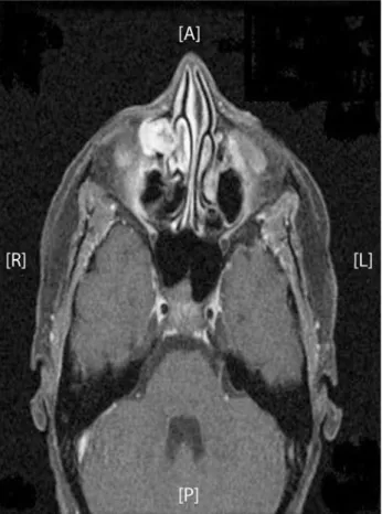
Figure 1. Solid-cystic lesion occupying the right lacrimal sac region consistent with a neoplastic process.
DISCUSSION
Epiphora is the most common clinical sign of lacrimal sac tumors(1-4).
These tumors may also present as masses in the canthal area. Sangui-neous discharge (spontaSangui-neous or on lacrimal sac irrigation), epistaxis, pain, and skin ulceration are all highly suspicious of malignancy.
Pathologically, lacrimal sac tumors can be divided into epithelial tumors, mesenchymal tumors, lymphomatous lesions, melanoma, and neuronal tumors. Inlammatory lesions are not true neoplasms
but appear as masses in the lacrimal sac with similar symptoms(1).
The most common malignant tumors afecting the lacrimal sac are of the epithelial type, with squamous subtype having the highest incidence within this group. ACC is an extremely unusual entity in the
lacrimal drainage system(1,2,4).
ACC is slow-growing(6,7) but has a tendency for perineural invasion
and spread to adjacent tissues, such as the bone(4,8). Local recurrence
is frequently observed several years after surgical excision. Although
a late inding, hematogenous metastases are possible and(7) most
commonly afect the lungs. Local lymph node involvement is rare(5-7).
From a histological standpoint, there are three types of ACC: cribriform, tubular, and solid. Cribriform pattern foci are generally cons-tant even when a diferent histological tumor type predominates. The cribriform pattern is the most common and associated with the best prognosis, whereas the solid type is less frequent but has a poorer
prognosis(3,7,8).
The most appropriate approach to the management of lacrimal sac tumors depends more on the tumoral size and the general
con-dition of the patient rather than the histological type(5). The
classi-cal treatment of malignant lacrimal sac tumors has been complete excision of the tumor and lacrimal drainage system, including the canaliculi and nasolacrimal duct, followed by radiotherapy and/or
adjuvant chemotherapy(1,9). At present, a multidisciplinary approach to
disease treatment is essential in obtaining the best clinical outcomes(11).
Ra m o s a, e ta l.
3 3 5
Arq Bras Oftalmol. 2016;79(5):333-5
Figure 2. Presence of basophilic mucoid material in pseudocystic areas (hematoxylin-eosin stain, ×20 times magniication).
surgery should always be evaluated individually, as exenteration
does not ensure a better prognosis(2) and causes substantial facial
dis igurement that may lead to severe psychological and psychiatric disorders.
Following appropriate treatment, patients must undergo life-long follow-up given the high rate of local recurrence of lacrimal
sac tumors(11). Because of the poor speciicity of clinical symptoms,
clinicians must have a high degree of suspicion to arrive at a prompt deinitive diagnosis. Lacrimal sac tumors should be considered in any pa-tient with chronic epiphora or dacryocystitis who does not respond to standard treatments. Radiological studies with DCG, Computed Tomography (CT)-DCG, CT, and magnetic resonance imaging are
essential in ruling the possibility of a lacrimal sac tumor(1,5). In any case,
pathological examinations are required for a deinitive diagnosis of
lacrimal sac tumor(4).
CONCLUSION
Lacrimal sac tumors should be considered in any case of epiphora or dacryocystitis that does not improve with dacryocystorhinostomy. Imaging studies are required to rule out the presence of a lacrimal drainage system tumor.
The inding of a mass or gelatinous tissue during dacryocystorhi-nostomy may require termination of the procedure or performance of
a biopsy and imaging studies to rule out the existence of neoplasia(11).
Ophthalmologists should be aware of the characteristic triad of primary lacrimal sac tumors, which includes the clinical indings of persistent dacryocystitis or epiphora, an irreducible mass, and abnor-mal dacrycystography. Delayed diagnosis may worsen the prognosis of this rare type of malignant tumor.
REFERENCES
1. Stefanyszyn MA, Hidayat AA, Jacob J, Flanagan JC. Lacrimal sac tumors. Ophthal Plast Reconstr Surg. 1994;10(3):169-84.
2. Parmar DN, Rose GE. Management of lacrimal sac tumors. Eye (Lond). 2003;17(5): 599-606.
3. Kincaid MC, Meis JM, Lee MW. Adenoid cystic carcinoma of the lacrimal sac. Ophthal-mology. 1989;96(11):1655-8.
4. Ni C, D’Amico DJ, Fan CQ, Kuo PK. Tumors of the lacrimal sac: a clinicopathological ana-lysis of 82 cases. Int Ophthalmol Clin. 1982;22(1):121-40.
5. Montalban A, Liétin B, Louvrier C, Russier M, Kemeny JL, Mom T, et al. Malignant lacrimal sac tumors. Eur Ann Otorhinolaryngol Head Neck Dis. 2010;127(5):165-72. 6. Chawla B, Kashyap S, Sen S, Bajaj MS, Pushker N, Gupta K, et al. Clinicopathologic
review of epithelial tumors of the lacrimal gland. Ophthal Plast Reconstr Surg. 2013; 29(6):440-5.
7. Lukšić I, Suton P, Macan D, Dinjar K. Intraoral adenoid cystic carcinoma: is the presence of perineural invasion associated with the size of the primary tumor, local extension, surgical margins, distant metastases, and outcome? Br J Oral Maxillofac Surg. 2014;52(3): 214-8.
8. Miller CL, Ofutt WN, Kielar RA. Adenoid cystic carcinoma of the antrum and epiphora. Am J Ophthalmol. 1977;83(4):582-6.
9. Parnell JR, Mamalis N, Davis RK, Flaharty PM, Anderson RL. Primary adenoid cystic car-cinoma of the lacrimal sac: report of a case. Ophthal Plast Reconstr Surg. 1994;10(2): 124-9.
10. Choussy O, Babin E, Delas B, Bailhache A, François A, Marie JP, et al. Les tumeurs malignes primitives des voies lacrymales. Ann Otolaryngol Chir Cervicofac. 2007; 124(6):309-13. 11. El-Sawy T, Frank SJ, Hanna E, Sniegowski M, Lai SY, Nasser QJ, et al. Multidisciplinary
management of lacrimal sac/nasolacrimal duct carcinomas. Ophthal Plast Reconstr Surg. 2013;29(6):454-7.