v. 49 – no.3 – jul./set. 2012 Arq Gastroenterol 223
ARQGA/1619
AR
TIGO ORIGINAL / ORIGINAL AR
TICLE
INTRODUCTION
Gastroesophageal relux disease (GERD) asso ciated or not with hiatal hernia is a highly prevalent disease, being responsible for 75% of esophageal di seases. Its incidence generally ranges from 10%40%,
depending on the severity of symptoms(7). Gastro
esophageal relux is caused by the abnormal relux of gastric contents backwards into the esophagus, resulting in symptoms and esophageal mucosal injury. Heartburn, regurgitation and dysphagia are conside red typical symptoms. Cough, chest pain, hoarseness and wheezing are some of the atypical symptoms(2, 7).
Treatment of gastroesophageal relux is divided into changes in lifestyle, medical and surgical treat ment. Surgical treatments at our disposal today are conventional open procedures, laparoscopic and
MINILAPAROSCOPIC FUNDOPLICATION.
Technical adaptations and initial
experience
Daniellson
DIMBARRE
1, Paula Marcelo de
LOUREIRO
1, Christiano
CLAUS
1,
Gustavo
CARVALHO
2, Pedro
TRAUCZYNSKI
1and Fabiano
ELIAS
3ABSTRACT – Context - Gastroesophageal relux diasease (GERD) is a highly prevalent disease. Treatment is divided into lifestyle
modiications, medical and surgical treatment. Surgical laparoscopy is the gold standard treatment. In the last decade, there were an extensive research on procedures, less aggressive than laparoscopy and with better esthetic results. Minilaparoscopy is “reemerging” as a safe, effective and with excellent cosmetic results in selected patients treated for gastroesophageal relux diasease. We present a serie of 27 patients treated for GERD by minilaparoscopic laparoscopy.Material - Between October 2009July 2011 a total of 27
patients underwent fundoplication by minilaparoscopy. It is used one 10mm trocar, a telescope of 30 degrees and four 3 mm trocars at regular positions. Regular surgical steps are done with no modiications. Cardiac tape, suture needles, and eventually extracting bag, gauze, are placed and taked out through the umbilical port. With these technical adjustments, we can perform the procedure safely and effectively, similarly to standard laparoscopic technique.Results - Of the 27 patients, 22 were female and 5 male. The average
body mass index was 25.5 kg/m2. Hiatal hernias were small (<3 cm) in 24 patients. Mean operative time was 60 minutes. In all cases the hiatoplasty was performed with simple or ‘x’ stiches of 2.0 Ethibond. There was no need for conversion to standard laparoscopy or open surgery. The length of hospital stay was less than or equal to 24 hours in all patients. In this series of patients there were no postoperative complications. We did not observe any complication of the surgical wound. There were no evidence of recurrence of symptoms or endoscopic changes. Conclusion - Hiatoplasty associated with fundoplication using minilaparoscopic instruments
is safe, feasible and effective. If compared to other “new access”, has a spectacular esthetic results. Can be done with only minor technical adjustments, for any experienced laparoscopic surgeon, and is perfectly adaptable to our inancial reality.
HEADINGS – Fundoplication. Laparoscopy. Surgical procedures, minimally invasive.
Declared conflict of interest of all authors: none.
1 Jacques Perissat Institute, Minimally Invasive Surgery Department, Positivo University, Curitiba, PR; 2 Pernambuco Federal University Minimally Invasive Surgery; 3 Santa Cruz Hospital Minimally Invasive Surgery.
Correspondence: Dr. Daniellson Dimbarre - Rua República Argentina, 210 - conjunto 1.006 - Água Verde - 80240-210 - Curitiba, PR, Brazil. E-mail: daniellsondimbarre@ hotmail.com
endoscopic procedures(7). Among them, the surgical
laparoscopy is the gold standard treatment, since the endoscopic procedures have not yet proved effec tiveness in the long term(7). Since 2004, after the irst
publication on NOTES(8, 14) less invasive techniques
with better cosmetic results than the already esta blished laparoscopy has emerged. In Brazil, Gus tavo Carvalho(3, 4, 5) shown very convincing results in
respect to cosmesis using instruments 2 and 3 mm, without the need for costly new equipment, or extensive learning curves of new methods such as laparoendos copic single site surgery (LESS) or NOTES(6, 11, 12, 13).
Dimbarre D, Loureiro PM, Claus C, Carvalho G, Trauczynski P, Elias F. Minilaparoscopic fundoplication. Technical adaptations and initial experience
224 Arq Gastroenterol v. 49 – no.3 – jul./set. 2012
METHODS
Between October 2009 July 2011 a total of 27 patients were selected for hiatoplasty with fundoplication by mini laparoscopy based on symptoms, endoscopy, SEED and manometry. Indications included patients without signii cant improvement of GERD symptoms after 6 months of treatment, at least, and/or complicated esophagitis. Contra indications to minilaparoscopy included patients with BMI >35 kg/m2, extensive abdominal scarring, hepatic steatosis
and prior history of diffuse peritonitis. All patients were informed in advance through the informed consent. There were no conlict of interests.
Surgical technique
We routinely used a 32 FR Fouchet. We prefer to use a Y vertical intraumbilical incision for better cosmesis. The 12 mm Hg pneumoperitoneum is established. It is used (1) 10 mm trocar, a telescope of 30 degrees, and (4) 3 mm trocars at regular positions.
The dissection of the gastrofrenical ligament, angle of Hiss, pars laccida, diaphragmatic pillars, esophagus, identii cation of the vagus nerve and the retrogastric space proceed the same way as the usual technique.
During repair, the use of 3 mm instruments leads us to some technical pitfalls. Cardiac tape, suture needles, and eventually extracting bag, gauze, are placed through the um bilical port. Another technicality is that after accomplishing the hiatoplasty, we do not cut the suture, being kept in the same location for further withdrawal inside a bag extractor.
Fundoplication is performed with ligation of short gastric vessels electively, posteriorly, with the use of ligatures or elec trocautery. In order to accomplish the gastrofundoplication, we use a suture longer than usual, so that we have only one set of needle suture. Suture and needles used are drawn into the bag extractor, which is takedout of the cavity through the trocar of 10 mm.
With these technical adjustments, we can perform the procedure safely and effectively, similarly to standard lapa roscopic technique, with cosmesis far superior than conven tional laparoscopy.
RESULTS
Of the 27 patients, 22 were female and 5 male. The average BMI was 25.5 kg/m2. Hiatal hernias were small (< 3 cm) in
24 patients. The preoperative manometry was performed in 19 patients and SEED in 3 patients. Mean operative time was 60 minutes. In all cases the hiatoplasty was performed with simple or ‘x’ stiches of 2.0 Ethibond. In the irst 17 cases we used a portal of 5 mm in the left hypochondrium for placement and removal of needles with 2 mm instruments and use of conventional needle holder.
After the development of the 3 mm needle holder, we began to use 3 mm instruments in all ports beyond the 10 mm port. There were no intraoperative complications. In two patients we changed from a port of 3 mm for a 5 mm port.
In one case, because the 3 mm instrument from the left hand of the surgeon has broken and in another case was identiied in the inspection of the cavity, a liver with severe steatosis and we have decided to replace the 3 mm instrument for a 5 mm retractor to avoid iatrogenic liver damage. There was no need for conversion to standard laparoscopy or open surgery. The length of hospital stay was less than or equal to 24 hours in all patients. In this series of patients there were no postoperative complications. Six patients underwent endoscopy between the 3 and 6 months after surgery and
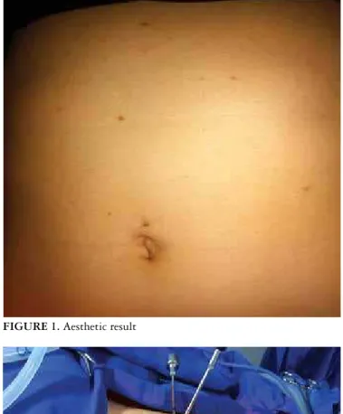
FIGURE 1. Aesthetic result
Dimbarre D, Loureiro PM, Claus C, Carvalho G, Trauczynski P, Elias F. Minilaparoscopic fundoplication. Technical adaptations and initial experience
v. 49 – no.3 – jul./set. 2012 Arq Gastroenterol 225
endoscopic fundoplication, and the rear view is exactly the same as conventional laparoscopic fundoplication.
We did not observe any complication of the surgical wound. There were no evidence of recurrence of symptoms or endoscopic changes.
DISCUSSION
GERD with or without hiatal hernia, occurs due to sev eral mechanisms including incompetence of the LES or the mechanisms of antirelux barrier. Several studies demons trate an important role in the hiatoplasty for the deinitive correction of GERD(2, 7). Due to this fact, less aggressive
endoscopic treatments than laparoscopic fundoplication has no wide acceptance in the surgical community worldwide. After the birth of NOTES, followed by LESS, there were a extensive research on procedures less aggressive than lapa roscopy and with better aesthetic results(8, 14).
Based on the experience of several authors with minilapa roscopy and the development of new materials(1, 3, 6, 9, 10, 11, 12, 13, 14),
we have performed 27 gastrofundoplications using mini instru ments. Of the 27 patients, 23 were female and 4 male. The ave rage BMI was 25.5 kg/m2. Hiatal hernias were small (< 3 cm)
in 21 patients. The preoperative manometry was performed in 20 patients and radiographs in 25. Mean operative time was 60 minutes. There were no intraoperative complications. There was no need for conversion to standard laparoscopy or open surgery. The length of hospital stay was less than or equal to 24 hours in all patients. There were no postoperative complications. Six patients underwent endoscopy between
the 3rd and 6th months after surgery, the rear view of the fun
doplication is exactly the same as conventional laparoscopic fundoplication. There were no complications of the surgical wound and no recurrences.
Using the same steps as the already established lapa roscopy, with small technical adjustments imported from laparoscopic cholecystectomy and inguinal hernioplasty, we performed hiatoplasty and fundoplication, with excellent cosmetic results in selected patients. With this technique, we do not add any disposable or expensive equipment other than a kit of 3 mm permanent instruments. This approach does not require any long learning curve, because the movements are the same as conventional laparoscopy.
If needed, we do not hesitate to enlarge the incision and change for a instrument of conventional laparoscopy. Thus, there is no additional risk to the method.
There are no longterm studies that demonstrate effec tiveness of pure endoscopic techniques. However, nowadays, we are able to perform the fundoplication and hiatoplasty with the same principles already established, following the same technical steps, as safely and with excellent cosmetic results. No need to relearn a completely new technique in surgery as LESS, with its poor ergonomic conditions and with huge inancial and time investments as NOTES tech niques. Hiatoplasty associated with fundoplication using minilaparoscopic instruments is safe, feasible and effective. If compared to other “new access”, has a spectacular esthetic results. Can be done with only minor technical adjustments, for any experienced laparoscopic surgeon, and is perfectly adaptable to our inancial reality.
Dimbarre D, Loureiro PM, Claus C, Carvalho G, Trauczynski P, Elias F. Fundoplicatura por minilaparoscopia. Adaptações técnicas e experiência inicial. Arq Gastroenterol. 2012,49(3):2236.
RESUMO – Contexto - A doença do reluxo gastroesofágico (DRGE) é uma doença altamente prevalente. O seu tratamento é dividido em modiicações
de estilo de vida, tratamento médico e cirúrgico. A cirurgia laparoscópica é o tratamento padrãoouro. Nas últimas décadas houve uma extensa pesquisa sobre procedimentos menos agressivos do que a laparoscopia e com melhores resultados estéticos. A minilaparoscopia vem “reemergindo” como método seguro, eicaz e com excelentes resultados estéticos em pacientes selecionados, tratados para DRGE. É apresentada uma série de 27 pacientes tratados para a DRGE por minilaparoscopia. Métodos Entre outubro de 2009 e julho de 2011, o total de 27 pacientes foi submetido a fun
doplicatura por videominilaparoscopia. Foram utilizados um trocarte de 10 mm, um telescópio de 30 graus e quatro trocarteres de 3 mm nas posições regulares. Os passos cirúrgicos são feitos sem modiicações, de maneira habitual. Fita cardíaca, agulhas de sutura e, eventualmente, saco extrator e gaze são colocados e retirados através do portal umbilical. Com esses ajustes técnicos, podese realizar o procedimento de forma segura e eicaz, semelhantemente à técnica laparoscópica padrão. Conclusão - Hiatoplastia associada à fundoplicatura laparoscópica, utilizandose de instrumentos
minilaparoscópicos é método seguro, viável e eicaz. Se comparado a outros “novos acessos”, tem resultado estético espetacular. Pode ser realizado com apenas pequenos ajustes técnicos, por qualquer cirurgião experiente em laparoscopia e é perfeitamente adaptável a nossa realidade inanceira.
Dimbarre D, Loureiro PM, Claus C, Carvalho G, Trauczynski P, Elias F. Minilaparoscopic fundoplication. Technical adaptations and initial experience
226 Arq Gastroenterol v. 49 – no.3 – jul./set. 2012
REFERENCES
1. Blinman T. Incisions do not simply sum. Surg Endosc. 2010 [Epub ahead] doi:10.1007/s004640090854z.
2. Cappel, MS. Clinical presentation, diagnosis, and management of gastroesopha geal relux disease. Med Clin North Am. 2005;89:24391.
3. Carvalho GL, Cavazzola LT. Can mathematic formulas help us with our patients. Surg Endosc. 2010 [Epub ahead of print]. doi: 10.1007/s0046401010653. 4. Carvalho GL, Silva FW, Ramos CHC. Colecistectomia minilaparoscópica sem
utilização de endoclipes: técnica e resultados em 719 casos. Rev Bras Videocir. 2007;1:5–11.
5. Carvalho GL, Silva FW, Silva JS, de Albuquerque PP, Coelho R de M, Vilaça TG, Lacerda CM. Needlescopic clipless cholecystectomy as an efficient, safe, and costeffective alternative with diminutive scars: the first 1,000 cases. Surg Laparosc Endosc Percutan Tech. 2009;19:368–72.
6. Cheah WK, Lenzi JE, So JB, Kum CK, Goh PM. Randomized trial of needles copic versus laparoscopic cholecystectomy. Br J Surg. 2001;88:45–7.
7. DeVault, TD, Castell, BT. Guidelines for the diagnosis and treatment of gastro esophageal relux disease. Updated guidelines for the diagnosis and treatment of gastroesophageal relux disease. 2008.
8. Ko CW, Kalloo AN. Peroral transgastric abdominal surgery. Chin J Dig Dis. 2006;7:67–70.
9. Mamazza J, Schlachta CM, Seshadri PA, Cadeddu MO, Poulin EC. Needlescopic surgery: A logical evolution from conventional laparoscopic surgery. Surg Endosc. 2001;15:1208–12.
10. Perissat J. Laparoscopic cholecystectomy: the European experience. Am J Surg. 1993;165:444–9.
11. Reardon PR, Kamelgard JI, Applebaum B, Rossman L, Brunicardi FC. Feasi bility of laparoscopic cholecystectomy with miniaturized instrumentation in 50 consecutive cases. World J Surg. 1999;23:128–31.
12. Sarli L, Costi R, Sansebastiano G, Minilaparoscopic cholecystectomy vs lapa roscopic cholecystectomy. Surg Endosc. 2001;15:6148.
13. Schwenk W, Neudecker J, Mall J, Bohm B, Muller JM. Prospective randomized blinded trial of pulmonary function, pain, and cosmetic results after laparoscopic vs. microlaparoscopic cholecystectomy. Surg Endosc. 2000;14:3458.
14. Zorron R, Maggioni LC, Pombo L, Oliveira AL, Carvalho GL, Filgueiras M. NOTES transvaginal cholecystectomy: preliminary clinical application. Surg Endosc. 2008;22:542–7.