DOI: 10.1590/0004-282X20130126
ARTICLE
Experimental model of intracranial
hypertension with continuous
multiparametric monitoring in swine
Modelo Experimental de Hipertensão Intracraniana com
monitorização multiparamétrica contínua em suínos
Almir Ferreira de Andrade1, Matheus Schmidt Soares1, Gustavo Cartaxo Patriota2,
Alessandro Rodrigo Belon2, Wellingson Silva Paiva1, Edson Bor-Seng-Shu1, Marcelo de Lima Oliveira1,
Clarissa Nóbrega Nascimento1, Gustavo Sousa Noleto1, Aderaldo Costa Alves Junior2, Eberval Gadelha
Figueiredo1, José Pinhata Otoch2, Manoel Jacobsen Teixeira1
Intracranial hypertension (IH) develops in approximately 50% of all patients with severe traumatic brain injury (TBI) and is more common in patients with intracranial hematoma (ICH)1,2. herefore, it is very important to identify a suitable
animal model to study and understand the pathophysiology
of refractory IH to develop efective treatments.
Many models of neurosurgical experiments in small ani-mals, such as rats and cats, have been developed; however, compared to humans, they have smaller brain volumes and more distinct behaviors3,4. While there are existing porcine
models, they simulate ICH by infusing autologous blood into the brain tissue and do not replicate intracranial lesions3–6.
1Division of Neurosurgery, University of São Paulo Medical School, São Paulo SP, Brazil;
2LIM 26, Experimental Surgery Laboratory, University of São Paulo Medical School, São Paulo SP, Brazil.
Correspondence: Wellingson Paiva; Rua Eneas Aguiar 255 / 4080; 0543-010 São Paulo SP - Brasil; E-mail: [email protected]
Conflict of interest: There is no conflicts of interest to declare.
Received 04 March 2013; Received in final form 24 May 2013; Accepted 31 May 2013. ABSTRACT
Objective: Intracranial hypertension (IH) develops in approximately 50% of all patients with severe traumatic brain injury (TBI). Therefore, it is very important to identify a suitable animal model to study and understand the pathophysiology of refractory IH to develop effective treatments. Methods: We describe a new experimental porcine model designed to simulate expansive brain hematoma causing IH. Under anesthesia, IH was simulated with a balloon insufflation. The IH variables were measured with intracranial pressure (ICP) parenchymal moni-toring, epidural, cerebral oximetry, and transcranial Doppler (TCD). Results: None of the animals died during the experiment. The ICP epidural showed a slower rise compared with parenchymal ICP. We found a correlation between ICP and cerebral oximetry. Conclusion: The model described here seems useful to understand some of the pathophysiological characteristics of acute IH.
Keywords: experimental model, intracranial hypertension, transcranial doppler sonography.
RESUMO
Objetivo: A hipertensão intracraniana (HIC) ocorre em até 50% de todos os pacientes com traumatismo cranioencefálico (TCE). Por isso, é importante estabelecer um modelo animal adequado para estudar a fisiopatologia da HIC refratária, com a perspectiva de desenvolver trata-mentos eficazes. Métodos: Os animais foram submetidos a um protocolo padrão de anestesia. A hipertensão intracraniana foi estabelecida através de insuflação de um balão. As variáveis HIC foram medidas com a pressão intracraniana (PIC) do parênquima, oximetria, epidural e doppler transcraniano. Resultados: A PIC epidural apresentou elevação mais lenta, comparada com a PIC parenquimal. Houve correlação entre a PIC e a oximetria cerebral. O registro da PIC, oximetria e índice de pulsatilidade foi realizado em todos os animais sem dificuldade.
Conclusão: O modelo descrito parece ser útil para a compreensão de algumas características fisiopatológicas na HIC aguda.
Here, we describe a new experimental model designed to simulate expansive brain hematoma causing IH by using an
infusion pump to progressively inlate an intracerebral bal
-loon; the lesion can be easily relieved via cuf delation.
METHODS
his protocol was approved by the Research Ethics Com
mittee of the Hospital das Clinicas – University of Sao Paulo Medical School.
Animals
We obtained 2-month old crossbred Landrace and Duroc pigs weighing approximately 18–20 kg from a private farm lo-cated in Suzano, Brazil; they were delivered to the University of Sao Paulo Veterinary School and transported to the medi-cal school on the day of the experiment.
Anesthesia protocol
Prior to surgery, pigs were fasted for 12 h but had free access to water. We then co-administered intramuscu-lar ke tamine (Ketamin-S®
, Cristália) at a dose of 15 mg/kg and xylazine (Anasedan®
, Ceva) at a dose of 2 mg/kg as a preanes thetic. Once intravenous (IV) access was obtained, anes thesia was induced with propofol (1% Provine®
, Claris)
at a dose of 5 mg/kg. he animals also received an initial IV
vo lume of 20 ml/kg physiological saline (NaCl 0.9%) to com
-pensate for volume loss due to fasting, and luid support was
con ti nued throughout at a rate of 5 ml/kg/h. Anesthesia was maintained with IV propofol (1% Provine®
) at a dose of 5-10 mg/kg/h, and IV fentanyl was given for analgesia (Fentanest®
, Cristália) at a starting dose of 5 μg/kg followed by continuous IV infusion of 0.08-0.15 mg/kg/min.
After endotracheal intubation, the animals were me cha -nically ventilated by controlled volume (Fan Dixtal®
5010), tidal volume (VT) of 10 ml/kg, fraction of inspired oxygen (FiO2) of 0.40, and positive end expiratory pressure (PEEP) of
5 cmH2O. he ventilatory parameters were adjusted to main
-tain partial pressure of carbon dioxide (PaCO2) between 35
and 40 mmHg, partial pressure of oxygen (PaO2) between
100 and 150 mmHg, and blood pH between 7.35 and 7.45. To
assess ventilation adequacy, we continuously measured i
-nal pressure of endtidal carbon dioxide (EtCO2), peripheral
hemoglobin saturation by pulse oximetry (SpO2), and
arte-rial blood gas samples (0.3 ml). he right femoral artery was
catheterized for invasive monitoring of mean blood pressu-re (MBP). Six arterial blood gas samples wepressu-re obtained after additional interventions to ensure maintenance of phy sio-logical parameters. We also collected 5-ml serum samples to measure ubiquitin-C and beta amyloid precursor protein (SAA) levels. Hemodynamic data were collected and mea-sured with a multiparameter Dixtal Monitor®
2020.
Animal core temperature was maintained between 37°C and 39°C with the use of a blanket and previously heated maintenance solutions.
Experimental model preparation
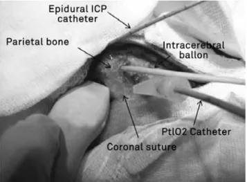
An “L”-shaped skin incision was made at the sagittal midline to expose the coronal and sagittal sutures (Figure 1), and we made two 3-mm trephinations to install the balloon and multiparameter oximetry catheter. Two trephinations were also made in the middle fossa; the posterior one was for the intracranial pressure (ICP) epidural catheter (micro-sensor-type microchip, Neurodur®
; Raumedic, Germany),
and the anterior one was used as a window for the transcra-nial Doppler (TCD) transducer. A trephination located 1-cm lateral to the metopic suture and anterior to the coronal su-ture allowed installation of a multiparameter catheter cere-bral tissue oximetry sensor (microsensor-type microchip, Neurovent-PTiO®
; Raumedic) 1.5cm deep in the frontal lobe.
A trepanation located 1-cm lateral to the sagittal suture and 1-cm posterior to the coronal suture was performed to in-troduce a pediatric 8-French bladder catheter to a depth of 2 cm in the parietal lobe for accessing the parietal subcorti-cal white matter.
Algorithm experiment
hrough the continuous infusion pump, the cuf (balloon) of the pediatric catheter was progressively illed with 0.9% saline solution over 15 min. In Group A, we infused 4 ml sa
-line; in Group B, an additional 3 ml was infused over 15 min at 1 h after the irst infusion to simulate a rebleed; and in Group C, 7 ml was infused over 15 min. his luid system was
Figure 1. Figure of experimental model during the procedure. We performed two 3-mm trephinations with a balloon and multiparameter oximetry catheter (PtiO2). Two trephinations were also made in the middle fossa; the posterior one was for the intracranial pressure (ICP) epidural catheter (microsensor-type microchip, Neurodur®; Raumedic, Germany), and the
tested with the inal volume of 0.9% saline solution prior to
insertion into the parietal lobe. After complete catheter
ins-tallation and balloon inlation, the model was maintained
without intervention for 1 h until animal physiological pa-rameters were stabilized.
he average weight of an adult human brain is 1350 g6,
and those of the pigs used (2 months and 20 kg) were 75 g, which corresponds to 5.5% of the weight of the human
brain. hus, the 4ml volume in Group A corresponded to a
lesion of 72.7 ml in the adult human brain. In the other two
groups, the inal volume of 7 ml corresponded to a lesion of
127.3 ml.
After the irst hour of calibration and parameter stabiliza
-tion, the balloon was inlated by using the continuous infu -sion pump (Braun B Infusomat compact®
) over 15 min. After 1.5 h, 3% hypertonic saline solution 3% (5.3 ml/kg) was ad-ministered. After 30 min, the pigs underwent surgery, and the
balloon was delated. he experiment ended after an addi
-tional hour during which the physiological parameters were observed. At each intervention, we performed a neurological assessment of the pupils and duplex with a Doppler appa-ratus (SonoSite – Micromax model) by using a sector trans-ducer of 4–8 mHz through the right temporal trephination
over the intracerebral artery according to the color low tech
-nique, followed by Doppler blood low velocity measurement
(Figure 2).
At the end of the experiment, the animals were sacrii
ced via an IV overdose of propofol (20 mg/kg) and fentanyl (10 mg/kg) followed by 40 ml 19.1% potassium chloride
so-lution. he brain was then surgically removed, weighed on a
high-precision balance, and sectioned to exclude the possi-bility of other cerebral bleeds.
Animal disposal
he pigs were placed in white plastic Biohazard bags with labels that clearly identiied the origin, content, and respon
-sible researcher. hey were then transported to the hospital
to be incinerated.
RESULTS
We have tested this pilot model in six animals and were able to identify correlations between progressive ICP
increa-ses and balloon inlation (Table 1). In a preliminary analysis of data from the irst six animals, we observed stable syste
mic parameters, including blood pressure, arterial blood
gases, heart rate, and oxygen saturation. We veriied that
parenchymal pressure rises and falls faster compared with epidural pressure. None of the animals died during the ex-periment, and there were no complications, which indicates that our method is safe. We clinically evaluated the animals throughout and monitored their pupillary responses to each
phase of the experiment. In all animals, we veriied pupillary
response with anisocoria following increased ICP after
bal-loon inlation, as well as reversal of this pattern when the bal
-loon was delated; this indicated a pattern of brain compla -cency and a clinical response to uncal herniation, similar to what is observed in the human brain. We also found a cor-relation between ICP and cerebral oximetry. Our results sug-gest that this is a reproducible experimental model.
DISCUSSION
In this study, we developed a new experimental model of ICH in pigs with lesion based on the Monro-Kellie doctrine1,
which posits that the intracranial content is incompressible and has a constant volume. In the presence of ICH, the vas-cular space may decrease by 50%; intracellular space could shrink to decrease total brain volume, and cerebrospinal
luid (CSF) production and absorption might decrease and
increase, respectively.
Kim et al.10 recently reported that changes in intracranial
venous, arterial, and CSF compartments could be mathema-tically estimated using serial analysis, allowing the calcu-lation of a cerebral complacency index (CCI) as a
correla-tion coeicient of changes in compartments. he authors
Figure 2. SonoSite - Micromax model using a sector transducer of 4–8 mHz through the right temporal trephination over the intracerebral artery according to the color flow technique, followed by Doppler blood flow velocity measurement. A – before balloon isufflation, with normal cerebral flow; B – cerebral flow decrease after balloon insufflation; C – cranial ultrasonography to confirm balloon position and insufflation.
mentioned that a negative CCI represents a physiological doctri ne of Monro-Kellie that illustrates the volumetric com-pensation between CSF and arterial compartments, whereas
positive values relect doctrine disorders by increasing the
volumes of both compartments. Clinical observations in-dicate that the A waves (plateau) and arterial hypertension were associated with negative CCI, and positive CCI was ob-served in refractory IH.
he skull is a semirigid structure with low elasticity that
gradually decreases with age, as demonstrated by engi neering experiments using strain gauges11–13. Like any subs tance, bone
tissue has the physical property of elasticity. When ICP in-creases, it causes micrometric deformations of the skull bones. According to the principles of the Monro-Kellie doc-trine, there is an initial equilibrium between intracranial compartments, and dysfunction appears after intracerebral
expansions. Groups A and C involved diferent volumes of conti nuous expansion, whereas Group B underwent dis -continuous expansion to simulate lesion re-expansion. All animals were closely observed for changes for 1.5 h after the start of the expansion before a mock clinical intervention was performed (3% hypertonic saline solution). After 30 min,
a mock surgical intervention was made (balloon delation). he animals were observed for an additional hour before the
experiment ended with the sacriicing of the animals and re
-moval of the brain for macroscopic and histological analyses.
he model allows the determination of a decrease in intra
-cranial compliance by refractory IH.
Other previous studies have also used experimental swine models. Wagner et al.5 described a model with blood
infusion in a balloon, but this method was not satisfactory because hematoma size was highly variable, and the mo del showed poor reproducibility. Shi et al.4 developed another
model of intracranial hemorrhage in 24 pigs and studied his-tological changes in brain tissue after autologous blood infu-sion. However, that investigation did not assess acute cere-brovascular features.
IH parameters were evaluated by TCD through changes in systolic, diastolic, and MBP, in addition to the pulsatility index measured immediately before and after each inter-vention. A sudden change in mean MBP leads to a
simulta-neous change in cerebral blood low initially, but it also trig -gers a number of other responses14.hus, the assessment of
cerebral hemodynamics in the acute phase of these
condi-tions may contribute to more efective planning of thera -peutic strategies for reducing secondary brain lesions15. In
our model, it was possible to evaluate cerebrovascular va-soreactivity with TCD without problems in all tested
ani-mals. Additionally, the volume infused was conirmed by
ultrasonography.
Regarding ICP measurement, epidural sensor placement
for ICP monitoring has been discussed for many years as an important option because it has fewer complications com-pared to intraparenchymal pressure monitoring16–18. In our
study, the pressures were similar in animals with the balloon
uninlated. However, ICP latency was larger in epidural sys
-tem. he existence of a signiicant drift and latency of epi
-dural pressure system compared with parenchymal pressure
system was irst described by Powell and Crockard19. Raabe
et al.20 also described this latency. he authors conclu ded
that these drifting periods could be found during throu ghout
the entire measurement. he biomechanical conditions in the epidural space can inluence pressure distribution
to the sensor and interfere with epidural pressure system measurements16,20.
he model described here seems useful for understan
ding some important pathophysiological characteristics of acute IH.
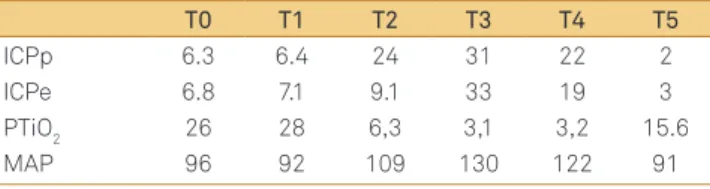
Table 1. Average measures of intracranial variable during experiment.
T0 T1 T2 T3 T4 T5
ICPp 6.3 6.4 24 31 22 2
ICPe 6.8 7.1 9.1 33 19 3
PTiO2 26 28 6,3 3,1 3,2 15.6
MAP 96 92 109 130 122 91
ICPp: intracranial pressure (parenchymal); ICPe: intracranial pressure (epi -dural); PTiO2: cerebral oximetry; MAP: middle arterial pressure; T0: baseline measures; T1: before balloon insufflation; T2: after balloon insufflation, T3: 1.5 hour after balloon insufflations; T4: after saline solution infusion; T5: after balloon deflation.
References
1. Andrade AF, Paiva WS, Amorim RL, Figueiredo EG, Rusafa Neto E, Teixeira MJ. The pathophysiological mechanisms following traumatic brain injury. Rev Assoc Med Bras. 2009;55(1):75-81.
2. Miller JD. Traumatic brain swelling and edema. In: Cooper PR (Ed). Head Injury. 3rd ed. New York: Morgan Hill, 1993:331-354.
3. Ryska O, Pantoflicek T, Laszikova E, Prazak J, Koblihova E, Ryska M. Artificial liver support system reduces intracranial pressure more effectively than bioartificial system: an experimental study. Int J Artif Organs 2012;35:503-510.
4. Shi Y, Li Z, Zhang S, et al. Establishing a model of supratentorial hemorrhage in the piglet. Tohoku J Exp Med 2010;220:33-40.
5. Wagner KR, Xi G, Hua Y, et al. Lobar intracerebral hemorrhage model in pigs: rapid edema development in perihematomal white matter. Stroke 1996;27:490-497.
7. Friess SH, Ralston J, Eucker SA, Helfaer MA, Smith C, Marguiles SS. Neurocritical care monitoring correlates with neuropathology in a swine model of pediatric traumatic brain injury. Neurosurgery 2011;69:1139-1147.
8. Adams RD, Victor M. Principles of Neurology. 3rd ed. New York: McGraw-Hill, Inc., 1985.
9. Kanter MJ, Narayan RK. Management of head injury. Intracranial pressure monitoring. Neurosurg Clin N Am 1991;2:257-265.
10. Kim DJ, Czosnyka Z, Kasprowicz M, et al. Continuous monitoring of the Monro-Kellie doctrine: is it possible? J Neurotrauma 2012;29:1354-1363.
11. Geeraerts T, Merceron S, Benhamou D, Vigué B, Duranteau J. Non-invasive assessment of intracranial pressure using ocular sonography in neurocritical care patients. Intensive Care Med 2008;34:2062-2067.
12. Thees C, Scholz M, Schaller M D C, et al. Relationship between intracranial pressure and critical closing pressure in patients with neurotrauma. Anesthesiology 2002;96:595-599.
13. Panerai RB. The critical closing pressure of the cerebral circulation. Med Eng Phys 2003;25:621-632.
14. Bor-Seng-Shu E, Kita WS, Figueiredo EG, et al. Cerebral
hemodynamics: concepts of clinical importance. Arq Neuropsiquiatr 2012;70:352-356.
15. Dagal A, Lam AM. Cerebral blood flow and the injured brain: how should we monitor and manipulate it? Curr Opin Anaesthesiol 2011;24:131-137.
16. Eide PK. Comparison of simultaneous continuous intracranial pressure (ICP) signals from ICP sensors placed within the brain parenchyma and the epidural space. Med Eng Phys 2008;30:34-40.
17. Bruder N, N’Zoghe P, Graziani N, Pelissier D, Grisoli F, François G. A comparison of extradural and intraparenchymatous intracranial pressures in head injured patients. Intensive Care Med 1995;21:850-852.
18. Paiva WS, de Andrade AF, Amorim RL, Figueiredo EG, Matushita H, Teixeira MJ. Intracranial pressure monitoring in children with fulminant hepatic failure. Rev Neurol 2009;48:134-136.
19. Powell MP, Crockard HA. Behavior of an extradural pressure monitor in clinical use. J Neurosurg 1985;63:745-749.