ARTICLE
DOI: 10.1590/0004-282X20130119
Irregularly shaped lacunar infarction: risk
factors and clinical significance
Infartos lacunares com morfologia irregular: fatores de risco e significado clínico
Chao Feng1, Cai-Yu Zhang2, Yu Xu3, Ting Hua3, Xue-Yuan Liu1, Min Fang1
Lacunar infarcts are small infarcts with a diameter of 15–20 mm; they occur in the distributions of small penetra ting arteries such as arteries in the thalamus, gangliocap sular regions, corona radiate, and brainstem1,2. he primary
pathogenesis of small lacunar infarctions is the occlusion of small penetrating arteries resulting from hypertensive lipo
hyalinosis or ibroid necrosis2,3. Most lacunar infarcts appear
as round, regular focal lesions, whereas other lacunar infarcts appear as irregularly shaped lesions4.Some lacunar infarcts
even appear from the merging of several small lesions5.
Few studies have investigated the shapes of lacunar in farction; thus, little information is known about irregularly shaped lacunar infarctions such as their risk factors, patho
logical mechanisms, and clinical signiicance. A very inte
resting question is whether the irregular shape of a lacunar
infarction is just a coincidence and has no clinical signii
cance or whether the irregular shape represents a diferent
pathogenesis from regularly shaped lacunar infarctions and
has some inluence on the clinical symptoms and prognosis of
stroke. We hypothesized that lacunar strokes with irre gularly
1Department of Neurology, Shanghai Tenth People’s Hospital of Tongji University, Shanghai, China;
2Department of Neurology, Weihai Woman and Children’s Hospital, Weihai, Shangdong, China;
3Department of Radiology, Shanghai Tenth People’s Hospital of Tongji University, Shanghai, China.
Correspondence: Xue-Yuan Liu and Min Fang; Department of Neurology, Shanghai Tenth People’s Hospital of Tongji University; Middle Yanchang Road Number
301 / Zhabei District; Shanghai, China; E-mail: [email protected] (X.-Y. Liu); [email protected] (M. Fang)
Conflict of interest: There is no conlict of interest to declare.
Financial support: This study was supported by grants from the National Natural Science Foundation of China (No. 81000492, No. 30971029, and
No. 81171163).
Received: 06 April 2013. Received in inal form: 10 May 2013; Accepted: 17 May 2013.
ABSTRACT
Objective: Our study focused on acute lacunar infarct shapes to explore the risk factors and clinical signiicance of irregularly shaped lacunar infarctions. Methods: Based on the shape of their acute lacunar infarct, patients (n=204) were classiied into the “regular” group or “irregu-lar” group. The characteristics of the lacunar infarction were compared between the regular and irregular groups, between patients with and without neurological deterioration, and between patients with different modiied Rankin scale (mRS) scores. The risk factors for irregularly shaped lacunar infarctions, neurological deterioration, and high mRS scores were identiied. Results: Blood pressure variability (BPV) was an independent risk factor for irregularly shaped lacunar infarction. Infarction size, prevalence of advanced leukoaraiosis, and irregularly shaped lacunar infarcts were independent risk factors for higher mRS scores. Conclusions: The irregularly shaped lacunar infarcts were cor-related with BPV. Irregularly shaped lacunar infarctions and leukoaraiosis may be associated with unfavorable clinical outcomes.
Keywords: blood pressure variability, lacunar infarction, lesion shape.
RESUMO
Objetivo: Estudar as diferentes formas dos infartos lacunares agudos, investigando os fatores de risco e o signiicado clinico daqueles com morfologia irregular. Métodos: Os 204 pacientes com infartos lacunares agudos foram classiicados em dois grupos: aqueles com morfologia regular e aqueles com morfologia irregular. Foram estudadas as características dos dois grupos e caracterizados os fatores de risco para infartos irregulares, deterioração neurológica e altos escores da escala de Rankin modiicada. Resultados: Variabilidade da pressão arterial é fator de risco independente para infartos lacunares irregulares. Tamanho do infarto, prevalência de leucoaraiose e formato irregular dos infartos lacunares são fatores de risco independentes para escores mais elevados na escala de Rankin modiicada. Conclusões: Variabi-lidade da pressão arterial está relacionada ao formato irregular dos infartos lacunares agudos. Este tipo de infarto e a leucoaraiose podem estar relacionado a desfechos clínicos desfavoráveis.
shaped infarcts have diferent pathogeneses, clinical symp toms, and prognoses in comparison to lacunar strokes with regularly shaped infarcts. In this study, we evaluated the
risk factors, severity of neurological deicits, prognosis, and
pathogeneses of irregularly shaped lacunar infarctions.
METHODS
General information
his study was based on consecutive patients who atten
ded the Department of Neurology of Tenth People’s Hospital (Shanghai, China) from January 2012 to September 2012
within the irst 24 hours after stroke onset and were diag
nosed as having an acute lacunar infarction. he identiied
infarcts had a diameter of less than 20 mm (as determined by magnetic resonance imaging [MRI]) and were categorized as
small vessel occlusions, based on the Classiication of Stroke
System6. Patients who underwent thrombolytic therapy, pa
tients with previous physical disabilities, and patients who could not be followed up were excluded from the study. Two hundred four patients were enrolled in this study.
Patients were divided into two groups (i.e. the regular group and the irregular group), based on the shape of their
infarctions, as determined by MRI. he shape of an infarc tion was considered “regular” when the infarction was ovoid, spheroid, or sticklike, and the shape of an infarction was considered “irregular” when the infarction was slablike with multiple angles or was composed of multiple shapes or con glomerated beads4,5 (Figure).
he blood pressure was measured after the patients were admitted in the hospital. he ambulatory blood pressure was
monitored during the irst 24 hours of hospitalization. he 24
hour systolic blood pressure standard deviation (24hSBPSD)
was recorded to indicate blood pressure variation (BPV). he following levels were tested: blood glucose, hemoglobin A1c (HbA1c), and blood lipids, which included triglyceride (TG),
total cholesterol (TC), lowdensity lipoprotein (LDL), and highdensity lipoprotein (HDL). On days 1 and 14 of hospi
talization, the severity of neurological deicits was evalua ted
in accordance with the National Institute of Health stroke
scale (NIHSS). An increased NIHSS score on day 14 was re garded as neurological deterioration. Before hospital dis charge, the patients were administered individualized reha
bilitation treatment plans by a rehabilitation therapist. After
three months, a telephone interview was conducted by using
a modiied Rankin’s scale (mRS) to evaluate the clinical out
comes of each patient. All procedures were approved by the
Ethics Committee of the Shanghai Tenth People’s Hospital of Tongji University (Shanghai, China) and were performed with the written consent of the patients if they had no im pairment in cognitive function and were capable of writing or from the guardians of the patients if a patient had impaired consciousness, cognitive function, or writing ability.
MR protocols
Patients were imaged by using a 3.0T MRI scanner (Siemens 3.0T Magnetom Verio, Medical Solutions, Er
langen, Germany) with a standard eightchannel phased
ar ray head coil. T1weighted images (repetition time/echo time=2000/9), T2weighted images (repetition time/echo
time=6000/94), luidattenuated inversion recovery (FLAIR) images (repetition time/echo time=8500/94), and difusion
weighted images (DWI) (motionprobing gradients in three directions with a b factor of 1000 s/mm2) were obtained from
the axial plane with a slice thickness of 5.5 mm.
MR images review and criteria
Images were analyzed by two experienced radiologists
who were blinded to the clinical information. An acute la
cunar infarct was deined as a hypointense focal lesion with
a diameter of 3–20 mm in T1weighted images and as a hy
perintense focal lesion in T2weighted, FLAIR, and DWI ima
ges1,2. he shape of the infarct was evaluated in accordance
with the criteria described previously. he longest diameter
of the infarcts was measured from DWIs. Leukoaraiosis was
also evaluated from the FLAIR images and scored as grade 0
to grade 3, based on the Fazekas scale7. Grades 2 and 3 were
regarded as evidence of advanced leukoaraiosis (adLA). he
discrepancy of the shape of the infarct and leukoaraiosis score was resolved by visual consensus.
Statistical analysis
Data were analyzed by using SAS 9.2 software. he Chi
square test and Student t test were used to compare the risk Figure. Regularly and irregularly shaped lacunar infarctions.
(A and B) Irregularly shaped lacunar infarctions on diffusion-weighted images (DWI). (C and D) Regularly shaped lacunar infarctions on DWI.
A
C
B
factors between patients with regular infarctions and pa tients with irregular infarctions and between patients with neurological deterioration and patients without neurologi
cal deterioration. he analysis of variance (ANOVA) and Chi
square test were used to compare risk factors of patients with
diferent mRS scores. Logistic regression models were cons
tructed to identify independent risk factors for irregular in farctions, neurological deterioration, and a high mRS score.
A p<0.05 indicated statistical diference.
RESULTS
In this study, 138 patients had regular infarctions and 66 patients had irregular infarctions. Compared to the patients in the regular group, the patients in the irregular group had a larger infarction size, higher diastolic blood pressure (DBP),
and higher 24hSBPSD. he other factors did not show a sta
tistical signiicance (Table 1). A logistic regression model was
constructed to identify the possible risk factors of irregularly shaped lacunar infarctions. Information on age, sex, smoking, systolic blood pressure (SBP) and DBP after hospital admis
sion, 24hSBPSD, HbAIc, TG, TC, LDL, and HDL were added to the model. he results showed that only 24hSBPSD is an
independent risk factor for irregularly shaped lacunar infarc tions (OR=1.64; 95%CI=1.26–21.4; p<0.001).
Twenty (9.8%) patients had neurological deterioration.
he proportion of patients with irregularly shaped lacunar
infarctions was higher in patients with neurological dete rioration than in patients without neurological deterioration (p<0.01). Logistic regression was used to identify possible risk
factors for neurological deterioration. Age, sex, smoking, size
of infarct, prevalence of hypertension, diabetes, dyslipidemia,
adLA, and irregularly shaped lacunar infarctions were adde d to the model. After adjusting for confounding factors, irre gularly shaped lacunar infarction was identiied as an inde pendent risk factor for neurological deterioration (OR=4.54, 95%CI=1.4214.58, p=0.01).
Early prognosis of lacunar infarction was evaluated with the mRS. During the threemonth follow up, no patients died or showed severe physical disabilities, required cons tant nursing care and attention, or became bedridden and incon
tinent. hus, the mRS score ranged from 0 to 4. he ANOVA results showed that age, prevalence of diabetes, adLA, irre gu lar infarctions, SBP, HbAIc, and infarct size difered among pa
tients with diferent mRS scores (p<0.05) (Table 2). Logistic
regression was used to identify the risk factors for higher
mRS scores. Age, sex, smoking, size of infarct, prevalence of hypertension, diabetes, dyslipidemia, adLA, and irregular
ly shaped lacunar infarction were added to the model. he results showed that infarct size, prevalence of adLA, and
irregu lar infarction were independent risk factors for higher mRS scores (Table 3).
DISCUSSION
he present study focused on the shape of lacunar in farcts and analyzed the risk factors and clinical features of
irregularly shaped lacunar infarctions. he following re sults were obtained: (1) irregularly shaped lacunar infarc tions were prevalent in 32.3% of the sample population; (2) the 24hSBPSD is the only independent risk factor for irregu larly shaped lacunar infarction; (3) the irregular shape of a lacunar infarction is associated with a larger infarction size, neurological deterioration, and unfavorable early prognosis; and (4) leukoaraiosis is associated with an unfavorable early prognosis of lacunar infarction.
A recent study on the shape of acute lacunar infarcts re ports that 32.4% of these infarcts are conglomerated bead shaped infarcts5. In the current study, the range of irregularly
shaped infarcts was greater than the range of conglomera ted beadshaped infarcts. However, the proportion of irregu larly shaped infarcts in this study was not higher than the re ported proportion of conglomerated beadshaped infarcts.
he relatively obscure standard used in the evaluation of the
shape of infarcts may be the primary reason for the observed
similarities in proportion. To date, no deinite standard exists for classifying the shapes of infarcts. hus, visual consensus used in diferent studies remains the only method of classi
fying infarcts on the basis of their shape.
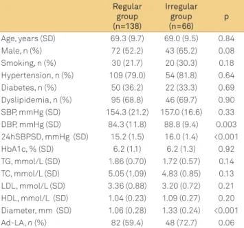
Table 1. Baseline characteristics of patients with regular and irregular infarctions.
Regular group (n=138)
Irregular group (n=66)
p
Age, years (SD) 69.3 (9.7) 69.0 (9.5) 0.84
Male, n (%) 72 (52.2) 43 (65.2) 0.08
Smoking, n (%) 30 (21.7) 20 (30.3) 0.18
Hypertension, n (%) 109 (79.0) 54 (81.8) 0.64 Diabetes, n (%) 50 (36.2) 22 (33.3) 0.69 Dyslipidemia, n (%) 95 (68.8) 46 (69.7) 0.90 SBP, mmHg (SD) 154.3 (21.2) 157.0 (16.6) 0.33 DBP, mmHg (SD) 84.3 (11.8) 88.8 (9.4) 0.003 24hSBPSD, mmHg (SD) 15.2 (1.5) 16.0 (1.4) <0.001
HbA1c, % (SD) 6.2 (1.1) 6.2 (1.3) 0.92
TG, mmol/L (SD) 1.86 (0.70) 1.72 (0.57) 0.14 TC, mmol/L (SD) 5.05 (1.09) 4.83 (0.85) 0.13 LDL, mmol/L (SD) 3.36 (0.88) 3.20 (0.72) 0.21 HDL, mmol/L (SD) 1.04 (0.23) 1.09 (0.27) 0.20 Diameter, mm (SD) 1.06 (0.28) 1.33 (0.24) <0.001
Ad-LA, n (%) 82 (59.4) 48 (72.7) 0.06
Table 2. Characteristics of patients at different mRS scores.
mRS=0 (n=39)
mRS=1 (n=83)
mRS=2 (n=46)
mRS=3 (n=21)
mRS=4
(n=15) p
Age, years (SD) 65.7±9.4 68.8±9.5 70.4±9.5 70.7±9.7 74.4±8.2 0.03
Male, n (%) 21 (53.9) 48 (57.8) 27 (58.7) 14 (66.7) 5 (33.3) 0.36
Smoking, n (%) 8 (20.5) 22 (26.5) 11 (23.9) 8 (38.1) 1 (6.7) 0.27
Hypertension, n (%) 32 (82.1) 62 (74.7) 38 (82.6) 16 (76.2) 15 (100.0) 0.22
Diabetes, n (%) 7 (18.0) 33 (39.8) 18 (39.1) 5 (23.8) 9 (60.0) 0.02
Dyslipidemia, n (%) 30 (76.9) 58 (69.9) 31 (67.4) 12 (57.1) 10 (66.7) 0.62
SBP, mmHg (mean±SD) 158.1±20.3 152.2±21.1 154.2±16.3 153.5±22.2 169.4±11.8 0.03
DBP, mmHg (mean±SD) 82.8±10.5 84.7±12.0 88.9±9.3 85.9±13.5 89.3±9.4 0.07
24hSBPSD, mmHg (mean±SD)
15.2±1.4 15.6±1.5 15.6±1.5 15.1±1.9 15.4±1.3 0.50
HbA1c,% (mean±SD) 5.7±0.7 6.2±1.2 6.3±1.2 6.1±1.4 6.7±1.7 0.04
LDL, mmol/L (mean±SD) 3.36±0.79 3.39±0.84 3.25±0.86 3.20±0.80 3.06±0.89 0.59
Diameter, mm (mean±SD) 1.12±0.29 1.09±0.25 1.26±0.30 1.27±0.30 1.30±0.34 <0.001
Ad-LA, n (%) 13 (33.3) 54 (65.1) 32 (69.6) 17 (81.0) 14 (93.3) <0.001
Irregular shape, n (%) 2 (5.1) 19 (22.9) 28 (60.9) 9 (42.9) 8 (53.3) <0.001
24hSBPSD: 24-hour systolic blood pressure standard deviation; AdLA: advanced leukoaraiosis; DBP: diastolic blood pressure; HbAlc: hemoglobin Alc; LDL: low-density lipoprotein; mRS: modiied Rankin Scale; SBP: systolic blood pressure; SD: standard deviation.
Table 3. Risk factors for the higher mRS scores.
OR 95%CI p
Diameter, +1 mm 4.34 1.57–12.0 0.005
Ad-LA (1 vs. 0) 2.41 1.15–5.05 0.02
Irregular shape (1 vs. 0) 3.28 1.74–6.18 <0.001 AdLA: advanced leukoaraiosis; CI: conidence interval; OR: odds ratio.
Hypertension is a primary independent risk factor for lacunar infarctions, including silent and acute infarctions2.
Hypertension, particularly longstanding hypertension, is the
primary reason for lipohyalinosis and ibroid necrosis, which
occlude the small arteries and thus result in lacunar infarc tions, especially infarcts with diameters less than 15–20 mm1.
In this study, most infarctions were associated with hyper tension. Similar prevalence rates of hypertension, SBP, and DBP were found in patients with regular and irregular infarc
tions. However, BPV was more signiicant in patients with ir regular infarctions. Blood pressure variability has been asso ciated with an increased risk of stroke8,9. his study showed
that BPV is also associated with irregularly shaped lacunar infarctions. Clinical and experimental studies prove that
longstanding hypertension with signiicant BPV may cause
severe endothelial dysfunction of the small blood vessels10,11,
thereby resulting in the breakage of the bloodbrain barrier and enlargement of Virchow–Robin spaces12. herefore, BPV
may be associated with the severity of small vessel disease (SVD) such as the severity of leukoaraiosis13,14. In this study,
the severity of leukoaraiosis showed a weak association with irregular infarctions (p=0.06).
Leukoaraiosis reduces vascular density15 and cerebral
blood low16, and may result in infarct growth by preven
ting peripheral compensation during ischemic stress17. his
mechanism may be associated with the pathogenesis of ir regularly shaped infarcts, which had larger sizes than regu
lar infarcts in this study. he irregular shape of infarcts may
be caused by the asymmetric enlargement of the infarction. However, an intrinsic SVDrelated mechanism may be more
signiicant in irregular infarctions than in regular infarctions.
Previous studies prove a correlation between leukoa raiosis and unfavorable clinical outcomes of ischemic stro ke18. his association may be related to a dysfunctional
neu ronal network19 and enlarged infarction size due to leu
koaraiosis17,18. In this study, a similar correlation was ob
served between irregularly shaped lacunar infarctions and clinical outcomes. Neurological deterioration on day 14 and outcomes after 3 months were independent of leukoaraiosis and infarct size. It is reasonable to assume that a large infarc tion would be associated with unfavorable clinical outcomes. However, the association between the shape of an infarct and clinical outcome remains puzzling. Because of infarction en largement, it is possible that the size of the infarct in irregu larly shaped infarctions may be even larger after 1 or 2 weeks than the shape determined via MRI within 24 hours after
stroke onset. Other factors, such as inlammation20, which
was not considered in this study, may also be related to this phenomenon.
he primary strengths of this study include its prospec tive study design, use of various statistical methods, and use of 24hour ambulatory blood pressure monitoring that could
supply accurate BPV. he primary limitations of this study in
clude the limited sample size and nonspeciic standards for identifying diferent infarct shapes, which may be inluenced
References
1. Fisher CM. Lacunes: small, deep cerebral infarcts. Neurology 1965;15:774-784.
2. Moran C, Phan TG, Srikanth VK. Cerebral small vessel disease: a review of clinical, radiological, and histopathological phenotypes. Int J Stroke 2012;7:36-46.
3. Pantoni L. Cerebral small vessel disease: from pathogenesis and clinical characteristics to therapeutic challenges. Lancet Neurol 2010;9:689-701.
4. Herve D. Shape and volume of lacunar infarcts: a 3D MRI study in cerebral autosomal dominant arteriopathy with subcortical infarcts and leukoencephalopathy. Stroke 2005;36:2384-2388.
5. Ryu DW, Shon YM, Kim BS, Cho AH. Conglomerated beads shape of lacunar infarcts on diffusion-weighted MRI: what does it suggest? Neurology 2012;78:1416-1419.
6. Ay H, Benner T, Arsava EM, et al. A computerized algorithm for etiologic classiication of ischemic stroke: the Causative Classiication of Stroke System. Stroke 2007;38:2979-2984.
7. Fazekas F, Chawluk JB, Alavi A, Hurtig HI, Zimmerman RA. MR signal abnormalities at 1.5 T in Alzheimer’s dementia and normal aging. AJR 1987;149:351-356.
8. Shimbo D, Newman JD, Aragaki AK, et al. Association between annual visit-to-visit blood pressure variability and stroke in postmenopausal women: data from the Women’s Health Initiative. Hypertension 2012;60:625-630.
9. Rothwell PM, Howard SC, Dolan E, et al. Prognostic signiicance of visit-to-visit variability, maximum systolic blood pressure, and episodic hypertension. Lancet 2010;375:895-905.
10. Diaz KM, Veerabhadrappa P, Kashem MA, et al. Relationship of visit-to-visit and ambulatory blood pressure variability to vascular function in African Americans. Hypertens Res 2012;35:55-61.
11. Eto M, Toba K, Akishita M, et al. Reduced endothelial vasomotor function and enhanced neointimal formation after vascular injury in a rat model of blood pressure lability. Hypertens Res 2003;26:991-998.
12. Klarenbeek P, van Oostenbrugge RJ, Lodder J, Rouhl RP, Knottnerus IL, Staals J. Higher ambulatory blood pressure relates to enlarged Virchow-Robin spaces in irst-ever lacunar stroke patients. J Neurol 2013;260:115-121.
13. Gunstad J, Cohen RA, Tate DF, et al. Blood pressure variability and white matter hyperintensities in older adults with cardiovascular disease. Blood Press 2005;14:353-358.
14. Xiong YY, Mok V. Age-related white matter changes. J Aging Res 2011;201:1-13.
15. Moody DM, Thore CR, Anstrom JA, Challa VR, Langefeld CD, Brown WR. Quantiication of afferent vessels shows reduced brain vascular density in subjects with leukoaraiosis. Radiology 2004;233:883-890.
16. O’Sullivan M, Lythgoe DJ, Pereira AC, et al. Patterns of cerebral blood low reduction in patients with ischemic leukoaraiosis. Neurology 2002;59:321-326.
17. Heistad DD, Mayhan WG, Coyle P, Baumbach GL. Impaired dilatation of cerebral arterioles in chronic hypertension. Blood Vessels 1990;27:258-262.
18. Arsava EM, Rahman R, Rosand J, et al. Severity of leukoaraiosis correlates with clinical outcome after ischemic stroke. Neurology 2009;72:1403-1410.
19. Yamanouchi H, Sugiura S, Tomonaga M. Decrease in nerve ibres in cerebral white matter in progressive subcortical vascular encephalopathy of Binswanger type. An electron microscopic study. J Neurol 1989;236:382-387.