798
DOI: 10.1590/0004-282X20130125 ARTICLE
Tokuhashi Scoring System has limited
applicability in the majority of patients
with spinal cord compression secondary
to vertebral metastasis
A Escala de Tokuhashi possui aplicabilidade limitada na maioria dos pacientes
com compressão medular secundária à metástase vertebral
Matheus Fernandes de Oliveira, Breno de Amorim Barros, Jose Marcus Rotta, Ricardo Vieira Botelho
Chronic degenerative diseases and cancer have been high lighted as major causes of morbidity and mortality in aging populations1–3. Up to 40% of cancer patients will deve lop skeletal metastases; the spine, because of its size, conti
guity, and rich vascularization, is the primary afected bone
site1–4. Among patients who develop spinal metastases, only 5–10% develop epidural spinal cord compression, and 10% of those patients will be symptomatic1–7 (Figure 1).
Although there are numerous scales and questionnaires that attempt to adequately manage these patients, little
attention is given to the real applicability of these tools and
their prognostic implications. he Tokuhashi Scoring System (TSS)8,9 is a widely used prognostic tool and has a signiicant predictive value in comparison to other scales10,11.
he TSS is complex and consists of six areas: the clinical-oncological pattern represented by the Karnofsky score (KS),
other neurological measurements (Frankel scale), and four other areas that depend on knowledge of the primary tumor
site and other data for rating the tumor stage (extra spinal
bone metastases, spinal metastases, and metastases of major
Department of Neurosurgery, Hospital do Servidor Público Estadual de São Paulo, IAMSPE, São Paulo SP, Brazil.
Correspondence: Ricardo Vieira Botelho; Av. Dr. Altino Arantes 390 / apto 81 / Vila Clementino; 04042-002 São Paulo SP - Brasil; E-mail: [email protected]
Conflict of interest: There is no conflict of interest to declare.
Received 07 February 2013; Received in final form 04 July 2013; Accepted 11 July 2013.
ABSTRACT
Spine is the primary bone site affected by systemic metastasis. Although there are scales that attempt to manage these patients, their real applicability is unknown. The Tokuhashi Scoring System (TSS) is a widely used prognostic tool. At the time of treatment, the data necessary to complete TSS may be incomplete, making its application impossible. Objective: To evaluate the number of TSS scores completed by the time the clinical therapeutic decision was made. Methods: From July 2010 to January 2012, we selected patients who were diagnosed with spinal metastases. Results: Sixty spinal metastasis patients (21 female, 39 male) were evaluated between July 2010 and January 2012. At the time of the treatment decision, only 25% of the patients had completed the TSS items. Conclusion: In the majority of patients with vertebral metastasis, TSS variables cannot be applied.
Keywords: neoplasm metastasis, spinal diseases, prognosis.
RESUMO
A coluna vertebral é o sítio ósseo mais acometido na doença neoplásica metastática. Embora haja escalas que buscam normatizar o trata-mento destes pacientes, sua real aplicabilidade é incerta. A Escala de Tokuhashi (TSS) é uma ferramenta prognóstica vastamente empre-gada. No momento do tratamento, os dados necessários ao preenchimento da escala podem estar incompletos, tornando sua aplicação inviável. Objetivo: Avaliar o número de TSS completos até a tomada de decisão terapêutica. Métodos: De Julho de 2010 a Janeiro de 2012, selecionamos pacientes diagnosticados com metástases espinhais. Resultados: Sessenta pacientes foram avaliados durante o período; destes, 21 eram mulheres e 39, homens. Até a tomada de decisão, foi possível completar os itens da TSS em apenas 25% dos pacientes.
Conclusão: Na maioria dos pacientes com metástases espinhais, a TSS não pôde ser aplicada.
799
Matheus Fernandes de Oliveira et al. Tokuhashi scoring: vertebral metastasis
internal organs). Each domain generates a speciic score and
determines a sum that guides the treatment toward conser
vative, palliative, and/or excisional. he proportion of pa
tients who have suicient data for the TSS (to determine
uti-lity until the decisionmaking)12,13 is unknown.
OBJECTIVE
To evaluate the percentage of patients with suicient data to apply TSS prediction to treatment decision making.
METHODS
Patients with a known or suspected diagnosis of spinal me tastases who were consecutively admitted to the Hospital do
Servidor Público Estadual de São Paulo (HSPE) were eva luated with the TSS from July 2010 to January 2012. he pro ject was approved by the Research and Ethics Committee of HSPE.
he patients received complete neurological examina
tions, and the Frankel scale and the following TSS items were collected (if available) until therapeutic decisions were made:
neurological and general clinical condition (as described on
the KS), the number of bony extra-spinal metastases (mea
sured with skeletal scintigraphy with Technetium-99), the
number of metastases in the vertebral bodies (measured by
a neuroaxis magnetic resonance image [MRI] of the entire
spine and/or scintigraphy of the skeleton), the number of me tastases in important internal organs (measured by a chest
and abdominal computed tomography [CT[ scan), breast and gynecological evaluations, and a speciic search for the
primary cancer sites.
he decision making consisted of performing surgery,
ra diotherapy or biopsy.
Statistics
he numerical data are described as mean±standard de
viation. he categorical data are presented as percenta ges. Stu
-dent’s ttests were used for the paired and unpaired groups as
appropriate. he signiicance level was established as p<0.05.
RESULTS
Sixty spinal metastasis patients were evaluated from July 2010 to January 2012. Of these patients, 21 were female and 39 were male. he average age was 60.52±11.69 for women and 63.20±10.54 for men. here was no statistically signii
cant diference between the ages of the groups (p>0.05).
Among the 60 patients, only 2 were asymptomatic, both
of whom were referred from the oncology department after an active search for metastasis. Eighteen patients presented with spinalpain: 23 because of neurological deicits and 17 because of both pain and neurological deicits.
Only 15 patients (25%) completely fulilled the TSS.
All of the patients were neurologically (Frankel scale) and
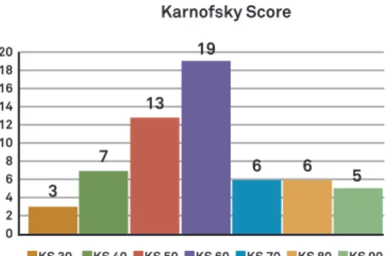
clinically (KS) evaluated. Neurologically, 6 patients presented with complete deicit (Frankel A), 3 with Frankel B, 23 with Frankel C, 12 with Frankel D, and 16 with Frankel E (Figure 2). he KS in our sample varied from 30 to 90. hree patients (5%) presented with a KS of 30, 7 patients with 40, 13 patients with 50, 19 patients with 60, 6 patients with 70, 6 patients
with 80, and 5 patients with 90 (Figure 3).
Fifteen of the 60 patients (25%) were evaluated with bone
scintigraphy. All 15 showed spinal uptake; 5 of these patients
presented with difuse skeletal uptake; and 3 presented with
skull, sternum, and rib uptake.
Until the therapeutic decision was made, no patient was
given an entire neuroaxis MRI evaluation due to time cons-traints and MRI availability. In contrast, all of the patients
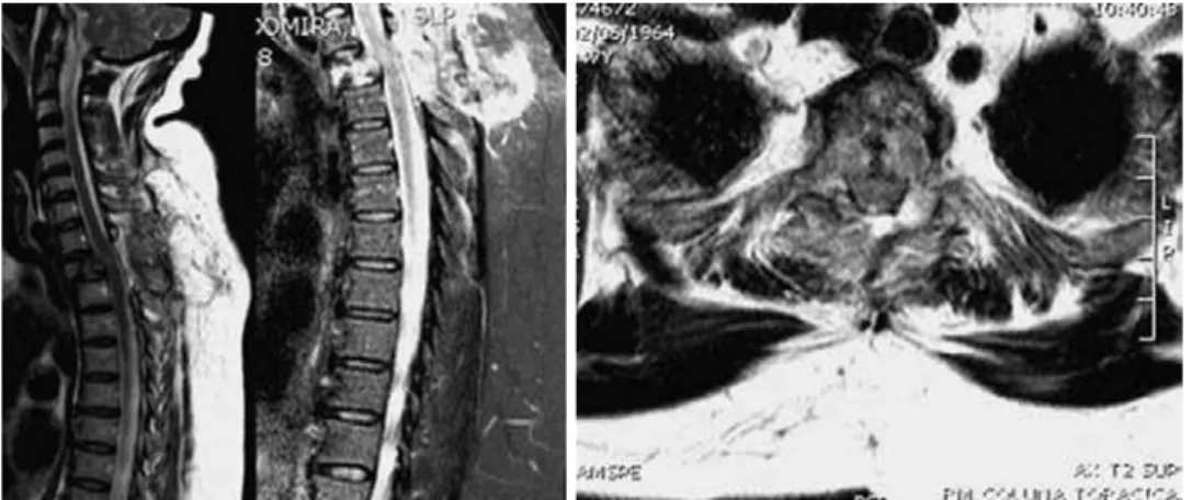
Figure 1. Left: MRI images of a 47-year-old female patient with no previous oncological history who presented with acute paraparesis
800 Arq Neuropsiquiatr 2013;71(10):798-801
underwent local spine MRIs directed at the spinal cord com pression site.
In this study, 78% of spinal metastases were localized in
the thoracic spine, 41% in the lumbar spine, 13% in the cervi cal spine, and 10% in the sacral spine.
Before the treatment decision was made, only 25% of pa tients had undergone bone scintigraph y, 30% had received thoracic and abdominal tomography, and no patient had a
complete spine MRI evaluation.
In the search for metastases in important internal organs,
only 18 patients were evaluated by a thoracic or abdominal
CT scan. In ive of these cases, only difuse lymphadenopathy
was noted; pulmonary nodules were observed in four cases, liver metastasis in three, and pleural thickening in one.
hirty-nine of the 60 (65%) patients had received a histo
pathological diagnosis prior to admission. Of these diagnoses,
11 were in the breast, 11 were in the prostate, and 5 were in the lung; 4 patients had multiple myeloma, 3 had colon cancer, and
2 had non-Hodgkin’s lymphoma. Bladder, kidney, and larynx
cancer were reported by one patient each (Figu re 4).
he TSS was not completely fulilled in 75% of patients (Table); rather, the decisions concerning conservative, pallia
tive, and excisional treatment were based on clinical, neuro logical, and imaging data.
Fifty-ive percent of patients were treated conservatively
with radiotherapy; 33% underwent a decompressiononly ap
proach, decompression was added to a spine ixation in 5%, 4%
only underwent a diagnostic percutaneous biopsy, and an an terior decompression approach was used in 3% of cases.
DISCUSSION
he TSS is a tool used to determine survival prognosis. he extent and complexity of disease management depends on the patient’s prognosis and life expectancy; patients with life expectancies less than 6 months are treated conserva
tively or with palliative surgery, a life expectancy of 6 to 12 months involves palliative surgery, and the expectation for patients with a life expectancy greater than 12 months is ex cision surgery12,13.
Prognostic prediction scales should be based on accessi
ble data. Metastatic disease is a predictor of poor outcome in
cancer patients; in particular, metastatic disease has a high
potential of mortality, morbidity, and deinitive neurological deicits, such as paraplegia and paraparesis. Time is the main
factor involved; delayed clinical or surgical treatments may
generate irreversible neurological deicits7.
In our sample, most of the patients who sought treatment
for symptomatic spinal metastases presented with few neu
rological deicits, and they were healthy enough for surgery. A full evaluation of all the TSS variables requires sophisti
cated imaging diagnostic tools with expensive, complex, time consuming, and not always readily available sources. Only a
quarter of our patients were able to completely satisfy the re
quirements for the TSS before the decision-making process.
Figure 3. Patient’s clinical status at admission (Karnofsky
Score – KS).
Table 1. The percentage of patients able to fulfill each TSS
item at the time of decision making.
TSS item Completed (%)
General condition (Karnofsky) 100%
Neurological status (Frankel) 100%
Primary cancer site 65%
Metastases in important internal organs 30%
Metastases in vertebral bodies 25%
Bone metastases in extra-spinal sites 25%
Figure 2. Neurological status of patients at admission,
presented as the Frankel scale.
Frankel Scale
Frankel A
Frankel B
Frankel C
Frankel D
Frankel E 16
6 3
23
23
Figure 4. Primary cancer sites at admission.
Primary Cancer Site
Unknown
Breast
Prostate
Lung
Multiple Myeloma
Colon
Non Hodgkin Lymphoma
Bladder
Kidney
Larinx 21
11 11
5 4
3 2 1 1 1
Karnofsky Score
20 18 16 14 12 10 8 6 4 2 0
13
7
3
KS 30 KS 40 KS 50 KS 60 KS 70 KS 80 KS 90 19
6 6
801
Matheus Fernandes de Oliveira et al. Tokuhashi scoring: vertebral metastasis
Many other systems to predict the prognosis and guide de
cision making are available, such as the Sioutos, Tomita, Van der Linden, and Bauer scores. he TSS has been the most wide
ly accepted and used scoring system, and it has signiicant pre dictive value14–18. However, every score, even with its particu larities, requires complete evaluation of the systemic disease,
including a histological diagnosis. Several authors have
demons-trated that the primary tumor site is the most important prog nostic factor for patient survival, and this is well accepted19.
However, in vertebral metastasis with spinal cord compres sion, there is an urgent need for decisionmaking, and the histo logical diagnosis and tumor staging data require time that is not available before treatment7. he histological diagnosis of prima
ry cancer requires a prior sample for analysis, sample ixation,
slide preparation, staining, and immunohistochemical analysis
for a deinitive diagnosis. his could take 2 to 5 days using stan dard services. A bone scintigraphy study protocol also demands
several hours for completion. CT scans of the vital organs and MRI require fasting time and the availability of equipment.
Although the apparatuses for diagnosis are present in the study hospital, clinical and neurological conditions and the
speciic protocols of radiological studies do not allow for full
patient assessment before surgery. Furthermore, complete evaluations sometimes require the use of more than one
device, which undoubtedly dramatically increases the time required to complete the tests.
he urgent treatment of vertebral metastasis still remains
paramount to protect spinal cord vitality1,2,6,8,11,14–17. Cancer
staging data were absent in the majority of our cases until the moment of therapeutic intervention in the spinal cord compression cases.
hus, some authors maintain that the surgical decision
cri teria should be based on clinical and neurological disor ders instead of prognostic scales1,20. Our results support those views. Although diagnostic equipment was available, the neu rological status associated with patient health is urgent, and
the multiplicity of required tests prevents complete fulillment of the TSS before making a treatment decision. To our know-ledge, the applicability of the TSS in clinical conditions outside
research protocols has not been previously evaluated in the li
terature. herefore, our reasoning appears to be a novelty.
CONCLUSIONS
In the majority of patients with vertebral metastasis, TSS
variables were incomplete, and the system was not useful in guiding treatment types.
References
1. Kilbride L, Cox M, Kennedy CM, Lee SH, Grant R. Metastatic spinal cord compression: a review of practice and care. J Clin Nurs 2010;19:1767-1783.
2. Paton GR, Frangou E, Fourney DR. Contemporary treatment strategy for spinal metastasis: the “LMNOP” system. Can J Neurol Sci 2011;38:396-403.
3. Shiue K, Sahgal A, Chow E, et al. Management of metastatic spinal cord compression. Expert Rev Anticancer Ther 2010;10:697-708. 4. Bauer HC, Tomita K, Kawahara N, Abdel-Wanis ME, Murakami H.
Surgical strategy for spinal metastases. Spine (Phila Pa 1976) 2002;27:1124-1125.
5. Bauer HC, Wedin R. Survival after surgery for spinal and extremity metastases. Prognostication in 241 patients. Acta Orthop Scand 1995;66:143-146.
6. Bilsky MH, Lis E, Raizer J, Lee H, Boland P. The diagnosis and treatment of metastatic spinal tumor. Oncologist 1999;4:459-469.
7. Choi D, Crockard A, Bunger C, et al. Review of metastatic spine tumor classification and indications for surgery: the consensus statement of the Global Spine Tumour Study Group. Eur Spine J 2010;19:215-222.
8. Hessler C, Vettorazzi E, Madert J, Bokemeyer C, Panse J. Actual and predicted survival time of patients with spinal metastases of lung cancer: evaluation of the robustness of the Tokuhashi score. Spine (Phila Pa 1976) 2011;36:983-989.
9. Tokuhashi Y, Matsuzaki H, Oda H, Oshima M, Ryu J. A revised scoring system for preoperative evaluation of metastatic spine tumor prognosis. Spine (Phila Pa 1976) 2005;30:2186-2191.
10. Tokuhashi Y, Matsuzaki H, Toriyama S, Kawano H, Ohsaka S. Scoring system for the preoperative evaluation of metastatic spine tumor prognosis. Spine 1990;15:1110-1113.
11. Hirabayashi H, Ebara S, Kinoshita T, et al. Clinical outcome and
survival after palliative surgery for spinal metastases: palliative surgery in spinal metastases. Cancer 2003;97:476-484.
12. Itshayek E, Yamada J, Bilsky M, et al. Timing of surgery and radiotherapy in the management of metastatic spine disease: a systematic review. Int J Oncol 2010;36:533-544.
13. Leithner A, Radl R, Gruber G, et al. Predictive value of seven preoperative prognostic scoring systems for spinal metastases. Eur Spine J 2008;17:1488-1495.
14. Papastefanou S, Alpantaki K, Akra G, Katonis P. Predictive value of Tokuhashi and Tomita scores in patients with metastatic spine disease. Acta Orthop Traumatol Turc 2012;46:50-56.
15. Polly DW Jr, Chou D, Sembrano JN, Ledonio CG, Tomita K. An analysis of decision making and treatment in thoracolumbar metastases. Spine (Phila Pa 1976) 2009;34(22 Suppl):S118-S127.
16. Putz C, Wiedenhöfer B, Gerner HJ, Fürstenberg CH. Tokuhashi prognosis score: an important tool in prediction of the neurological outcome in metastatic spinal cord compression: a retrospective clinical study. Spine (Phila Pa 1976) 2008;33:2669-2674.
17. Tomita K, Kawahara N, Kobayashi T, Yoshida A, Murakami H, Akamaru T. Surgical strategy for spinal metastases. Spine (Phila Pa 1976) 2001;26:298-306.
18. Ulmar B, Huch K, Naumann U, et al. Evaluation of the Tokuhashi prognosis score and its modifications in 217 patients with vertebral metastases. Eur J Surg Oncol 2007;33:914-919.
19. Wibmer C, Leithner A, Hofmann G, et al. Survival analysis of 254 patients after manifestation of spinal metastases: evaluation of seven preoperative scoring systems. Spine (Phila Pa 1976) 2011;36:1977-1986.