903
Arq Neuropsiquiatr 2012;70(11):900-904
Intracerebral microbleeds in sepsis:
susceptibility-weighted MR imaging findings
Micro-hemorragias intracerebrais na sepse: achados de imagem na RM ponderada em
susceptibilidade magnética
Diogo Goulart Corrêa1, Luiz Celso Hygino Cruz Júnior2, Paulo Roberto Valle Bahia3, Emerson Leandro Gasparetto2,3
1MD; Medical Resident of the Department of Radiology, Universidade Federal do Rio de Janeiro (UFRJ), Rio de Janeiro RJ, Brazil; 2MD; Neuroradiologist at Clínica de Diagnóstico por Imagem/Diagnósticos da América S/A (CDPI/DASA), Rio de Janeiro RJ, Brazil; 3MD, Ph.D; Assistant Professor of Radiology (Neuroradiology), UFRJ, Rio de Janeiro RJ, Brazil.
Correspondence: Diogo Goulart Corrêa; Rua Rodolpho Paulo Rocco 255 / Cidade Universitária / Ilha do Fundão; 21941-913 Rio de Janeiro RJ - Brasil; E-mail: [email protected]
Conflict of interest: There is no conflict of interest to declare.
Received 15 April 2012; Received in final form 17 May 2012; Accepted 24 May 2012
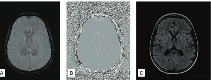
We present two patients with sepsis and intracerebral micro-bleeds. he irst case is a nine years old girl who presented visual hallucinations, tremors in the limbs, and an episode of generalized tonic-clonic seizure in the 12th day of an otherwise successfully
treatment of a pulmonary sepsis. Brain magnetic resonance im-aging (MRI) showed numerous small rounded foci of decreased signal intensity on susceptibility-weighted imaging (SWI) spread throughout the brain, predominantly in the corpus callosum (Fig 1), which had high signal intensity on the phase map of SWI, suggesting blood deposits. he remaining conventional MRI se-quences were normal. he patient and her mother denied any history of head trauma. During hospitalization, platelets counts, partial thromboplastin time, prothrombin time, and international normalized ratio were always normal.
he second patient is a 40 years old woman treating a septic shock of urinary origin for three weeks, who present-ed generalizpresent-ed tonic-clonic seizures. SWI showpresent-ed linear low signal intensity on the cortex surface, mainly in frontal lobes, and multiple foci of low signal intensity on the subcortical
white matter and cerebellum, which had high signal inten-sity on the phase images of SWI, suggesting areas of subarach-noid hemorrhages in the frontal lobes and microbleeds into the subcortical white matter and cerebellum (Fig 2). During hospitalization, D-dimer was normal. Although she had some altered values in platelets count (100,000/mm3, was the lower
value), prothrombin time (worst INR value was 2.3), and par-tial thromboplastin time, due to sepsis, she did not developed disseminated intravascular coagulation.
he typical imaging features of intracerebral microbleeds are small foci of decreased signal intensity on gradient-recalled echo T2* and/or SWI on MRI, usually without correspondence on others sequences1. Generally, microbleeds are related with
hemorrhagic transformation of an ischemic stroke, recurrence of spontaneous intracerebral bleeding, cerebral autosomal domi-nant arteriopathy with subcortical infarcts and leukoencepha-lopathy, cerebral amyloid angiopathy and trauma1. here are few
studies correlating intracerebral microbleeds with infective en-docarditis2, but none with other causes of sepsis.
Fig 1. Susceptibility-weighted imaging (A) and phase images of susceptibility-weighted imaging (B) show multiple foci of low signal intensity on genu and splenium of the corpus callosum and subcortical white matter on susceptibility-weighted imaging, with high signal intensity on the phase images of susceptibility-weighted imaging, suggesting blood deposits. FLAIR image (C) on the same position shows no abnormalities.
904 Arq Neuropsiquiatr 2012;70(11):900-904
1. Koennecke HC. Cerebral microbleeds on MRI Prevalence, associations, and potential clinical implications. Neurology 2006;66:165-171. 2. Klein I, Iung B, Labreuche J, et al. Cerebral microbleeds are frequent in
infective endocarditis: a case-control study. Stroke 2009;40:3461-3465. 3. Lundy DJ, Trzeciak S. Microcirculatory dysfunction in sepsis. Crit Care
Clin 2009;25:721-731.
4. Mittal S, Wu Z, Neelavalli J, Haacke EM. Susceptibility-weighted imaging: technical aspects and clinical applications, part 2. AJNR Am J Neuroradiol 2009;30:232-252.
5. Conforto AB, Lucato LT, Leite CC, Evaristo EF, Yamamoto FI, Scaff M. Cerebral microbleeds and intravenous thrombolysis: case report. Arq Neuropsiquiatr 2006;64:855-857.
References
Fig 2. Susceptibility-weighted imaging showing the posterior fossa (A) and at the skull convexity (B) demonstrate multiple foci of low signal intensity on the subcortical white matter and cerebellum, suggesting microbleeds, and linear low signal intensity on the surface of the frontal cortex, compatible with subarachnoid hemorrhage. Post-contrast-T1 weighted image on the same position as A (C) and FLAIR image on the same position as B (D) show no abnormalities. The rest of the exam did not show any sign of aneurysms or meningitis.
A
B
D
C
Histopathological analysis of these microbleeds shows focal hemosiderin deposition, which can be an evidence of microangiopathy1. Furthermore, there is evidence of
mi-crocirculation dysfunction, including the cerebral, due to sepsis3. Then, we hypothesize that endothelium
dysfunc-tion, generated by sepsis of any origin, and may be a cause of intracerebral microbleeds.
Gradient-recalled echo T2* and SWI are especially sen -sitive for detection of these kind of hemorrhages. hese se-quences have been recently incorporated into the daily practice, but they are not always done in a sepsis scenario, because the majority of these patients are clinically unstable and needs a fast exam. hus, the presence of intracerebral microbleeds in patients with sepsis may be more common than we currently suppose. Probably, this type of bleeding is not being detected on these patients because the more sensi-tive sequences are not being performed.
SWI is more sensitive than gradient-recalled echo T2* in detecting size, number, volume, and distribution of hemor-rhagic lesions4. hen, if SWI is available, this sequence should
be performed, even in detriment of gradient-recalled echo T2* in order to save time.
Furthermore, intracerebral microbleeds are a frequent inding in brain MRI, even in healthy people. Widespread use of gradient-recalled echo T2* and/or SWI has increased their detection in several diseases, in which generate great con-cern for clinical management, such as in ischemic stroke5,
spontaneous intracerebral bleeding, cerebral amyloid angi-opathy and trauma1. In this meaning, the importance of