Arq Neuropsiquiatr 2008;66(1):111-113
111
Migrating intraventricular cysticercosis
Magnetic resonance imaging indings
Antônio Luiz Eiras de Araujo
1,2, Rosana Souza Rodrigues
1,2, Edson Marchiori
2,
Ricardo Andrade Pinheiro
1, Marlo Flores
1, José Ricardo Duarte Alves
1, Emerson L. Gasparetto
2neurocisticercose intraventricular Migrada: acHados de ressonÂncia MagnÉtica
1Hospital Barra D’Or; 2Department of Radiology of the Federal University of Rio de Janeiro, Rio de Janeiro RJ, Brazil.
Received 24 July 2007, received in inal form 29 October 2007. Accepted 30 November 2007.
Dr. Emerson L. Gasparetto – Rua Lopes Trovão 88 / 1702B - 24110-071 Niterói RJ - Brasil.
Neurocysticercosis (NCC) is the most common par-asitic infestation affecting the central nervous system (CNS)1,2. It is frequent cause of seizures and
hydrocepha-lus in adults from endemic regions, including Brazil3,4. Brain
parenchymal involvement occurs in 60 to 92% of patients with NCC, but intraventricular (IV) lesions are seen in only 7 to 20% of cases1,5,6. Although NCC is usually self-limited
by the life cycle of the parasite, intraventricular involve-ment is often more dificult to manage due to obstruc-tion of cerebrospinal luid (CSF) pathways and ependy-mal inlammation1,2.
Intraventricular neurocysticercosis (IVNCC) can pro-duce mechanical obstruction of CSF flow anywhere in the ventricular system, resulting in hydrocephalus or fo-cal neurologifo-cal deicits. The symptoms can also result from ependymal inlammation associated with cyst de-generation, or from diffuse meningitis causing commu-nicating hydrocephalus1,2,7. The intraventricular cysts may
circulate freely throughout the CSF pathways or become
attached to the ependyma anywhere in the ventricles, but their predilection is for the occipital horn of the lateral and fourth ventricles1,4,7. Although the obstruction of the
CSF low is a common cause of symptoms in patients with IVNCC, reports demonstrating the migrating cysts causing hydrocephalus are extremely rare8,9.
We report a case of intraventricular neurocysticerco-sis presenting with hydrocephalus due to third ventricle obstruction secondary to migrating cysticercus.
case
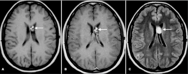
A 29-year-old female patient presented with acute onset of severe headache and nausea. The neurological and ophthal-mologic examinations were unremarkable. She referred a his-tory of neurocysticercosis two years before, which was treated with albendazole. At that time, the MR imaging showed multiple well-deined round lesions in the frontal horn of the left lateral ventricle, which had high signal on FLAIR, T1- and T2-weighted images, showing no signiicant enhancement after contrast ad-ministration. These lesions were occasioning mild compression
Arq Neuropsiquiatr 2008;66(1)
112
Migrating intraventricular cysticercosis Araujo et al.
on the septum pellucidum. The third and lateral ventricles had normal size, with no signals of hydrocephalus (Fig 1).
Laboratorial investigation, routine blood chemistry indings were normal. The CSF revealed 94 white blood cells with 58% segmented neutrophils and 36% lymphocytes, protein level was 47 mg/dL, and glucose level was 52 mg/dL. The CSF was negative for VDRL, toxoplasmosis and HIV antibodies, and the CSF culture was sterile. The CSF immunoluorescence and ELISA tests were negative for cysticercosis antibodies.
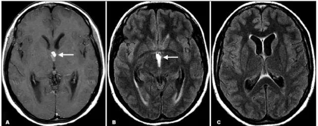
The new MR imaging demonstrated a third ventricle multi-loculated mass with high signal on FLAIR, T1- and T2-weighted images, showing mild ring enhancement after contrast admin-istration. The lesion was obstructing the CSF low, resulting in mild hydrocephalus and discrete ependimary transudation (Figs 2 and 3). No scolex or additional ventricular and parenchymal lesions were seen.
Because the worsening symptoms, she underwent a surgery for evacuation of the third ventricle lesion. During the surgery, several cysts were removed. However, because the severe arach-noidal and ependymal scarring, it was not possible to remove
the lesion completely. The histological examination conirmed the diagnosis of racemosus cysticercosis.
The patient gave informed consent for the publication of the case.
discussion
Cysticercosis is the most common parasitic infestation of the CNS, and it is caused by Taenia solium’s invasion in its larval stage. The CNS involvement occurs in 60 to 90% of patients with cysticercosis4-6. Brain parenchymal
involvement occurs in 60 to 92% of cases, but IV NCC oc-curs in only 7 to 20% of patients1,4,6.
The IV NCC is frequently caused by Cysticercus cel-lulosae, however Cysticercus racemosus can also infect the ventricular system1,4. The lesions are more commonly
seen in the IV ventricle (54-64%), followed by the III ven-tricle (23-27%), the lateral venven-tricles (11-14%) and Sylvi-us aqueduct (9%)1,2,4. Intraventricular cysts may become
symptomatic at the time of implantation secondary to
Fig 2. Axial T1-weighted post-gadolinium (A) and FLAIR (B and C) MR images show a third ventricle multiloculated mass show-ing mild rshow-ing enhancement after contrast administration (arrow). The lesion was obstructshow-ing the CSF low (arrow), resultshow-ing in mild hydrocephalus and discrete ependimary transudation.
Arq Neuropsiquiatr 2008;66(1)
113 Migrating intraventricular cysticercosis Araujo et al.
obstruction of CSF low, with consequent hydrocepha-lus and the symptoms and signs of increased intracranial pressure. When involution begins, the inlammatory reac-tion around a dead or dying cyst produces ependymitis, scarring, obstruction, and ventriculitis1,2,4,7.
Reports demonstrating intraventricular migrating neu-rocysticercosis are rare8,9. Thomas et al.9 reported a
10-year-old boy, who presented with headache and vomiting of one-week duration. The MR imaging showed marked hydrocephalus, and a cystic lesion isointense to CSF, with a thin wall and a small nodule (scolex) in the left temporal horn. The contrast images performed 20 minutes later re-vealed migration of the cyst to the occipital horn.
In this report, we present a rare case of migrating in-traventricular cysticercosis obstructing the third ventricle, presenting with headache and nausea secondary to hy-drocephalus. The patient referred a previous diagnosis of neurocysticercosis, and the prior MR imaging showed le-sions suggestive of NCC at the frontal horn of the left lat-eral ventricle. The follow-up MR imaging demonstrated a multiloculated enhancing mass in the third ventricle, with secondary dilatation of the lateral ventricles. No other lesion were seem Although a surgical approach was per-formed, the lesion could not be completely removed due to severe arachnoidal and ependymal scarring.
In conclusion, intraventricular neurocysticercosis is an uncommon form of the disease, which can present as migrating cysts resulting in CSF low obstruction and hydrocephalus. The surgical treatment can be indicated, but complete resection of the cysts can be dificult due to the arachnoidal and ependymal scarring secondary to the lesions.
reFerences
1. Citow JS, Johnson JP, McBride DQ, Ammirati M. Imaging features and surgery-related outcomes in intraventricular neurocysticercosis. Neu-rosurg Focus 2002;12:e6.
2. Cuetter AC, Andrews RJ. Intraventricular neurocysticercosis: 18 consec-utive patients and review of the literature. Neurosurg Focus 2002;12:e5. 3. Agapejev S. Clinical and epidemiological aspects of neurocysticercosis
in Brazil: a critical approach. Arq Neuropsiquiatr 2003;61:822-828. 4. Amaral L, Maschietto M, Maschietto R, et al. Unusual manifestations
of neurocysticercosis in MR imaging: analysis of 172 cases. Arq Neu-ropsiquiatr 2003;61:533-541.
5. Montemor MR Netto, Gasparetto EL, Faoro LN, et al. Neurocysticerco-sis: a clinical and pathological study of 27 necropsied cases. Arq Neu-ropsiquiatr 2000;58:883-889.
6. Teitelbaum GP, Otto RJ, Lin M, et al. MR imaging of neurocysticerco-sis. Am J Roentgenol 1989;153:857-866.
7. Pfuetzenreiter MR, Avila-Pires FD. Clinical manifestations in patients with computerized tomography diagnosis of neurocysticercosis. Arq Neuropsiquiatr 1999;57:653-658.
8. Rangel-Guerra RA, Herrera J, Elizondo G, Gonzalez-Morantes J. Neu-rocysticercosis. Arch Neurol 1988;45:492.