Letters to the Editor
Radiol Bras. 2015 Jul/Ago;48(4):263–270
267
Bruno Niemeyer de Freitas Ribeiro1, Tiago Medina Salata2, Lívia de Oliveira Antunes2, Edson Marchiori3 1. Instituto Estadual do Cérebro Paulo Niemeyer, Rio de Janeiro, RJ, Brazil. 2. Hospital Casa de Portugal / 3D Diagnóstico por Imagem, Rio de Janeiro, RJ, Brazil. 3. Universidade Federal do Rio de Janeiro (UFRJ), Rio de Janeiro, RJ, Brazil. Mailing Address: Dr. Bruno Niemeyer de Freitas Ribeiro. Instituto Estadual do Cérebro Paulo Niemeyer – Ser-viço de Radiologia. Rua do Rezende, 156, Centro. Rio de Janeiro, RJ, Brazil, 20231-092. E-mail: [email protected]. bones. Desmoplastic fibromas may occur at any age range,
al-though its higher incidence is observed at the first three decades of life(1–3,6). Despite conflicting data, it seems there is no
predi-lection for sex(2,6). Local recurrence is frequently observed in cases
where complete resection is not.Clinically, the patients are either asymptomatic or may present with pain, edema, joint effusion and pathological fracture(1–6). The differential diagnosis should
con-sider rhabdomyosarcoma, fibrosarcoma, giant cell tumor, among others. Despite the imaging methods usefulness in the lesion delimitation, the diagnosis is histopathological.
At MRI, most lesions present with iso/hyposignal on T1-weighted images and low signal intensity on T2-T1-weighted im-ages(1,3–6), but there are reports of lesions with hypersignal on
T2-weighted images(1–3,6). The enhancement may be variable, and
according to some authors, such variation may be a result of the cellular content of the lesion(3,4). In the present case, there was
homogeneous iso/hyposignal on T1-weighted images and subtle hypersignal on T2-weighted images, with foci of low signal inten-sity. After gadolinium injection, marked contrast enhancement, with noticeable perineural dissemination through the third divi-sion of the trigeminal nerve were observed. Such aspects on T2-weighted sequences, and the presence of perineural dissemina-tion are not commonly observed as compared with the typical imaging pattern described at MRI.
Reports on diffusion in desmoplastic fibromas were not found in the literature. In the present case, areas of diffusion restriction were not observed. Recent studies highlight the use of diffusion-weighted imaging in the evaluation of head and neck lesions, showing that apparent diffusion coefficient < 1.22 × 10⁻³
mm2 /s are suggestive of malignancy(7). In the present case, the value for
apparent diffusion coefficient was 1.45 × 10⁻³ mm2
/s, corroborat-ing the previously described findcorroborat-ings.
The authors conclude that the diagnosis of desmoplastic fi-bromas should be considered in patients under the age of 30 pre-senting with tumor particularly located in the mandible, and that such a hypothesis cannot be ruled out in case of less noticeable foci of hyposignal on T2-weighted images.
REFERENCES
1. Woods TR, Cohen DM, Islam MN, et al. Desmoplastic fibroma of the mandible: a series of three cases and review of literature. Head Neck Pathol. 2015;9:196–204.
2. Nedopil A, Raab P, Rudert M. Desmoplastic fibroma: a case report with three years of clinical and radiographic observation and review of the literature. The Open Orthopaedics Journal. 2013;7:40–6.
3. Kim OH, Kim SJ, Kim JY, et al. Desmoplastic fibroma of bone in a toe: radiographic and MRI findings. Korean J Radiol. 2013;14:963–7. 4. Kang DM, Juhng SK, Sohn YJ, et al. Imaging findings of desmoplastic
fibroma rarely involving the clavicle: case report. Korean J Radiol. 2014; 15:130–3.
5. Frick MA, Sundaram M, Unni KK. Imaging findings in desmoplastic fibroma of bone: distinctive T2 characteristics. AJR Am J Roentgenol. 2005;184:1762–7.
6. Moorjani V, Stockton V. Desmoplastic fibroma with perineural extension. AJR Am J Roentgenol. 2005;185:1498–9.
7. Gonçalves FG, Ovalle JP, Grieb DFJ, et al. Diffusion in the head and neck: an assessment beyond the anatomy. Radiol Bras. 2011;44:308–14.
http://dx.doi.org/10.1590/0100-3984.2014.0135
Creutzfeldt-Jakob dementia
Demência por doença de Creutzfeldt-Jakob
Dear Editor,
A 72-year-old woman with rapidly progressive dementia, be-havioral changes and apraxia of gait for seven months, extrapyra-midal signs and diffuse myoclonus. Electroencephalography
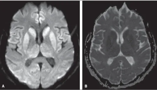
dem-onstrated periodic electric activity with high amplitude acute phase waves diffusely distributed over the cortex. The cerebrospinal fluid was normal. Magnetic resonance imaging (MRI) was performed (Figure 1).
The association of clinical, radiological, electroencephalic or cerebrospinal fluid findings (presence of 14-3-3 brain protein in diseased patient for less than two years – absent in this case),
Figure 1.A: Axial magnetic resonance imaging of the skull demonstrating foci of hypersignal at diffusion-weighted sequences in the heads of the caudate nuclei, putamina, thalami and medial occipitotemporal gyri. B: At the ADC mapping, the low signal intensity in the same region confirms the diffusion restriction.
Letters to the Editor
Radiol Bras. 2015 Jul/Ago;48(4):263–270
268
allows for a probable diagnosis of Creutzfeldt-Jakob dementia (CJD). The differential diagnosis is made with other diseases as-sociated with dementias, as follows: a) Alzheimer’s disease, that does not present with alterations observed at diffusion-weighted images; b) vascular dementia, with multiple infarcts, but with ab-normalities at diffusion-weighted images only in cases of recent infarcts, and without diffuse cortical involvement; c) other en-cephalopathies that present alterations restricted to the cortex at diffusion-weighted images (such as MELAS – mitochondrial myopathy, encephalopathy, lactic acidosis and stroke-like episodes – a genetic metabolic disease occurring in younger patients); venous hypertensive encephalopathy and chronic herpes simplex encephalitis.
CJD is a subacute spongiform encephalopathy that presents with rapidly progressive dementia, and is the most frequent among rare prion diseases. Myoclonus, pyramidal, extrapyramidal and cerebellar signs are associated. Psychiatric symptoms are observed at the first six months; and progressive immobility, cortical blind-ness, dysphagia and mutism constitute late signs of the disease. Death generally occurs one year after the symptoms onset, since there is no therapy to prevent a fatal outcome(1). The disease is
subdivided into sporadic (85% of cases), familial, iatrogenic and a less common, recently described variant related to epidemic bo-vine spongiform encephalopathy(2).
Like in other acute encephalopathies, the 14-3-3 brain pro-tein may be present in the cerebrospinal fluid. Encephalography may demonstrate periodic activity composed of three-phase high-frequency waves over attenuated background activity.
At MRI, hyperintense signal is observed in the basal ganglia, putamen and later in the cerebral cortex on T2-weighted and FLAIR sequences(1,3). Such alterations suggest CJD, more than
any other dementia disorder(4).
At early stages of the disease, however, conventional imag-ing studies may present normal results. Diffusion-weighted se-quences may favor an early diagnosis, demonstrating abnormal hyperintense foci even before the appearance of electroencepha-lographic alterations, and should be performed in case of suspi-cion of CJD(1,4,5). Diffusion restriction is observed, probably in
association with the cytotoxic edema secondary to spongiform degeneration and to accumulation of abnormal cytoplasmic vacu-oles. As the disease progresses, global atrophy is observed and, in general, this is the only alteration depicted at computed tomogra-phy(1,4,6).
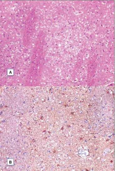
Histopathological analysis (Figure 2A) demonstrated spongiform alterations with variable intensity in the neuropil, markedly in the caudate nucleus, putamen, and in the region CA1 of the hippocampus, moderately in the frontal and temporal cor-tices, and slightly in the parietal and occipital cortices. Immuno-histochemical analysis demonstrated gliosis (Figure 2B). Such results are compatible with a definite diagnosis of CJD. The le-sions distribution, the absence of similar cases in the family, and the absence of a known infectious source are compatible com the sporadic presentation of the disease(4,7). Such a diagnosis is
relevant for controlling the transmission and to rule out treatable causes of dementia(1,8).
REFERENCES
1. Macfarlane RG, Wroe SJ, Collinge J, et al. Neuroimaging findings in human prion disease. J Neurol Neurosurg Psychiatry. 2007;78:664–70. 2. Venneti S. Prion diseases. Clin Lab Med. 2010;30:293–309. 3. Meissner B, Kallenberg K, Sanchez-Juan P, et al. MRI lesion profiles in
sporadic Creutzfeldt-Jakob disease. Neurology. 2009;72:1994–2001.
Fabiano Reis1, Ana Laura Gatti Palma1, Ricardo Schwingel1, Hélio Henrique Jorge Torres1, Mariana Mari Oshima1, Luciano Souza Queiroz1, Fábio Rogério1
1. Universidade Estadual de Campinas (Unicamp), Campinas, SP, Brazil. Mailing Address: Dr. Fabiano Reis. Faculdade de Ciências Médicas, Universidade Estadual de Campinas, Departamento de Radiologia. Rua Tessália Vieira de Camargo, 126, Cidade Universitária Zeferino Vaz. Caixa Postal: 6111. Cam-pinas, SP, Brazil, 13083-887. E-mail: [email protected].
http://dx.doi.org/10.1590/0100-3984.2014.0109
4. Ukisu R, Kushihashi T, Tanaka E, et al. Diffusion-weighted MR imag-ing of early-stage Creutzfeldt-Jakob disease: typical and atypical manifes-tations. Radiographics. 2006;26 Suppl 1:S191–204.
5. Shiga Y, Miyazawa K, Sato S, et al. Diffusion-weighted MRI abnormalities as an early diagnostic marker for Creutzfeldt-Jakob disease. Neurology. 2004;63:443–9.
6. Manners DN, Parchi P, Tonon C, et al. Pathologic correlates of diffusion MRI changes in Creutzfeldt-Jakob disease. Neurology. 2009;72:1425– 31.
7. Head MW. Human prion diseases: molecular, cellular and population biology. Neuropathology. 2013;33:221–36.
8. Vitali P, Maccagnano E, Caverzasi E, et al. Diffusion-weighted MRI hyperintensity patterns differentiate CJD from other rapid dementias. Neurology. 2011;76:1711–9.
Figure 2.A: Section of the caudate nucleus demonstrating abundant small, optically empty vacuoles in the gray matter. The characteristic architecture of the caudate nucleus is highlighted by parallel white matter tracts through the gray matter (100× hematoxylin-eosin staining). B: GFAP immunohistochemistry dem-onstrating reactive astrocytes, a finding compatible with spongiform encephal-opathy (400×).
A