106 Copyright © 2015 The Korean Movement Disorder Society
LETTER TO THE EDITOR
http://dx.doi.org/10.14802/jmd.15013 / J Mov Disord 2015;8(2):106-107 pISSN 2005-940X / eISSN 2093-4939
Received: April 9, 2015 Revised: April 22, 2015 Accepted: April 22, 2015
Corresponding author: Myung Sik Lee, MD, Department of Neurology, Gangnam Severance Hospital, Yonsei University College of Medicine, 211 Eonju-ro, Gangnam-gu, Seoul 135-720, Korea / Tel: +82-2-2019-3322 / Fax: +82-2-3462-5904 / E-mail: [email protected]
cc his is an Open Access article distributed under the terms of the Creative Commons Attribution Non-Commercial License (http://creativecommons.org/ licenses/by-nc/3.0) which permits unrestricted non-commercial use, distribution, and reproduction in any medium, provided the original work is properly cited.
JMD
Parkinsonism and Dementia Associated with
Giant Virchow-Robin Spaces
Myung Sik Lee,1 Cheol Hyung Lyoo,1 Tae Sub Chung2
1Departments of Neurology and 2Radiology, Gangnam Severance Hospital, Yonsei University College of Medicine, Seoul, Korea
Virchow-Robbin space (VRS) denotes dilated subarachnoid space along the penetrating arteries to the level of capillaries. Giant VRSs (GVRS), deined as greater than 1.5 cm, are found in the basal ganglia along the lenticulostriate arteries, pons and midbrain along the collicular arteries and high cerebral convex-ity along the medullary arteries.1,2
CASE HISTORY
Most patients with GVRSs in the high cerebral convexity ex-hibit no neurological deicits,1,3-5 including motor and sensory
evoked potential studies.3 We present a patient who developed
parkinsonism and dementia associated with GVRSs. Positron emission tomography (PET) studies showed normal striatal do-pamine transporter uptake, but reduced glucose metabolism in the cerebral cortex and right thalamus.
A 64-year-old man developed bradykinesia and memory dis-turbances. On neurological examination, the patient was found to have a masked face. Glabellar and snouting reflexes were present. Speed and amplitude of inger and foot tapping were reduced bilaterally and were pronounced on the let side. Mus-cle tone was mildly increased in all four limbs tested. On pull tests, the patient stabilized after 3 to 4 backwards steps. He stood on widened base and walked with mildly reduced stride and cadence. he patient’s Uniied Parkinson’s Disease Rating Scale (UPDRS) total motor score was 15. here were no abnor-malities on cerebellar function tests. Mini-Mental State Exami-nation (MMSE) score was 27. Neuropsychological tests, howev-er, showed impairments (< 15 percentile for age and sex matched controls) in immediate and delayed recall of verbal and visual subjects, attention, confrontational naming, generative naming,
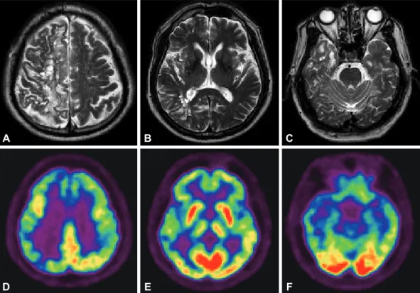
visuospatial function, and inhibitory control. T2 weighted brain magnetic resonance imaging (MRI) studies showed scattered high signal intensity and multiple round and septate cystic le-sions, mainly in the right parietal, frontal and temporal white matters (Figure A, B, and C). MR cerebral angiography showed no abnormalities. [18F]-FP-CIT PET studies showed normal
striatal uptake. [18F]-deoxyglucose PET studies showed
hypo-metabolism, predominantly involving the right thalamus and the right parietal, frontal and temporal cortical areas (Figure D, E, and F). On follow-up examination 4 years ater onset, there was worsening of parkinsonian motor deicits, particularly gait disturbances and postural instability, and further cognitive dys-functions. His UPDRS total motor score was 27 and MMSE score was 20. Follow-up brain MRI studies showed no signii-cant changes.
DISCUSSION
Giant Virchow-Robbin spaces in the high cerebral convexity are characterized by clustered sharp demarcated round, oval, or linear cystic lesions. On brain MRI studies, they did not enhance and are iso-intense with cerebrospinal luid.2,3 In approximately
80% of reported cases, GVRSs involved one cerebral hemi-sphere.1-5
Giant Virchow-Robbin spaces in the high cerebral convexity are usually found incidentally4,5 or during the evaluation for
non-specific neurological symptoms (e.g., fainting, dizziness, hearing impairment,1 headache, or visual change2). Rarely,
pa-tients may present with dementia.2 he patient reported herein
Parkinsonism-Dementia Associated with VR Spaces Lee MS, et al.
www.e-jmd.org 107
MR tractography studies of patients with GVRSs showed markedly reduced ibers in the afected ce-rebral hemisphere.4 Therefore, GVRSs and white
matter lesions in our patient may have disrupted cortical eferent ibers to the basal ganglia and afer-ent fibers to the motor-related cerebral cortices. The parkinsonism seen in our patient seemed to share a common pathogenic mechanism with the parkinsonism associated with Binswanger disease. Alternatively, right thalamic hypometabolism, rep-resenting reduced basal ganglia or cortical inputs to the thalamus, may also have contributed to the asymmetric parkinsonism of our patient.
In our patient, cortical glucose hypometabolism and consequent cognitive dysfunctions seemed to be associated with loss of aferent ibers to the cerebral cortex or cortical neuronal loss associated with ret-rograde axonal degeneration. Our patient demon-strated that GVRSs in the high cerebral convexity are
not always clinically silent but may cause progressive parkinsonism and dementia.
Conflicts of Interest
he authors have no inancial conlicts of interest.
REFERENCES
1. Ogawa T, Okudera T, Fukasawa H, Hashimoto M, Inugami A, Fujita H, et al. Unusual widening of Virchow-Robin spaces: MR appearance. AJNR Am J Neuroradiol 1995;16: 1238-1242.
2. Salzman KL, Osborn AG, House P, Jinkins JR, Ditchield A, Cooper JA, et al. Giant tumefactive perivascular spaces. AJNR Am J Neuroradiol 2005;26:298-305.
3. Ugawa Y, Shirouzu I, Terao Y, Hanajima R, Machii K, Mo-chizuki H, et al. Physiological analyses of a patient with ex-treme widening of Virchow-Robin spaces. J Neurol Sci 1998;159:25-27.
4. Wong SH, Das K, Puthuran M, Lecky B, White R. Giant Virchow-Robin spaces: functional magnetic resonance im-aging and tractography. Arch Neurol 2010;67:768-769. 5. Tseng HS, Ho CS, Chiu NC. Multiple giant Virchow-Robin
spaces. Pediatr Neurol 2013;49:143.
Figure 1. T2-weighted brain magnetic resonance imaging studies show mixture of high signal intensity lesions and numerous round and septate cystic lesions in the (A) right high cerebral convexity and (B and C) temporal lobes. [18F]-deoxyglucose PET studies show diffuse cerebral cortical hypometabolism, predominantly in the (D) bilateral
frontal, right parietal (E) right thalamic and (F) right temporal cortices. PET: positron emission tomography. A
D
B
E
C