Case Report
Key words
Takotsubo cardiomyopathy; echocardiography; myocardial contraction.
This report presents the late follow-up of a case of Takotsubo cardiomyopathy with good clinical outcome and improved left ventricular global systolic function. However, there was persistence of significant regional longitudinal systolic dysfunction evaluated using a new echocardiographic technique (speckle tracking), with corresponding measures of strain (S) and strain rate (SR). We emphasize the importance of this new method to monitoring this cardiomyopathy, since it identifies patients with persistent systolic dysfunction who will possibly benefit from maintenance of clinical therapy.
Two-Dimensional Strain in Takotsubo Cardiomyopathy
Carlos Bellini G. Gomes e Gustavo J. Veras
Clínica Medicar - Hospital São Carlos, Fortaleza, CE - BrazilMailing address: Carlos Bellini G. Gomes •
Rua Dr. José Lourenço, 938 - Meireles - 60160-110 - Fortaleza, CE - Brazil E-mail: [email protected], [email protected]
Manuscript received June 28, 2009; revised manuscript received September 11, 2009, manuscript accepted December 23, 2009.
laparoscopic cholecystectomy and cholangiography under general anesthesia. The surgery went well, and in the recovery room, the patient developed acute pulmonary edema and significant hemodynamic instability, requiring support with vasoactive drugs.
ECG showed sinus tachycardia without acute ischemic changes. Chest radiography revealed severe pulmonary venous congestion. Bedside echocardiogram showed cardiac chambers with normal dimensions, akinesia of left ventricular (LV) septal anterior, apical, anteroapical and inferoapical walls, severe LV systolic dysfunction (ejection fraction = 30% by the Simpson method), indirect signs of pulmonary arterial hypertension (systolic pulmonary artery pressure = 58 mmHg). Cardiac enzymes were high (CPK = 3,019/CKMB, mass = 90.4/ troponin I = 2.46). This picture suggested the possibility of coronary artery disease (CAD), and the patient underwent coronary angiography, which revealed no coronary lesions. Ventriculography showed apical and anterior akinesia, with preserved basal contractility, which characterizes the classic form of stress-induced cardiomyopathy or Takotsubo syndrome. The patient was admitted to the ICU, with progressive clinical improvement. She was discharged, and after one week, we performed a new echocardiogram that showed partial improvement of LV contractility. Apical hypokinesia persisted, but with an EF estimated at 59% and stage I LV diastolic dysfunction. Another echocardiogram was performed 15 days after the picture has shown complete resolution of segmental dyssynergy, with full recovery of global LV contractility (EF = 76% Teichholz and 70% Simpson), and normal pulmonary pressures, as well.
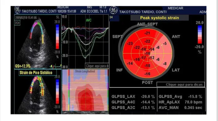
The patient was clinically well, under use of angiotensin-converting enzyme (ACE) [Check my insertion] and beta-blockers. She had recently returned to the clinic for echocardiography after 20 months of the event described. ECG revealed normal global and segmental LV contractility (EF = 72% Teicholz and 69% Simpson), as well as normal diastolic function (E/A ratio = 1.23, E/E´= 9). Considering the offline analysis of two-dimensional images obtained (apical sections of 4, 3 and 2 chambers), we could assess the LV regional systolic function, with the measures of longitudinal myocardial strain (S) in various segments, as shown in Table 1. There was a significant reduction in the rates of myocardial deformation in previously affected segments, mainly in the anterior and lateral regions (medial-basal), with significant reduction in overall strain (-15.8%) (Bull’s eye image - to the right of Figure 1).
Discussion
Takotsubo cardiomyopathy is traditionally characterized by the complete resolution of ventricular dysfunction4,5, with
Introduction
The stress-induced cardiomyopathy, also known as Takotsubo cardiomyopathy, is a syndrome characterized by a segmental left ventricular dysfunction that affects mainly the apical region1. It is triggered by several factors such as emotional or psychological stress2, major surgeries and severe systemic diseases. It presents a variable clinical picture ranging from chest pain and dyspnea to cardiogenic shock with ECG changes and elevation of myocardial necrosis markers, with no significant coronary disease at angiography, though. It is characterized by the rapid resolution of the picture, with improved ventricular function and, in most cases, complete recovery of left ventricular dyssynergy, with very good prognosis3. New echocardiographic methods have been applied in the evaluation of regional myocardial contractility, especially the two-dimensional strain. We report a case in which this new echocardiographic tool was used to further scrutinize ventricular function in late follow-up.
Case report
A 55-year-old hypertensive woman with normal preoperative exams, including electrocardiogram (ECG), echocardiogram (ECHO) and myocardial perfusion scintigraphy underwent
Case Report
Gomes & Veras Two-dimensional strain in Takotsubo cardiomyopathy
Arq Bras Cardiol 2010; 95(2): e35-e37 improvement in echocardiographic parameters, especially
an improved subjective analysis of segmental contractility and global systolic function. Echocardiography plays a key role in diagnosis, since it identifies segmental contractility alterations and other possible findings, such as obstruction in LV outflow tract, mitral valve failure and associated diastolic dysfunction. This examination also quantifies the LV global systolic function. Moreover, it allows following up ventricular function, identifying the complete resolution of changes in a short period. However, conventional echocardiography has the limitation of assessing only the contractility in its radial direction. The complex geometric arrangement of the myocardial fibers is widely known. It determines systolic mechanisms in several directions: radial (thickness), longitudinal (shortening) and circumferential (torsion).
New echocardiographic techniques have emerged in recent years, enabling the assessment of regional contractility through quantitative methods. These techniques include the two-dimensional strain through the speckle tracking method. The two-dimensional strain is a new ultrasound method used to measure the strain and myocardial strain rate6. It assesses motility by measuring the displacement of several
Table 1 - Longitudinal systolic function of left ventricle by measuring peak systolic two-dimensional strain in the relevant segments eval
Anterior Lateral Inferior Septal
Segments Basal Medial Apical Basal Medial Apical Basal Medial Apical Basal Medial Apical Strain (%) -5 -6 -10 -8 -6 -8 -14 -16 -16 -23 -22 -17
Figure 1 -To the left, analysis of systolic function by the two-dimensional strain method in the LV segments assessed (two-chamber apical section), where we observe
a signiicant reduction in peak systolic strain in anterior and lateral LV segments (red, blue and pink curves in curve chart). On the right, the polar map (Bull’s eye) representing the peak systolic strain of various LV segments, with a clear regional systolic dysfunction in the anterior and lateral segments with global strain of –15.8%.
acoustic points (speckles) naturally present in different interfaces of the myocardium, in a conventional two-dimensional image. Each point in a given region is followed over several frames throughout the cardiac cycle. These points are then tracked, and by means of calculations performed by specific software applications, the image is spatially and temporally processed, whereby these elements are recognized and selected6. The individual displacement of each point (speckle) represents the local tissue movement. By tracking these points, strain and strain rate are measured after a careful automatic analysis of the image quality. The diagnostic information of each trace is presented in a colored parametric form (combination of colors to data digitally collected), representing different quantitative indices of myocardial contractility. Thus, normal peak systolic strain is shown in red, dyskinetic areas or with very low systolic strain in dark blue, for example. Longitudinal and circumferential strains showed negative curves representing systolic shortening, while radial strain is positive. The following are considered normal values of longitudinal peak systolic strain: -20 to -22% (+/- 2.2%)7.
Few reports have applied this method to this important entity, increasingly diagnosed in our country. There is usually
Peak systolic strain
Case Report
Gomes & Veras
Two-dimensional strain in Takotsubo cardiomyopathy
Arq Bras Cardiol 2010; 95(2): e35-e37
a complete resolution of systolic dysfunction in a period of 2 to 4 weeks from the initial event, with improvement in left ventricular ejection fraction8. Burri et al9 monitored some patients with this cardiomyopathy and demonstrated through this new technique a significant reduction in regional systolic function, with a significant improvement over time.
Conclusion
This case addressed the application of two-dimensional strain in Takotsubo cardiomyopathy, and identified significant changes in regional contractility even after a follow-up period. A number of questions were raised about this method in treating this cardiomiopathy, such as the possibility of being a partially reversible disease, rather than the traditional definition of its complete resolution. Hence, the longitudinal two-dimensional strain technique was useful in monitoring
affected patients, since it is able to identify those that evolve with continuing changes in contractility and possibly require permanent treatment and careful follow-up.
Potential Conflict of Interest
No potential conflict of interest relevant to this article was reported.
Sources of Funding
There were no external funding sources for this study.
Study Association
This study is not associated with any post-graduation program.
References
1. Tsuchihashi K, Ueshima K, Uchida T, Oh-Mura N, Kimura K, Owa M, et al. Transient left ventricular apical ballooning without artery coronary stenosis: a novel heart syndrome mimicking acute myocardial infarction. J Am Coll Cardiol. 2001; 38 (1): 11-8.
2. Wittstein IS, Thiemann DR, Lima JA, Baughman KL, Schulman SP, Gerstenblith G, et al. Neurohumoral features of myocardial stunning due to sudden emotional stress. N Engl J Med. 2005; 352 (6): 539-48.
3. Merli E, Sutcliffe S, Gori M, Sutherland GG. Tako-tsubo cardiomyopathy: new insights into the possible underlying pathophysiology. Eur J Echocardiogr. 2006; 7 (1): 53-61.
4. Gianni M, Dentali F, Grandi AM, Sumner G, Hiralal R, Lonn E. Apical ballooning syndrome or takotsubo cardiomyopathy: A systematic review. Eur Heart J. 2006; 27 (13): 1523-9.
5. Kumar S, Mostow N, Grimm RA. Quick resolution of takotsubo cardiomyopathy: a brief review. Echocardiography. 2008; 25 (10): 1117-20.
6. Leitman M, Lysyansky P, Sidenko S, Shir V, Peleg E, Binenbaum M, et al. Two-dimensional strain: a novel software for real-time quantitative echocardiographic assessment of myocardial function. J Am Soc Echocardiogr. 2004; 17 (10): 1021-9.
7. Kang SJ, Lim HS, Choi BJ, Choi SY, Hwang GS, Yoon H, et al. Longitudinal strain and torsion assessed by two-dimensional speckle tracking correlate with the serum level of tissue inhibitor of matrix metalloproteinase-1, a marker of myocardial fibrosis, in patients with hypertension. J Am Soc Echocardiogr. 2008; 21 (8): 907-11.
8. Wittstein IS, Thiemann DR, Lima JAC, Baughman KL, Schulmann SP, Gerstenblith G, et al. Neurohumoral features of myocardial stunning due to sudden emotional stress. N Engl J Med. 2005; 352 (6): 539-48.
9. Burri MV, Navin CN, Lloyd SG, Hsiung MC, Dod HS, Beto RJ, et al. Assessment of systolic and diastolic left ventricular and left atrial function using vector velocity imaging in Takotsubo cardiomyopathy. Echocardiography. 2008; 25 (10): 1138-44.