Case 4 - 77-Year-Old Female Patient with Heart Failure, Normal Left
Ventricular Systolic Function and Signs of Restrictive Cardiopathy
Mônica Samuel Ávila, André Cogo Dalmaschio, Luiz Alberto Benvenuti
Instituto do Coração (InCor) HC-FMUSP, São Paulo - BrazilMailing address: Vera D. Aiello •
InCor - Av. Dr. Enéas de Carvalho Aguiar, 44 - 05403-000 - São Paulo, SP E-mail: [email protected]
Key words
Heart failure, ventricular dysfunction, amyloidosis, restrictive cardiomyopathy.
Section Editor: Alfredo José Mansur ([email protected])
Associated Editors: Desidério Favarato ([email protected]) Vera Demarchi Aiello ([email protected])
The laboratory assessment on June 2008 showed: total cholesterol - 157 mg/dl, HDL-cholesterol - 34 mg/dl, LDL-cholesterol - 107 mg/dl, triglycerides - 83 mg/dl, TSH - 0.595 mUI/ml, blood glucose - 92 mg/dl, aspartate aminotransferase (AST) - 29 UI/l and alanine-aminotransferase (ALT) - 28 UI/l.
The echocardiogram (June/2008) showed diameter of aorta: 28 mm, left atrium: 43 mm, left ventricular diastolic diameter: 45 mm, left ventricular systolic diameter: 32 mm, left ventricular ejection fraction (LVEF): 56%, septal and posterior wall thickness: 11 mm. The patient presented mild pericardial effusion and the pulmonary artery systolic pressure was estimated at 61 mmHg.
The coronary angiography (June 23, 2008) did not disclose obstructive coronary lesions. In July 2008, the patient was re-admitted to the hospital to undergo investigation of dyspnea on exertion, associated with right pleural effusion and lower-limb edema.
The ECG (July 26, 2008) disclosed sinus rhythm, with a mean HR of 80 bpm, low-voltage QRS complex in the frontal plane, SÂQRS > - 60°, backwards (Figure 1)
The perfusion scintigraphy and pulmonary ventilation (July 28, 2008) were not suggestive of pulmonary thromboembolism. The patient presented concurring hypoperfusion and hypoventilation in the right middle and lower lobes, a finding suggestive of atelectasis, in addition to indirect signs of pleural effusion on the same side.
The abdominal ultrasonography (US) (July 28) disclosed homogenous hepatomegaly, inferior vena cava dilatation, bilateral pleural effusion and pericardial effusion. The anti-nuclear factor test was positive for a dilution of up to 1/1280 with a nucleolar pattern.
A diagnostic thoracentesis of the pleural effusion was performed, which was compatible with transudate. The patient was released from the hospital and referred to outpatient treatment.
During an outpatient consultation, in August 2008, the patient complained of dyspnea on moderate exertion. At the time, she received 20 mg of enalapril, 40 mg of furosemide, 25 mg of spironolactone, 100 mg of acetylsalicylic acid and 25mg of levothyroxine. The patient presented symptom worsening and was brought to the hospital once again (September 2, 2009).
At physical examination, HR was 93 bpm and BP was inaudible; she presented diffuse respiratory rhonchi, arrhythmic heart sounds and no heart murmur. Abdominal assessment was normal and she presented +++/4+ lower-limb edema.
A 77-year-old female patient presented for clinical treatment with a history of ongoing dyspnea on exertion, which had intensified 6 months before and evolved to dyspnea at rest, with orthopnea and lower-limb edema (September 2008).
The patient had presented precordial pain approximately 20 years before and was submitted to a coronary angiography and angioplasty. Moreover, she was aware she had arterial hypertension and hypothyroidism.
In April 2008, she presented worsening of the dyspnea, now present on minimal exertion and orthopnea. In addition, she presented a poorer general health status, loss of appetite, fever and mucous expectoration
She was diagnosed with decompensated heart failure and bronchopneumonia and received treatment with antibiotics and medication for heart failure, with symptom improvement. Laboratory assessment (April 2008) showed: hemoglobin - 12.8 g/dl, hematocrit -38.0% , 4,800 leukocytes/mm³ (with 69.0% of neutrophils, 18.0% of lymphocytes and 13.0% of monocytes), platelets - 134,000/mm³, creatinine - 0.8 mg/ dl, potassium - 3.2 mEq/l, sodium - 139 mEq/l and D-dimer - 1,339 ng/ml. The urinalysis showed proteinuria -3.35 g/l, leukocytes - 11,000/ml and red blood cells - 50,000/ml.
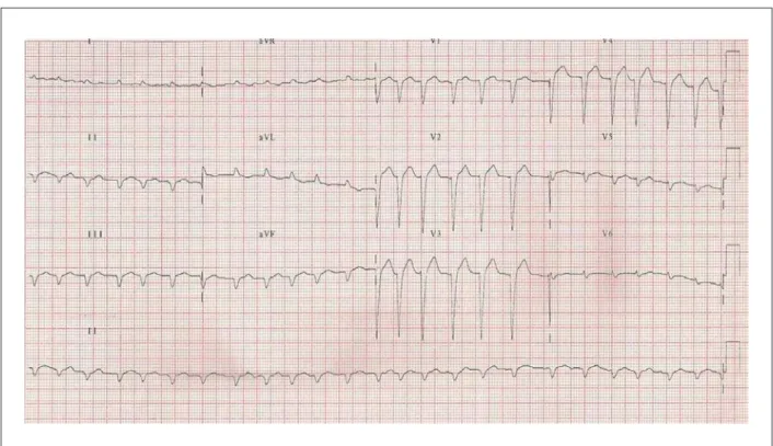
The ECG (September 2008) disclosed atrial fibrillation and low-voltage QRS complexes (Figure 2). The laboratory assessment disclosed leukocytosis, thrombocytopenia and increased creatinine levels (Table 1).
A new echocardiogram (September 1, 2008) disclosed left atrial diameter of 43 mm, left ventricular diastolic and systolic diameters of 40 and 25 mm, respectively and EF of 68%; septal thickness of 15 mm and posterior wall thickness of 14 mm. The right and left ventricles showed normal systolic function; the patient presented moderate mitral regurgitation and the pulmonary artery pressure was estimated at 40 mmHg.
The chest X-ray showed cardiomegaly and marked pleural effusion to the right.
The skull tomography (September 2, 2008) showed mild cortical and subcortical hypoattenuation to the left, compatible with acute ischemia. There were no fractures or bleeding.
The chest tomography (Sept. 2, 2008) disclosed moderate bilateral pleural effusion, larger on the right hemithorax. The radiographic image was described as having a “frostedground glass” appearance in the lower and medium lobes and lingula. There was right upper lobe consolidation.
Orotracheal intubation was necessary for ventilatory support, as well as intravenous noradrenalin administration, antibiotic therapy with ceftriaxone and clarithromycin, later substituted by imipenem, subsequently substituted by vancomycin and tazobactam and volume replacement. Two days later, the vasoactive drugs were reduced and HR remained at 106 bpm and BP at 113/70 mmHg. Right pleural paracentesis was carried out and 800 ml of citrine-yellow
fluid was removed.
Kidney function worsened and dialysis was indicated on the 5th day of hospitalization (Table 1). Kidney failure regression
was observed.
The cardiac magnetic resonance (MR) (Sept. 19, 2008) showed biatrial enlargement, ventricles with normal systolic function, left ventricular concentric hypertrophy, mitral regurgitation, mild pericardial effusion and small bilateral pleural effusion. It was not possible to interpret the delayed enhancement sequence, due to the incapacity of adjusting the screening parameters that could suggest deposition disease. The examination was difficult due to the clinical condition of the patient, who was unable to maintain respiratory pause (the multiple movement artifacts reduce the method’s sensitivity). Screening for urinary paraproteins (Sept. 9, with result on Sept. 23, 2008) disclosed lambda immunoglobulin of 3.9 mg/ dl (normal up to 1 mg/dl) and kappa immunoglobulin of 5.2 mg/dl (normal up to 1.4 mg/dl).
On the 18th day of hospitalization, the patient presented
arterial hypotension and kidney function worsening and was once again submitted to dialysis. The laboratory assessment is shown in Table 1.
The clinical picture developed with refractory arterial hypotension, cardiogenic shock and death (Sept. 29, 2008).
Clinical aspects
The case presents a 77-year-old female patient with hypothyroidism, coronary artery disease and systemic arterial
Figure 1 -ECG: Left axis deviation of QRS complex with upward delection (ASDB) and low-voltage QRS complex in the frontal plane and electrically inactive area in
Figure 2 -ECG: Atrial ibrillation, low voltage in the frontal area and electrically inactive area in the anterior wall.
hypertension, with a clinical picture of heart failure (HF), preserved ejection fraction (EF) and proteinuria. Among the signs and symptoms, the following are noteworthy: lower-limb edema, pleural and pericardial effusion, with significant worsening 6 months before death.
Among the possible hypotheses for such worsening are: ischemic equivalent, which was ruled out by the normal coronary angiography; pulmonary thromboembolism, which was not confirmed after the ventilation-perfusion scintigraphy and the evolution of the underlying disease. For the differential diagnosis, the patient was assessed for systemic diseases that can affect the heart and kidneys.
The triad of hypoalbuminemia (2.3g/dl), peripheral edema and proteinuria at nephrotic levels (3,35g/l) met the criteria for the diagnosis of nephrotic syndrome. There are several causes of nephrotic syndrome in adult patients; however, many of them are limited to the kidneys and are not applicable to the present case. A very important cause of nephrotic syndrome with cardiac involvement would be diabetes mellitus (DM); however, the patient did not present glycemic alterations suggestive of DM.
Systemic lupus erythematosus (SLE) can cause pleural and pericardial effusion, proteinuria and increase in the antinuclear factor. It main cardiac manifestations that generate heart failure (HF), such as myocarditis and Libman-Sacks endocarditis, do not cause diastolic HF, as seen in the present case. Constrictive pericarditis, another lupus manifestation, would not generate such high levels of pulmonary artery pressure, or ventricular wall thickening and there would not be pericardial alterations at the nuclear magnetic resonance. Moreover, the onset of symptoms after 70 years of age and the lack of hematological
and articular alterations did not indicate SLE, either1.
Schistosomiasis is another cause of cardiac involvement and nephrotic syndrome, with a predominance of manifestations in the right cardiac chambers, pulmonary hypertension and even cor pulmonale. There is, however, no left ventricular involvement, pericardial effusion or valve insufficiency, as observed in the patient.
The cardiac involvement, with symptoms of HF and preserved LVEF, suggests diastolic dysfunction. Such cardiac alteration is characterized by the modification of ventricular relaxation, impaired ventricular filling and/or increased filling pressures and a higher degree of dependence on the atrial contraction phase. The increased pressure in the left atrium and, consequently, in the pulmonary veins and capillaries, as well as the decrease in the ejected volume, explain the exertion intolerance and even the dyspnea at rest. Systemic arterial hypertension (SAH) with left ventricular hypertrophy (LVH), aortic stenosis with preserved LVEF, hypertrophic and restrictive cardiomyopathy, as well as coronary artery disease (CAD) are among the main causes of diastolic dysfunction.
The patient did not present aortic stenosis or criteria for the diagnosis of hypertrophic cardiomyopathy at the echocardiography; moreover, th coronary angiography had not disclosed arterial obstructions.
At the differential diagnosis of SAH with LVH and restrictive cardiomyopathy, it was noteworthy the frontal plane electrocardiographic low voltage, which favors deposition disease and consequent restriction to ventricular filling2. However, the
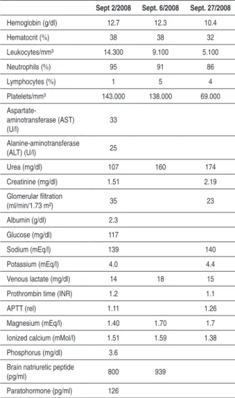
Table 1 - Laboratory assessment at the last hospital admission
Sept 2/2008 Sept. 6/2008 Sept. 27/2008
Hemoglobin (g/dl) 12.7 12.3 10.4
Hematocrit (%) 38 38 32
Leukocytes/mm³ 14.300 9.100 5.100
Neutrophils (%) 95 91 86
Lymphocytes (%) 1 5 4
Platelets/mm³ 143.000 138.000 69.000
Aspartate-aminotransferase (AST) (U/l)
33
Alanine-aminotransferase
(ALT) (U/l) 25
Urea (mg/dl) 107 160 174
Creatinine (mg/dl) 1.51 2.19
Glomerular iltration
(ml/min/1.73 m²) 35 23
Albumin (g/dl) 2.3
Glucose (mg/dl) 117
Sodium (mEq/l) 139 140
Potassium (mEq/l) 4.0 4.4
Venous lactate (mg/dl) 14 18 15
Prothrombin time (INR) 1.2 1.1
APTT (rel) 1.11 1.26
Magnesium (mEq/l) 1.40 1.70 1.7
Ionized calcium (mMol/l) 1.51 1.59 1.38
Phosphorus (mg/dl) 3.6
Brain natriuretic peptide
(pg/ml) 800 939
Paratohormone (pg/ml) 126
hypothesis of restrictive disease.
Among the diseases that can cause restrictive cardiomyopathy are the infiltrative ones (amyloidosis, sarcoidosis), deposition diseases (hemochromatosis and Fabry disease), endomyocardial diseases (endomyocardial fibrosis, anthracycline toxicity, radiation) and the idiopathic/ familial forms. Among them, the infiltrative diseases and Fabry disease cause kidney disease. Sarcoidosis is a systemic disease that might be associated with glomerulonephritis and renal interstitial disease, in addition to hypercalcemia. In both, proteinuria is uncommon at nephrotic levels.
The clinical picture associated with the complementary examinations described here suggests restrictive cardiopathy associated with nephrotic syndrome. The most likely hypothesis for such association is systemic amyloidosis.
Systemic amyloidosis is a group of diseases that present an extracellular deposition of insoluble fibrillar proteins, consisting of low molecular-weight subunits. Clinically, it is usually classified as primary (AL), secondary (AA), hereditary and age-associated (senile). AL amyloidosis is caused by the
deposition of proteins derived from light-chain fragments, in general a monoclonal immunoglobulin (80.0% of the cases). It can be isolated or in association with multiple myeloma (10.0% of the cases). AA amyloidosis can aggravate chronic diseases that course with recurring inflammation. The fibrillae consist of fragments of the amyloid protein A, an acute phase protein. There is also the hereditary amyloidosis (deposition of fibrillae derived from transthyretin), related to dialysis and the senile form (also deposition of transthyretin).
Amyloidosis AA shows cardiovascular involvement in only 5.0% of the cases and there is no reference to a systemic inflammatory process (although serological tests for hepatitis were not available)3.
The two subtypes of amyloidosis more probable for the case, therefore, would be amyloidosis AL and the senile type. There are important differences in the prognosis and evolution of these two subtypes: the mean survival after cardiovascular involvement is, respectively, 11 and 75 months4. Additionally,
the insidious course of the disease is the rule for senile amyloidosis, whereas the AL type shows rapid evolution of symptoms and a higher cardiac involvement.
In the absence of confirmation of plasmocyte dyscrasia, the differentiation between them is attained through immunohistochemical techniques5.
The cardiovascular involvement in cases of amyloidosis occurs in one-third of the patients. Right ventricular failure usually occurs with slight pulmonary edema, in spite of the high filling pressures. Therefore, hepatomegaly and lower-limb edema occur. Autonomic dysfunction can occur and that can generate postural hypotension and even syncope6.
Low voltage is the most common electrocardiographic alteration, predominating in the frontal plane. Other alterations include atrial fibrillation, conduction disorders and electrically inactive areas. However, high-degree atrioventricular blocks are uncommon2.
The echocardiogram is a noninvasive test that is important for the diagnosis of amyloidosis. The thickening of the LV wall with evidence of diastolic dysfunction is the earliest alteration and it can develop into restrictive cardiomyopathy7. Biatrial
enlargement and valve thickening can also occur.
Large pleural effusions, present in this case, occur in 1% to 2% of patients with amyloidosis. The differentiation between right pleural involvement and effusion secondary to cardiopathy is difficult to attain, considering the low sensitivity of the pleural biopsy and the absence of specific alterations at the imaging methods8.
The brain ischemia seen at the tomography could be caused by thromboembolic events, favored by the atrial fibrillation and the cardiac amyloidosis itself, which predisposes to the onset of intraventricular thrombi9,10.
Hypothyroidism can occur due to the amyloid infiltration in the gland; however, it is more common when concomitant to goiter, which was not described in the present case11.
The positive antinuclear antibody (ANA) has been described in the literature; however, it is not a common alteration12. The
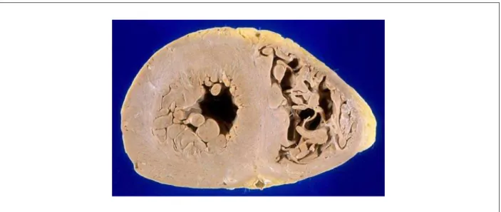
Figure 3 -Cross-section of heart ventricles. Observe the thickened walls and slightly yellowish color of the myocardium.
Finally, an increase in immunoglobulin light chain excretion was observed in the patient’s urine, with maintenance of the kappa/lambda ratio, with no description of a urinary monoclonal chain that would establish the diagnosis of amyloidosis13.
In summary, considering the picture of restrictive cardiomyopathy and nephrotic syndrome, in addition to the other clinical findings, one can hypothesize the patient presented systemic amyloidosis with renal and cardiac involvement. However, the complementary tests did not confirm this hypothesis, as the patient did not present monoclonal gammopathy or alteration in the kappa/lambda immunoglobulin ratio. The confirmation of the diagnosis of amyloidosis can be attained through a biopsy, which can be performed in the subcutaneous fat tissue (sensitivity of 65 to 80.0%)14 or endomyocardium (up to 97.0%)15, by
demonstrating amyloid deposition in tissues, classically stained by Congo red.
Dr. Mônica Samuel Ávila and Dr. André Cogo Dalmaschio
Diagnostic hypothesis
Systemic amyloidosis with cardiac and renal involvement. Other diagnoses: hypothyroidism, systemic arterial hypertension, acute brain ischemia.
Dr. Mônica Samuel Ávila and Dr. André Cogo Dalmaschio
Necropsy
There was around 2,500 ml of citrine-yellow fluid in the pleural cavities and pericardial effusion of the same aspect, with moderate volume. The heart weighed 510 g; the walls were thickened and the myocardium presented a more solid consistency than usual, in addition to a slightly yellowish color (Figure 3). The atriumsa were moderately dilated. The histological assessment of the myocardium showed
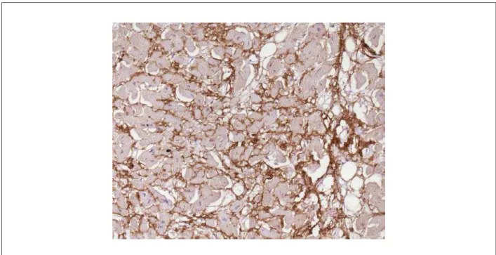
marked interstitial deposition of eosinophilic and amorphous material, which presented clear greenish polarization when stained by Congo red, characterizing amyloid substance deposition (Figure 4). The immunohistochemical screening of lambda chain immunoglobulins was strongly positive in these deposits (Figure 5), whereas the kappa chain screening was negative. Amyloid substance deposits were also found, albeit focally, in the endocardium, in the microcirculation vessel walls of several organs and in the tongue. The lungs weighed 748g and there was evidence of chronic passive congestion and focal areas of edema, as well as alveolar hemorrhage. An arterial branch with a subocclusive thromboembolus was identified in the lower lobe of the right lung at the start of thrombus organization phase and a small recent pulmonary infarction (Figure 6). The thyroid volume was enlarged and the gland presented multiple nodulations, fibrosis and calcification foci.
The bone marrow examination (sternum and iliac crest) did not show an abnormal number of plasmocytes. The immunohistochemical study showed weak cell labeling, probably due to the poor antigenic preservation of the material, but it was positive for both kappa and lambda immunoglobulin chains (polyclonal profile). There was a single adenomatous polyp in the colon, pedunculated, measuring 1.5 cm. The aorta presented slightmild atherosclerosis, as well as the coronary arteries. There were no encephalic lesions. The uterus and ovaries were not found (probable previous resection).
Dr. Luiz Alberto Benvenuti
Anatomopathological diagnoses
Primary amyloidosis with cardiac involvement (specific cardiomyopathy due to amyloid substance deposition); adenomatous goiter; isolated adenomatous polyp in the colon; pulmonary thromboembolism and recent pulmonary infarction (cause of death)16,17.
Figure 4 -Cardiac amyloidosis. Deposition of eosinophilic and amorphous (asterisks) material in the myocardial interstitium (A), presenting a clear greenish color after
staining with Congo red and seen under polarized light (B). HE, ´200 (A); Congo Red, ´400 (B).
Figure 5 -Strongly positive immunohistochemical reaction for immunoglobulin lambda chain in the heart interstitium (dark-brown color), corresponding to the areas of
amyloid substance deposition. Immunoperoxidase reaction for lambda chain, ´200.
Comments
The present case describes a 77-year-old female patient that presented progressive heart failure, with normal left ventricular ejection fraction and preserved ventricular systolic function; the latter was assessed by nuclear magnetic resonance around 10 days before the patient’s death.
It is noteworthy the fact that the patient presented proteinuria (3.35 g/l) and that the screening for urinary paraproteins showed an increase in the levels of kappa and lambda immunoglobulin chains. The necropsy showed that it
was a case of cardiac amyloidosis and medullary plasmocytosis was not detected. The amyloidosis can be classified as primary, hereditary, systemic senile, isolated atrial, reactional or secondary and dialysis-related.
of chronic inflammatory diseases. Thus, the differential diagnosis would be restricted to either primary amyloidosis (immunoglobulin light chain deposition) or the systemic senile form (normal transthyretin deposition).
In spite of the patient’s older age, which would favor the senile form, the presence of significant proteinuria pointed out to the primary form of the disease4. Interestingly, in the present
case, we observed the association between the amyloid deposition and the immunoglobulin lambda chain, of which immunohistochemical screening was strongly positive in the areas of myocardial amyloid deposition, showing it was the
primary form of amyloidosis. Although plasmocytosis was not detected in the bone marrow examination, it is not possible to rule out the possibility of some monoclonal gammopathy with slight expression or even localized plasmocytoma.
In the present case, the patient’s cardiac involvement is classified as specific cardiomyopathy, associated with amyloid substance deposition16,17.
Death occurred due to heart failure worsening, with thromboembolism and pulmonary infarction.
Dr. Luiz Alberto Benvenuti
References
1. Dember LM, Shepard JA, Nesta F, Stone JR. Case records of the Massachusetts General Hospital. Case 15-2005 - An 80-year-old man with shortness of breath, edema, and proteinuria. N Engl J Med. 2005; 352 (20): 2111-9.
2. Murtagh B, Hammill SC, Gertz MA, Kyle R, Tajik A, Grogan M. Electrocardiographic findings in primary systemic amyloidosis and biopsy-proven cardiac involvement. Am J Cardiol. 2005; 95 (4): 535-7.
3. Lachmann HJ, Goodman HJ, Gilbertson JA, Gallimore JR, Sabin CA, Gillmore JD, et al. Natural history and outcome in systemic AA amyloidosis. N Engl J Med. 2007; 356 (23): 2361-71.
4. Ng B, Connors LH, Davidoff R, Skinner M, Falk RH. Senile systemic amyloidosis presenting with heart failure: a comparison with light chain-associated amyloidosis. Arch Intern Med. 2005; 165 (12): 1425-9.
5. Arbustini E, Morbini P, Verga L, Merlini G. Concardi M, Porcu E, et al. Light and electron microscopy immunohistochemical characterization of amyloid deposits. Amyloid. 1997; 4: 157-70.
6. Chamarthi B, Dubrey SW, Cha K, Skinner M, Falk RH. Features and prognosis of exertional syncope in light-chain associated AL cardiac amyloidosis. Am J Cardiol. 1997; 80 (9): 1242-5.
7. Klein AL, Hatle LK, Burstow DJ, Taliercio CP, Seward JB, Kyle RA, et al. Comprehensive Doppler assessment of right ventricular diastolic function in cardiac amyloidosis. J Am Coll Cardiol. 1990; 15 (1): 99-108.
8. Celli, Rubinow A, Cohen AS, Brody JS. Patterns of pulmorary involnement in systemic amyloidosis. Chest. 1978; 74 (5): 543-7.
9. Zubkov AY, Rabinstein AA, Dispenzieri A, Wijdicks EF. Primary systemic amyloidosis with ischemic stroke as a presenting complication. Neurology. 2007; 69 (11): 1136-41.
10. Feng D, Edwards WD, Oh JK, Chandrasekaran K, Grogan M, Martinez MW, et al. Intracardiac thrombosis and embolism in patients with cardiac amyloidosis. Circulation. 2007; 116 (21): 2420-6.
11. Kimura H, Yamashita S, Ashizawa K, Yokoyama N, Naga-taki S. Thyroid disfunction in patients with amyloid goiter. Clin Endocrinol. 1997; 46 (6): 769-74.
12. Love WE, Miedler JD, Smith MK, Mostow EN, Cooper KD, Gilliam AC. The spectrum of primary cutaneous nodular amyloidosis: two illustrative cases. J Am Acad Dermatol. 2008; 58 (2 Suppl): S33-5.
13. Lachmann HJ, Gallimore R, Gillmore JD, Carr-Smith HD, Bradwell AR, Pepys MB, et al. Outcome in systemic AL amyloidosis in relation to changes in concentration of circulating free immunoglobulin light chains following chemotherapy. Br J Haematol. 2003; 122 (1): 78-84.
14. Andrews TR, Colon-Otero G, Calamia KT, Menkes DM, Boylan KB, Kyle RA. Utility of subcutaneous fat aspiration for diagnosing amyloidosis in patients with isolated peripheral neuropathy. Mayo Clin Proc. 2002; 77
Figure 6 -Thromboembolus at the initial stage of organization (asterisk) in medium-caliber pulmonary-artery branch in the lower lobe of the right lung (B). HE, ´25 (A)
(12): 1287-90.
15. Pellikka PA, Holmes DR Jr, Edwards WD, Nishimura RA, Tajik AJ, Kyle RA. Endomyocardial biopsy in 30 patients with primary amyloidosis and suspected cardiac involvement. Arch Intern Med. 1988; 148 (3): 662-6.
16. Richardson P, McKenna W, Bristow M, Maisch B, Mautner B, O’Connell J,
et al. Report of the World Health Organization/International Society and Federation of Cardiology Task Force on the Definition and Classification of cardiomyopathies. Circulation. 1996; 93 (5): 841-2.