Direct Ophthalmoscopy
versus
Detection of Hypertensive
Retinopathy: A Comparative Study
José Albuquerque de Figueiredo Neto, Guilherme Lima Palácio, Allison Nobrega dos Santos, Pamela Suelen Serra
Chaves, Germana Viana Gomes, Tassiane Soares Cabral
Universidade Federal do Maranhão, São Luiz, MA - Brazil
Mailing address: José Albuquerque de Figueiredo Neto •
Rua Rui Ribeiro Mesquita - Ed. Dom Gabriel, apto 402 - Calhau - 65075-260 - São Luiz, MA - Brazil
E-mail: [email protected], [email protected]
Manuscript received September 09, 2009; revised manuscript received November 23, 2009; accepted December 29, 2009.
Abstract
Background: Detection of hypertensive retinopathy (HR) with direct ophthalmoscopy is part of the assessment of hypertensive patients. Its use has been questioned because of its subjectivity and high interobserver variability.
Objective: To determine the prevalence of HR in hypertensive patients under outpatient monitoring, the correlation between diagnosis and ophthalmoscopy and angiography, and to correlate it with other target organ damages.
Methods: An observational, analytical and cross-sectional evaluation of 99 patients. Direct ophthalmoscopy and
angiography performed by two investigators independently. Classification of RH, as described by Keith, Wagener and Barker.
Results: The prevalence of HR of any grade was higher than 90.0% by both methods. On ophthalmoscopy, we observed
grade I abnormalities in 51.0%, grade II in 43.0%, with one patient with grade III HR. On angiography, we observed grade I abnormalities in 42.0% and grade II in 52.0%. We detected three patients with grade III HR, two of which were not detected by ophthalmoscopy. Observers’ agreement for the presence and severity of HR was poor with direct ophthalmoscopy and good with angiography. Renal dysfunction, ECG abnormalities (ventricular hypertrophy, pathological Q wave, repolarization abnormalities), and history of stroke were observed in 70.0%, 27.0% and 10.0% of patients, respectively. There was no relationship between the severity of HR and other target organ damages.
Conclusion: We observed a high prevalence of HR using both methods. Observers’ agreement for the diagnosis and determination of the severity of HR was better with angiography. In our sample, there was no association of the severity
of HR with other target organ damages. (Arq Bras Cardiol 2010; 95(2): 215-222)
Key words: Ophthalmoscopy; retina/injuries; hypertension.
patients untreated or uncontrolled, diagnosis made only with the DO and association of HR with mortality in studies where there was no control of other risk factors currently known, which may act with confusion factors8-11.
Recent studies using angiography12-17 showed high
reproducibility in the evaluation of microaneurysms, hemorrhages, arteriovenous crossing abnormalities, focal or generalized arteriolar narrowing. However, few studies have compared the sensitivity and reproducibility between DO and angiography in the detection of HR18.
Another issue in the study of HR is that some signs seem to be associated with certain clinical conditions19.
We performed this study because of the importance of hypertension as a cause of morbidity and mortality, lack of data on the prevalence of HR in our environment, the absence of studies comparing DO with angiography in diagnosis and the uncertainty regarding the usefulness of its detection in the evaluation of cardiovascular risk and associated target organ damages.
The objectives were to determine the prevalence of RH in patients monitored in a League of Hypertension of a University
Introduction
Hypertensive retinopathy (HR) is a condition characterized by a spectrum of retinal vascular signs that develop in people with hypertension1. Its detection with direct ophthalmoscopy
(DO) is part of the assessment of hypertensive patients2, but
its use has been questioned due to its high subjectivity and interobserver variability3.
The Seventh Joint National Committee on Prevention, Detection, Evaluation, and Treatment of High Blood Pressure and the V Brazilian Guidelines on Hypertension list HR as a marker of target organ damage. Therefore, it is a criterion for prescription of treatment4,5.
Hospital; to evaluate the correlation between DO and digital angiography (DA) in detecting RH; to study the association of HR with other target organ damages.
Methodology
Type of study
Analytical cross-sectional study conducted from June to December/2008 in a League of Hypertension of a University Hospital.
Sample
For convenience purposes, patients were selected as they came for routine medical appointments, observing the exclusion criteria of the study, namely:
a. Diabetes mellitus;
b. Media opacity (scarring, vitreous hemorrhage, cataract); c. Retinal detachment;
d. Lack of systemic clinical data in the medical records of the League of Hypertension.
We examined 131 consecutive patients. We excluded 32 patients: one, because of double evaluation, three because of media opacity on ophthalmological test, 6 due to insufficient data in their files and 22 have developed diabetes. We evaluated 99 patients.
Data collection
Performed by clinical and ophthalmological tests, namely: a. Clinical examinations - the patients underwent
systemic clinical examination for the presence of risk factors for cardiovascular disease and categorization of their blood pressure as for severity, according to the V Brazilian Guidelines on Hypertension4.
b. Routine laboratory tests - urinalysis, potassium and creatinine, fasting glucose, total cholesterol, LDL, HDL and triglycerides - in accordance with the V Brazilian Guidelines on Hypertension4.
c. 12-lead electrocardiogram assessed abnormalities consistent with left ventricular hypertrophy (Cornell criteria), and assessment of the presence of pathological Q wave and ventricular repolarization abnormality. The cleareance estimated of creatinine was calculated according to Cockcroft-Gault formula. Decreased renal function was defined as smaller than 60 ml/min.
Ophthalmologic examination was performed, including visual acuity, biomicroscopy and applanation tonometry. Direct ophthalmoscopy was also performed with coaxial ophthalmoscope 3.5 V (Welch Allyn, Skaneateles Falls, United States) and angiography with equipment CF60 UVi (Canon, Tokyo, Japan) with digital system Eye Q Pro (Canon, Tokyo, Japan) after pulilar dilation with 1.0% tropicamide eye drops. The images were obtained in the field of 60 degrees, centered on the macula.
E a c h p a t i e n t u n d e r w e n t f u n d o s c o p y b y t w o ophthalmologists, with similar experience and independently.
Angiographies were also interpreted by these physicians without prior knowledge of patients’ clinical conditions, independently. In a second step, a new interpretation was made by consensus. We performed classification of HR as described by Keith, Wagener and Barker6.
Statistical analysis
Data were analyzed using Epi Info 3.4.3 (2007). Quantitative variables were presented as averages and standard deviation. Qualitative variables were presented as frequencies and percentages.
As for fundus and angiography findings, we measured the Kappa coefficient of agreement among examiners. Agreement was considered poor in cases of Kappa coefficient smaller than 0.20; weak, between 0.21-0.40; moderate between 0.41-0.60; good between 0.61-0.80; and very good when it was higher than 0.80.
To test the association between risk factors and severity of HR, we used Fisher’s exact test. A significance level of 5.0% was adopted.
Ethical aspects
Data were collected after approval by the Research Ethics Committee (Process 33104-178/2007) and the patients signed a consent form.
Results
We evaluated 99 patients whose demographics are presented in Table 1.
Analysis of the presence of risk factors for cardiovascular diseases revealed that they had no risk factor in addition to hypertension in 9.1% of the cases, one or two factors in 45.5% and three or more risk factors or cardiovascular disease in 24.2%. (Table 2).
As for the time of diagnosis of hypertension, 42.4% of patients were younger than 6, 21.2% between 6 and 10, and 36.4% were older than 10. There was no association between the time of diagnosis and the presence of retinopathy (p > 0.05).
Staging of hypertension can be seen in Table 3.
The frequency of target organ damages is shown in Table 4. The prevalence of HR using DO and digital angiography, as well as the consensus of angiography by both observers are shown in Table 5.
The coefficient of agreement for the presence of HR with DO was poor, with Kappa equal to 0.22 CI 95% [0.38 to 0.83].
However, there was a good coefficient of agreement for the presence of HR with digital angiography, with Kappa value equal to 0.66 CI 95% [0.00 to 1.37].
When we evaluated observers’ coefficient of agreement in distinguishing grade I or grade II retinopathy through DO, this proved to be poor, with Kappa equal to 0.33 CI 95% [0.13 to 0.52].
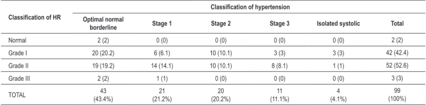
No relationship was found between the severity of HR and the stages of hypertension on examiners’ consensus through digital angiography (Table 6).
Renal dysfunction was present in 20.2% of the patients. No relationship was found between the degree of retinopathy and the severity of renal dysfunction (Table 7).
We analyzed the electrocardiograms of 90 patients and observed electrocardiographic abnormalities in 27.3% of them. No relationship was found between the degree of retinopathy and ECG abnormalities (Table 8).
History of stroke was present in 10 patients in the sample. However, no relationship was established between positive history of stroke and severity of HR.
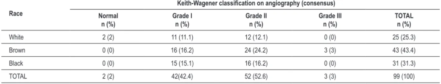
There was no association between race and severity of HR.
Table 1 - Demographic aspects and cardiovascular risk factors of the patients included in the research protocol of hypertensive retinopathy, from June to December/2008, HU-UFMA
Risk factors n (%)
Smokers
Yes 12 12.1
No 87 87.9
Dyslipidemic
Yes 62 62.6
No 37 37.4
HFDCV*
Present 68 68.7
Absent 31 31.3
Heart disease
Yes 20 20.2
No 79 79.8
CVA **
Yes 10 10.1
No 89 89.9
History of nephropathy
Yes 40 40.4
No 59 59.6
Hypercholesterolemia (> 200 mg/ml)
Yes 47 47.5
No 52 52.5
Hypertriglyceridemia (> 150 mg/ml)
Yes 61 61.6
No 38 38.4
*HFDCV - family history of cardiovascular disease, **CVA - cerebral vascular accident (stroke).
Table 2 - Staging of hypertension and target organ damage of patients included in the research protocol of hypertensive retinopathy, from June to December/2008, HU-UFMA
Variables n (%)
Hypertension stage
Optimal, normal or borderline 43 (43.4)
Stage I 21 (21.2)
Stage II 20 (20.2)
Stage III 11 (11.1)
Isolated systolic hypertension 04 (4.1)
Target organ damage n (%)
CVA 10 10.1
Nephropathy (clearance <
60 ml/min) 20 20.2
Hypertrophy or ischemia on
electrocardiogram 27 27.3
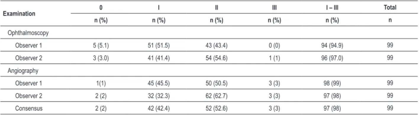
Table 3 -Prevalence of hypertensive retinopathy according to the classiication of Keith, Wagener and Barker, according to the method
diagnosed by observers 1 and 2 and consensus of angiography in the research protocol of hypertensive retinopathy, from June to December/2008, HU-UFMA
Examination 0 I II III I – III Total
n (%) n (%) n (%) n (%) n (%) n
Ophthalmoscopy
Observer 1 5 (5.1) 51 (51.5) 43 (43.4) 0 (0) 94 (94.9) 99
Observer 2 3 (3.0) 41 (41.4) 54 (54.6) 1 (1) 96 (97.0) 99
Angiography
Observer 1 1(1) 45 (45.5) 50 (50.5) 3 (3) 98 (99) 99
Observer 2 2 (2) 32 (32.3) 62 (62.7) 3 (3) 97 (98) 99
Table 4 -Classiication of hypertensive retinopathy according to the second stage of hypertension using the consensus among examiners in
digital angiography in the research protocol of hypertensive retinopathy, from June to December/2008, HU-UFMA
Classiication of HR
Classiication of hypertension
Optimal normal
borderline Stage 1 Stage 2 Stage 3 Isolated systolic Total
Normal 2 (2) 0 (0) 0 (0) 0 (0) 0 (0) 2 (2)
Grade I 20 (20.2) 6 (6.1) 10 (10.1) 3 (3) 3 (3) 42 (42.4)
Grade II 19 (19.2) 14 (14.1) 10 (10.1) 8 (8.1) 1 (1) 52 (52.6)
Grade III 2 (2) 1 (1) 0 (0) 0 (0) 0 (0) 3 (3)
TOTAL 43
(43.4%)
21 (21.2%)
20 (20.2%)
11 (11.1%)
4 (4.1%)
99 (100%)
Table 5 - Severity of hypertensive retinopathy and renal function in the research protocol of hypertensive retinopathy from June to December/2008, HU-UFMA
Renal function
Classiication of Keith, Wagener and Barker (1939) on angiography (consensus)
Normal n (%)
Grade I n (%)
Grade II n (%)
Grade III n (%)
TOTAL n (%)
Normal (CI > 90 ml/min) 0 (0) 15 (15.1) 15 (15.1) 0 (0) 30 (30.3%)
Light dysfunction (90 ml/min < CI < 30 ml/min) 2 (2) 16 (16.2) 29 (29.3) 2 (2) 49 (49.5%)
Moderate dysfunction (60 ml/min < CI <30 ml/min) 0 (0) 9 (9.1) 6 (6.1) 1 (1) 16 (16.2%)
Severe dysfunction (CI < 30ml/min) 0 (0) 2 (2) 2 (2) 0 (0) 4 (4%)
TOTAL 2 (2%) 42 (42.4%) 52 (52.6%) 3 (3%) 99 (100%)
Table 6 - Abnormalities on electrocardiogram and severity of hypertensive retinopathy in the research protocol of hypertensive retinopathy from June to December/2008, HU-UFMA
ECG
Keith-Wagener classiication on angiography (consensus)
Normal n (%)
Grade I n (%)
Grade II n (%)
Grade III n (%)
TOTAL n (%)
Normal 1 (1.1) 28 (31.1) 33 (36.7) 1 (1.1) 63 (70)
Abnormal 1 (1.1) 8 (8.9) 16 (17.8) 2 (2.2) 27 (30)
TOTAL 2 (2.2) 36 (40%) 49 (54.5) 3 (3.3) 90 (100%)
Table 7 - Relationship between history of stroke and severity of hypertensive retinopathy in the research protocol of hypertensive retinopathy from June to December/2008, HU-UFMA
CVA
Keith-Wagener classiication on angiography (consensus)
Normal n (%)
Grade I n (%)
Grade II n (%)
Grade III n (%)
TOTAL n (%)
Positive history 0 (0) 3 (3) 7 (7.1) 0 (0) 10 (10.1)
Negative history 2 (2) 39 (39.4) 45 (45.5) 3 (3) 89 (89.9)
TOTAL 2 (2) 42 (42.4) 52 (52.6) 3 (3) 99 (100)
Discussion
International guidelines consider the retinal findings differently. For the European Society of Hypertension and the European Society of Cardiology, grade III and IV HR as classified by Keith, Wagener and Barker, is regarded as an
associated clinical condition6,20.The International Society
Using angiography and standardized methodology for documenting and defining the various signs of HR, epidemiological studies have shown a prevalence ranging from 2-15.0% in the general population10,12,25-27.
The Beaver Dam Eye Study12, using angiography, evaluated
2,375 non-diabetics aged 43-86, finding a focal arteriolar narrowing in 14.0%, retinopathy in 8.0%, and pathological AV crossings in 2.0%. When hypertensives were excluded, there was focal arteriolar narrowing in 11.0%, retinopathy in 6.0% and pathological AV crossings in 2.0%. Therefore, it can be inferred that retinal microvascular abnormalities are common in nondiabetic populations, although they are more prevalent in patients with hypertension12.
In the Blue Mountains Eye Study27, 3,275 non-diabetic
individuals underwent angiography. The prevalence of retinopathy was 9.9%. The Atherosclerossis Risk in Communities (ARIC) Study25 studied 9,300 individuals older
than 49, without diabetes, using angiography. The prevalence of focal arteriolar narrowing was 6.0%, retinopathy of 3.0% and pathological AV crossing of 6.0%.
The higher prevalence of retinopathy observed in these studies, compared to the Framingham study, may be due to the higher sensitivity of photographic assessment techniques against ophthalmoscopy. However, these studies did not include the earliest sign of HR, the generalized arteriolar narrowing13.
In this study, the coefficient of agreement for the detection and assessment of the severity of HR using angiography was higher than that found using DO.
Cuspidi et al28 evaluated 197 patients with grade I
and II hypertension (73.0%) without pretreatment, using digital angiography, with two independent observers. The overall prevalence of retinal changes was 84.3% and 84.7% respectively.
In this study, there was no correlation of retinopathy with age, sex, time of diagnosis and stage of hypertension. Data from the Beaver Dam and ARIC study suggest that the prevalence of arteriolar narrowing and pathological AV crossing seem to be directly related with age. The prevalence of retinopathy was also age-dependent in the Blue Mountains study12,24,26.
The prevalence of retinopathy was higher in males, when adjusted for age, both in the Beaver Dam and in the ARIC study. However, in the Blue Mountains, no difference was found11,25,27.
Table 8 - Severity of hypertensive retinopathy according to race in the research protocol of hypertensive retinopathy from June to December/2008, HU-UFMA
Race
Keith-Wagener classiication on angiography (consensus)
Normal n (%)
Grade I n (%)
Grade II n (%)
Grade III n (%)
TOTAL n (%)
White 2 (2) 11 (11.1) 12 (12.1) 0 (0) 25 (25.3)
Brown 0 (0) 16 (16.2) 24 (24.2) 3 (3) 43 (43.4)
Black 0 (0) 15 (15.1) 16 (16.2) 0 (0) 31 (31.3)
TOTAL 2 (2) 42(42.4) 52 (52.6) 3 (3) 99 (100)
Detection, Evaluation and Treatment of Hypertension in the United States and the V Brazilian Guidelines on Hypertension consider the presence of any sign of retinopathy as target organ damage4,5.
In this study, using DO and considering the presence of any sign of retinopathy, we observed prevalence above 90.0%, with grade I abnormalities in 51.0% and grade II in 43%. One patient had grade III HR. No patient with grade IV was found. The most common findings were diffuse arteriolar narrowing and arteriolar wall opacification, and pathologic arteriovenous crossing. Considering only advanced damages (Grade III and IV), the prevalence of HR would be 1.0%.
In the Framingham Eye Study, with DO, in a general population aged 52-85, the prevalence of retinopathy was 0.8%, excluding patients with diabetes. The concept of retinopathy included only advanced forms such as retinal hemorrhages and exudates, without considering arteriolar wall abnormalities18.
McDonough, Garrison and Hames23, using DO, evaluating
2,210 people, found that 2.2% had retinopathy defined as the presence of microaneurysms, hemorrhages or exudates. On evaluation of earliest signs of retinopathy, 34.0% had generalized or focal arteriolar narrowing, and 13.2%, pathological AV crossings23.
Besharati et al24 studied 213 hypertensive patients with
DO and found a prevalence of 39.9%, considering any sign of retinopathy. In those with mild hypertension, the prevalence was 25.3%; in patients with moderate hypertension, 34.5%; and those with severe hypertension, prevalence was 84.6%. The most common findings were arteriolar narrowing (35.13%), pathologic arteriovenous crossing (17.12%) and cotton wool spots (9.0%)24.
In this study, the coefficient of agreement for the diagnosis and severity of HR showed a weak agreement, using DO.
Using DO in assessing hypertensive patients results in imprecision and interobserver variability. It is particularly imprecise in people with mild to moderate hypertension2,8.
In literature, it is reported that generalized arteriolar narrowing and pathologic arteriovenous crossings can be observed more commonly in patients with longer duration of diagnosed hypertension, representing signs of chronic vascular damage. On the other hand, focal arteriolar narrowing, retinal hemorrhages, microaneurysms and cotton wool spots are believed to be related to the current level of hypertension13,19.
The distribution of the severity of HR in this study, according to the classification of Keith, Wagener and Barker was presented as follows: Grade I - 42 patients (42.4%), grade II - 52 patients (52.6%), grade III - 3 patients (3%) and grade IV - no patient.
The high frequency of early signs of retinopathy, including grades I and II of Keith, Wagener, and Barker, and the low frequency of intense signs, grades III and IV, may be related to the duration of hypertension and treatment and regular control of blood pressure6.
In this study, renal dysfunction was observed in 20.2% of patients. However, no association was established between renal dysfunction and the presence and severity of HR. In the ARIC study, individuals with retinopathy (odds ratio 2.0; confidence interval 95.0%, 1.4-2.8), microaneurysms (OR, 2.0, CI 95.0%, 1.3 - 3.1), retinal hemorrhages (OR, 2.6, CI 95.0%, 1.6-4.0), cotton wool spots (OR 2.7, CI 95.0%, 1.6-4.8) and pathologic arteriovenous crossings (OR 1.4, CI 95.0%, 1.0-1.9) were more likely to develop renal dysfunction than individuals without retinal abnormalities11.
The study found electrocardiographic abnormalities consistent with left ventricular hypertrophy, presence of pathological Q waves and ventricular repolarization abnormalities in 27.0% of patients. Gillum29 noted that retinal
arteriolar narrowing detected by ophthalmoscopy determined two to six-fold increased likelihood of developing ischemic heart disease, after controlled analysis for hypertension, diabetes and hypercholesterolemia.
Duncan et al30 demonstrated that the presence of RH in
men doubled the chance of ischemic heart disease (relative risk 2.1; confidence interval 95%, 1.0 - 4.2). On the other hand, the ARIC study31 reported a correlation between generalized
arteriolar narrowing and ischemic heart disease in women (relative risk 2.2, confidence interval 95%, from 1.0 to 4.6) but not in men (relative risk 1.1, confidence interval 95%, 0.7 - 1.8).
The limitations of this study should be highlighted. The number of patients evaluated may have been insufficient to demonstrate the relationship between specific signs of HR
and target organ damage. The prevalence of abnormalities was higher than usually reported in the literature, and may have resulted from bias, given the subjectivity of defining abnormality in vascular diameters. Accurate methods of measurement may change these estimates. Another aspect is that the population of patients studied was of a League of Hypertension under regular clinical treatment and monitoring, which could explain the low frequency of more severe signs of HR.
The relevance of this study should also be emphasized. The scarcity of studies on the subject in our country and the region makes the familiarization of frequency of HR signs in the population studied to serve as a parameter for other studies. This study is also useful in showing the profile of fundus findings in patients under regular treatment, mostly classified in the early stages of HR. The high frequency signal casts doubt on its relevance in the risk stratification of hypertensive patients. These findings were not proportional to the frequency of other target organ damages. The work also is unique in comparing HR diagnosing techniques, once we do not find in literature and other studies comparing DO and angiography in detecting HR. Superior fundus findings open discussions on the role of this diagnostic technique in assessing hypertensive patients.
Conclusion
HR is frequent in hypertensive patients being treated in primary care programs. The diagnostic agreement among ophthalmologists is higher in the analysis of angiographies than on direct ophthalmoscopy. The lack of association between retinal abnormalities and other evidence of target organ damage may be related to the small sample size.
Potential Conflict of Interest
No potential conflict of interest relevant to this article was reported.
Sources of Funding
There were no external funding sources for this study.
Study Association
This article is part of the thesis of master submitted by Guilherme Lima Palácio, from Universidade Federal do Maranhão.
References
1. Walsh JB. Hypertensive retinopathy: description, classification, and prognosis. Ophthalmology. 1982; 89 (1): 1127-31.
2. World Health Organization. International Society of Hypertension Guidelines for the Management of Hypertension. J Hypertens. 1999; 17 (1): 151-83.
3. Maestri MM, Fuchs SC,Ferlin E, Pakter HM, Nunes G, Moraes RS, et al. Detection of arteriolar narrowing in fundoscopic examination: evidence of a low performance of direct ophthalmoscopy in comparison with a
microdensitometric method. Am J Hypertens. 2007; 20 (5): 501-5.
4. Chobanian AV, Bakris GL, Black HR, Cushman WC, Green LA, Izzo JR, et al. The seventh report of the Joint National Committee on prevention, detection, evaluation and treatment of high blood pressure: the JNC 7 report. JAMA. 2003, 19 (1): 2560-72.
arterial. Arq Bras Cardiol. 2007; 89 (3): e24-79.
6. Keith NM, Wagener HP, Barker NW. Some different types of essential hypertension: their course and prognosis. Am J Med Sci. 1974; 268 (6): 336-45.
7. Scheie HG. Evaluation of ophthalmic changes of hypertension and arteriolar sclerosis. AMA Arch Ophthalmol. 1953, 49 (1):117-38.
8. Dimmitt SB, West JN, Eames SM, Gibson JM, Gosling P, Littler WA. Usefulness of ophthalmoscopy in mild to moderate hypertension. Lancet. 1989, 1 (1): 1103-06.
9. Kagan A, Aureli E, Dobree J. A note on signs in the fundus oculi and arterial hypertension: conventional assessment and significance. Bull World Heath Organ.1966, 34 (6): 955-60.
10. Wong TY, Mitchell P. Hypertensive retinopathy. N Engl J Med. 2004; 351 (22): 2310-7.
11. Wong TY , Coresh J, Klein R, Muntner P, Couper DJ , Sharrett AR, et al. Retinal microvascular abnormalities and renal dysfunction: the atherosclerosis risk in communities study. J Am Soc Nephrol. 2004, 15 (1): 2469-76.
12. Klein R, Klein BE, Moss SE. The relation of systemic hypertension to changes in the retinal vasculature: the Beaver Dam Eye Study. Trans Am Ophthalmol Soci. 1997; 95: 329-50.
13. Wong TY, Klein R, Nieto FJ, Klein BE, Sharrett AR, Meuer SM, et al. Retinal microvascular abnormalities and 10-year cardiovascular mortality: a population-based case-control study. Ophthalmology. 2003, 110 (1): 933-40.
14. van Leiden HA ,Dekker JM, Moll AC ,Nijpels G,Heine RJ,Bouter LM, et al. Risk factors for incident retinopathy in a diabetic and nondiabetic population: the Hoorn study. Arch Ophthalmol. 2003, 121 (1): 245-51.
15. Couper DJ, Klein R, Hubbard LD, Wong TY, Sorlie PD, Cooper LS, et al. Reliability of retinal photography in the assessment of retinal microvascular characteristics: the Atherosclerosis Risk in Communities Study. Am J Ophthalmol. 2002, 133 (1): 78-88.
16. Wang JJ, Mitchell P, Leung H, Rochtchina E, Wong TY, Klein R. Hypertensive retinal vessel wall signs in a general older population: the Blue Mountains Eye Study. Hypertension. 2003, 42 (1): 534-41.
17. Hubbard LD, Brothers RJ, King WN, Clegg LX, Klein R, Cooper LS, et al. Methods for evaluation of retinal microvascular abnormalities associated with hypertensios/sclerosis in the Atherosclerosis Risk in Communities Study. Ophthalmology. 1999, 106(12): 2269-80.
18. Leibowitz HM, Krueger DE, Maunder LR, Milton RC, Kini MM, Kahn HA, et al. The Framingham Eye Study monograph: an ophthalmological and epidemiological study of cataract, glaucoma, diabetic retinopathy, macular degeneration, and visual acuity in a general population of 2631 adults,
1973-1975. Surv Ophthalmol. 1980; 24 (1): 335-610.
19. Sharrett AR, Hubbard LD, Cooper LS, Sorlie PD, Brothers RJ, Nieto FJ, et al. Retinal arteriolar diameters and elevated blood pressure: the Atherosclerosis Risk in Communities Study. Am J Epidemiol. 1999; 150 (1): 263-70.
20. Guidelines Committe. 2003 European Society of Hypertension/ European Society of Cardiology guidelines for the management of arterial hypertension. J Hypertens. 2003; 21 (1): 1011-53.
21. Whitworth JA; World Health Organization, International Society of Hipertension Wrinting Group. 2003 World Health Organization (WHO)/ International Society of Hypertension (ISH) statement on management of hypertension. J Hypertens. 2003; 21(1): 1983-92.
22. Williams B, Poulter NR, Brown MJ, Davis M, McInnes GT, Potter JF, et al. British Hypertension Society guidelines for hypertension management 2004 (BHS-IV): summary. BMJ. 2004, 328 (1): 634-40.
23. Mcdonugh JR, Garrinson GE, Hames CG. Blood pressure and hypertensive disease among negroes and whites: a study in Evans County, Georgia. Ann Intern Med. 1964, 61 (1): 208-28.
24. Besharati MR, Rastegar A, Shoja MR, Mayobi ME. Prevalence of retinopathy in hypertensive patients. Saudi Medical Journal. 2006, 27 (11): 1725-8.
25. Klein R, Sharret AR, Klein BE, Chambless LE, Cooper LS, Hubbard LD, et al. Are retinal arteriolar abnormalities related to atherosclerosis? The Atherosclerosis Risk in Communities Study. Arterioscler Thromb Vasc Biol. 2000, 20 (6): 1644-50.
26. Stolk RP, Vingerling JR, de Jong PT, Dielemans I, Hofman A, Lamberts SW, et al. Retinopathy, glucose, and insulin in an elderly population: the Rotterdam Study. Diabetes. 1995; 44 (1): 11-5.
27. Yu T, Mitchell P, Berry G, Li W, Wang JJ. Retinopathy in older persons without diabetes and its relationship to hypertension. Arch Ophthalmol. 1998, 116 (1): 83-9.
28. Cuspidi C, Meani S, Salerno M, Fusi V, Severgnini B, Valerio C, et al. Retinal microvascular changes and target organ damage in untreated essential hypertensives. J Hypertens. 2004, 22 (11): 2095-102.
29. Gillum RF. Retinal arteriolar findings and coronary heart disease. Am Heart J. 1991, 122 (1 Pt 1): 262-3.
30. Duncan BB, Wong TY, Tyroler HA, Davis CE, Fuchs FD. Hypertensive retinopathy and incident coronary heart disease in high risk men. Br J Ophthalmol. 2002; 86(1): 1002-6.