Pathophysiology and Diagnosis of
Vertebrobasilar Insuf
fi
ciency: A Review of the
Literature
Arlindo Cardoso Lima Neto
1,2Roseli Bittar
2Gabriel Scarabotolo Gattas
3Edson Bor-Seng-Shu
4Marcelo de Lima Oliveira
4Rafael da Costa Monsanto
1Luis Felipe Bittar
51Department of Otolaryngology, Banco de Olhos de Sorocaba
Hospital, Sorocaba, São Paulo, Brazil
2Department of Otoneurology, School of Medicine,
Universidade de São Paulo (USP), São Paulo, Brazil
3Department of Radiology, School of Medicine, USP, São
Paulo, Brazil
4Department of Neurology, School of Medicine, USP, São
Paulo, Brazil
5Department of Engineering, School of Engineering, USP, São
Paulo, Brazil
Int Arch Otorhinolaryngol 2017;21:302–307.
Address for correspondence Arlindo Cardoso Lima Neto, MD, PhD student, Department of Otolaryngology, Banco de Olhos de Sorocaba Hospital, Praca Nabek Shiroma 210, Sorocaba, São Paulo 18048210, Brazil (e-mail: [email protected]).
Introduction
The term“vertebrobasilar insufficiency”(VBI) is widely used in clinical practice. Thefirst authors to describe its clinical features were Kubic and Adams1 in 1946. In 1990, the
National Institute of Neurological Disorders and Stroke (NINDS) defined VBI as a transitory ischemia of the verte-brobasilar circulation, and that definition has remained since then.2
Keywords
►
vertigo
►
ischemia
►
stroke
►
vertebrobasilar
insuf
fi
ciency
►
dizziness
►
pathophysiology
Abstract
Introduction
Vertebrobasilar insuf
fi
ciency is de
fi
ned as transitory ischemia of the
vertebrobasilar circulation. Dizziness, vertigo, headaches, vomit, diplopia, blindness,
ataxia, imbalance, and weakness in both sides of the body are the most common
symptoms.
Objective
To review the literature regarding the three available diagnostic testing in
patients with dizziness complaints secondary to vertebrobasilar insuf
fi
ciency (VBI):
magnetic resonance angiography; transcranial Doppler ultrasound; and vertebrobasilar
deprivation testing.
Data Synthesis
We selected 28 studies that complied with our selection criteria for
appraisal. The most frequent cause of the hemodynamic changes leading to VBI is
atherosclerosis. The main clinical symptoms are dizziness, vertigo, headaches, vomit,
diplopia, blindness, ataxia, imbalance, and weakness in both sides of the body. Even
though arteriography is considered the most important exam to diagnose the disease,
the inherent risks of this exam should be taken into consideration. The magnetic
resonance angiography has been widely studied and is a good method to identify and
localize any occlusions and stenosis in both neck and intracranial great vessels.
Conclusion
Each patient with a suspected diagnosis of VBI should be individually
evaluated and treated, taking in consideration the pros and cons of each diagnostic
testing and treatment option.
received March 26, 2016 accepted August 23, 2016 published online October 26, 2016
DOI https://doi.org/ 10.1055/s-0036-1593448. ISSN 1809-9777.
Copyright © 2017 by Thieme Revinter Publicações Ltda, Rio de Janeiro, Brazil Systematic Review
The ischemia of the vertebrobasilar circulation clinically presents in one of two possible forms: (1) VBI, or (2) stroke related to the posterior circulation.3
More than 60% of patients diagnosed with VBI have at least one episode of dizziness during the course of the disease3and, in our experience, 25% of the elderly patients complain of imbalance secondary to VBI.4 Approximately 20% of the strokes involve the posterior cerebral circulation.5
Even though VBI is easily suspected when the patient presents with neurological impairment associated to the dizziness, cases presenting with mild imbalance could lead to a wrong diagnosis of other causes of vestibular dysfunction. VBI could cause discrete symptoms, which are related to the ischemia of the inner ear circulation, due to atherosclerosis of the vertebrobasilar arterial system.6
The objective of this study is to review the literature regarding the clinical features and diagnosis of VBI, highlight-ing the available diagnostic testhighlight-ing.
Review of the Literature
Methodology
We performed a review of the literature study based on a non-systematic database search. The search was performed from June to September, 2015. Inclusion criteria were original research, review, cross-section, case-control, case report, cohort, and clinical trial articles. Highly-cited articles pub-lished in peer-reviewed, high-impact journals referring to the pathophysiology, diagnosis, and available diagnostic testing for VBI were selected for appraisal. We excluded studies published in journals who were not peer-reviewed. Then, we read the selected articles in full and excluded articles that did not comply with our inclusion criteria.
Selected Manuscripts
Ourfinal study group included 24 studies, 14 of which were original research. Eleven of the studies were published before 2000, and 13 from 2000 to 2013 (►Fig. 1). Regarding the
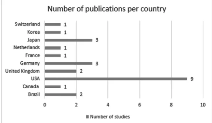
location where the studies were conducted, 12 were American, 8 were European, and 4 were Asian (►Fig. 2). The selected
manuscripts that were original research included 12 transver-sal studies (8 cross-sectional, 3 case-controls, 1 series of cases)
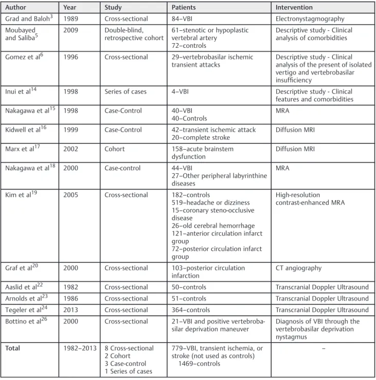
and two longitudinal studies (1 prospective and 1 retrospec-tive cohort). The original articles included three descriptions of clinical features of VBI, one demonstrated results of electro-nystagmography testing, one reported the validity of the vertebral deprivation nystagmus as a diagnostic tool, and 9 compared imaging studies in either controls or patients with VBI (5 studies using magnetic resonance imaging or magnetic resonance angiography, 1 performed angio-tomography in patients with VBI, and 3 reported on Doppler ultrasound results). From the remaining 10 studies,five were reviews, and the otherfive were anatomic descriptions. Demographic information of these studies are shown in►Table 1.
Discussion
Causes
The most frequent cause of the hemodynamic changes that lead to the development of VBI is atherosclerosis. Other common causes are: embolism, atherosclerosis of great ves-sels, and arterial dissection. Migraine,fibromuscular dyspla-sia, coagulopathies, and drug abuse are less frequent causes.7 Review studies investigating the epidemiology of VBI reported that the prevalence of atherosclerosis and blockage of the vertebrobasilar circulation differ according to the sex and age of the patients. It is more frequently observed in men after the fourth decade of life8; arterial hypertension, obesity, and smoking are considered to be the main risk factors.9
Clinical Features
Dizziness, vertigo, headaches, vomit, diplopia, blindness, ataxia, imbalance, and weakness in both sides of the body are the most common symptoms.7–9
Some authors advocate that weakness of the lower limbs associated to imbalance and ataxia of the legs, palsy of the oculomotor nerve, and/or oropharynx dysfunction are the most prevalent symptoms of VBI.7
Ischemia in the territory of the posterior circulation rarely causes only one symptom at a time through the course of the disease; in most cases, there is an association of different symptoms presenting at the same time.10–12
Grad and Baloh3, in a cross-section study involving patients with clinical diagnosis of VBI, stated that the
Fig. 1 Number of selected studies, categorized according to the decade of publication.
most frequent symptoms in this population were: visual symptoms (diplopia, visual hallucinations, deficits in the visual field, and blindness); drop attacks (sudden falls secondary to loss of tonus in the lower limbs, without loss of consciousness); and muscular incoordination and weakness. Mental confusion, headaches, hearing loss, paresthesia, dysarthria, and tinnitus are less frequent symptoms. Among their reported cases, almost 20% of the patients presented with peripheral nystagmus, and none showed nystagmus, suggesting a central origin. The symptoms usually started within a period that varied from 3 weeks to 3 years prior to thefirst consultation.
Isolated attacks of dizziness, or even the chronic imbalance that lasts more than 3 weeks are rarely associated with VBI, and are most commonly observed in diabetic patients.7
Descriptive studies published by Gomez6 and Grad and Baloh3 involving only patients with VBI (confirmed by impairment on the vertebrobasilar bloodflow in the angiogra-phy) stated that the dizziness complaints of these patients could begin within a time frame ranging from 4 weeks to 4 years, before neurologic signs are clinically observed. Furthermore, a review study comparing diagnostic testing for early diagnosis of occlusions in the posterior cerebral circulation reported angiog-raphy to be the most reliable test for this purpose.13
Table 1 Demographic information of the original research studies selected for appraisal
Author Year Study Patients Intervention
Grad and Baloh3 1989 Cross-sectional 84–VBI Electronystagmography
Moubayed and Saliba5
2009 Double-blind, retrospective cohort
61–stenotic or hypoplastic
vertebral artery 72–controls
Descriptive study - Clinical analysis of comorbidities
Gomez et al6 1996 Cross-sectional 29–vertebrobasilar ischemic
transient attacks
Descriptive study - Clinical analysis of the present of isolated vertigo and vertebrobasilar insufficiency
Inui et al14 1998 Series of cases 4–VBI Descriptive study - Clinical
features and comorbidities
Nakagawa et al15 1998 Case-Control 40–VBI
40–Controls
MRA
Kidwell et al16 1999 Case-Control 42–transient ischemic attack
20–complete stroke
Diffusion MRI
Marx et al17 2002 Cohort 158–acute brainstem
dysfunction
Diffusion MRI
Nakagawa et al18 2000 Case-control 44–VBI
27–Other peripheral labyrinthine
diseases
MRA
Kim et al19 2005 Cross-sectional 182–controls
519–headache or dizziness
15–coronary steno-occlusive
disease
26–old cerebral hemorrhage
121–anterior circulation infarct
group
72–posterior circulation infarct
group
High-resolution contrast-enhanced MRA
Graf et al20 2000 Cross-sectional 103–posterior circulation
infarction
CT angiography
Aaslid et al22 1982 Cross-sectional 50–controls Transcranial Doppler Ultrasound
Arnolds et al23 1986 Cross-sectional 51–controls Transcranial Doppler Ultrasound
Tegeler et al24 2013 Cross-sectional 364–controls Transcranial Doppler Ultrasound
Bottino et al26 2000 Cross-sectional 21–VBI and positive
vertebroba-silar deprivation maneuver
Diagnosis of VBI through the vertebrobasilar deprivation nystagmus
Total 1982–2013 8 Cross-sectional
2 Cohort 3 Case-control 1 Series of cases
779–VBI, transient ischemia, or
stroke (not used as controls) 1469–controls
Gomez6, in a cross-section descriptive study, suggested that some characteristics of dizziness might be typical in VBI patients: sudden dizziness spells, lasting 30 seconds to 15 minutes, starting after abruptly standing up or turning the head; also, it has no association with positional complaints, hearing loss, tinnitus, and aural fullness. An ongoing research conducted by our group, so far, failed to observe any specific characteristic of the dizziness in patients diagnosed with VBI. Nonetheless, the incidence of changes in other cranial nerves (visual complaints, by example) seems to be higher. Further-more, we observed a trend in the diagnosis of VBI in patients with one or more of the following conditions: (1) risk factors for cerebrovascular disease; (2) frequent episodes of vertigo that last minutes, waning within days or weeks; and/or (3) absence of hearing loss or tinnitus.
Other possible symptoms of the disease can be secondary to the lateral medullary syndrome, also known as Wallenberg syndrome. This entity is characterized by a stroke in the vertebral artery or PICA irrigation territories in the brainstem. Symptoms include difficulties swallowing, hoarseness, dizzi-ness, nausea and vomiting, nystagmus, and imbalance and gait incoordination.7
Pathophysiology of the VBI and the Vestibular Symptoms
Several anatomic studies reported the complexity of the irrigation pattern of the vestibular system, and suggested that fact to play a major role in the pathophysiologic mecha-nism of the VBI presenting with labyrinthic symptoms.7–9The
vestibular system is supplied by: (1) very small penetrating vessels coming from the basilar artery, supplying the vestib-ular nuclei; and (2) the internal auditory artery (IAA), origi-nated either from (a) the anterior-inferior cerebellar artery (80–85%); or (b) a vascular loop from the posterior-inferior cerebellar artery (PICA), which is a branch of the vertebral artery (15%). The PICA is a terminal vessel with very few collateral branches. The IAA irrigates the cochleovestibular nerve, the cochlea, and the posterior labyrinth.3,6Inui et al14 described in a report of four cases how the AICA emerge from the basilar artery, and the PICA from one of the vertebral arteries in 56% of the cases.
Considering that the labyrinthine branches are smaller and receive less collateral irrigation, it is possible that the laby-rinth should be more affected by atherosclerotic blockage of the vertebrobasilar arterial system. On the other hand, the cochlea receives collateral irrigation from the carotid artery, supplying adjacent portions of the petrous bone. This partic-ular feature prevents the cochlea from suffering ischemic symptoms in the case of a vascular insufficiency.6
Moubayed and Saliba,5 in a double-blind retrospective cohort, studied the prevalence of positional dizziness and imbalance, plus clinical and electronystagmographyfindings in two groups: one with changes in the vertebral arteries in magnetic resonance angiography (stenotic or hypoplastic vertebral arteries) and one without any changes. The authors observed no differences between the two groups. These results suggest that the clinical symptoms are triggered by occlusion or obstruction in smaller vessels, in more distal regions.
Isolated episodes of vertigo could arise from transient ischemia of the vestibular labyrinth, due to the characteristics of its terminal circulation without collateral vessels.3The fact that the posterior labyrinth integrates information bilaterally in a more active way than the cochlea also explains why VBI causes more vestibular than cochlear symptoms. Thus, the vestibular system could be more sensitive in detecting differences in the action potential secondary to ischemia.6
The ischemia could affect both peripheral and central structures of the vestibular system. Moubayed and Saliba5 describe two mechanisms through which the ischemic epi-sodes could cause isolated dizziness spells: (1) decrease in the blood flow to the vestibular nuclei or to the root of the entrance zone of the vestibulo-cochlear nerve; and (2) direct ischemia of the labyrinth.
Diagnosis of VBI by Neuroimaging
The patients with a suspected diagnosis of transient ischemic attacks or vertebrobasilar strokes should undergo neuroimag-ing tests. Even though arteriography is considered to be the most important exam for this purpose, the risks of performing this test in patients with VBI should be taken in consideration. Many patients choose not to take this test after reading the risks of the procedure in the informed consent terms. The main complications of the arteriography are: local complications of the arterial catheters; regional low bloodflow; and stroke.15
The magnetic resonance angiography (MRA) has been widely studied and it is considered to be a good method to identify and localize any occlusions and stenosis in both neck and intracranial great vessels.15–17Nakagawa et al15,18
com-pared patients with VBI to non-diseased controls and patients with other peripheral labyrinthine diseases and reported some findings to be characteristic in patients VBI: focal atherosclerosis decreasing the lumen of the basilar artery (mainly when close to the exit of the AICA), and diffuse narrowing in the origin of the vertebral artery with decreased distal bloodflow. In one of those studies, Nakagawa et al18 reported that stenosis of the basilar artery was a significant
finding among patients with the diagnosis of VBI when compared with age-matched controls. Nonetheless, the sur-prisingly high incidence of anomalies found by the authors (30%) with high levels of statistical significance (p¼0.003)
may raise concerns about the inclusion and exclusion criteria of the patient selection in the study.
Moubayed and Saliba5evaluated patients with positional dizziness and imbalance regarding the morphology of the vertebral arteries, risk factors for stroke involving the poste-rior circulation, and evolution of the symptoms over time in patients with VBI compared with controls. They concluded that 85.7% of the patients with dizziness and at least 3 risk factors for stroke have morphologic abnormalities in the vertebral arteries.
headaches, tinnitus, seizures, movement disorders, anxiety, dementia, traumatic head injuries, or symptoms of transitory ischemia; (3) patients with occlusion or stenosis of the coronary arteries; (4) patients with previous intracranial bleeding; (5) patients with stroke of the anterior circulation; and (6) patients with stroke related to the posterior circula-tion. The prevalence of stenosis increased gradually accord-ingly to the severity of the disease, and the results were statistically significant (p<0.0001 for every performed
com-parison). The authors also consider the proximal region of the vertebral arteries to be the place with a higher prevalence of occlusion of the posterior circulation. They also state that more studies focusing on understanding the natural history of these lesions and their evolution over time are still needed.
A small number of publications dedicated on evaluating posterior circulation; however, studies in healthy patients points toward angio-tomography and MRA to reach similar sensibility and specificity levels, especially when studying the basilar artery.20The advantage of the angio-tomography is that it provides less false-positive results, due to the smaller influence of the slower bloodflow in some narrow portions of the posterior circulation on the results. Other studies in healthy subjects reported the sensibility of the magnetic resonance angiography, angio-tomography, and transcranial Doppler to be 93.9%, 100%, and 70.2%, respectively, while the specificity rate was 94.8%, 95.2%, and 97.7%.21
The transcranial Doppler is a cheap, pain-free, and non-invasive test, which is capable of measuring the speed and direction of the bloodflow from the proximal areas of the great intracranial arteries. Thefirst clinical application of this exam was described in 1982.22 In a report named “The vascular diagnosis guidelines,”published by the American Academy of Neurology, the authors report the Doppler as a high sensitivity (50–80%) and high specificity (80–96%) exam.13
Another important piece of information given by the Doppler is the pulsatility index (PI), which is the relation between the measured speed of the bloodflow during the systolic pulse and thefinal diastolic pulse. Studies in the general healthy popula-tion demonstrated that the lower this relapopula-tion, the greater the resistance of the adjacent microvasculature. This index is highly predictive of early hemodynamic intracranial changes.23
It is known that the speed of the intracranial arterial blood
flow decreases and PI increases in older male patients, even in those without any cerebrovascular diseases. The opposite phenomenon is observed in female patients: the speed increases and PI decreases. It has also been demonstrated that there were no differences among different ethnic groups, regarding the blood flow speed and PI of the posterior circulation.24Such facts should be taken into consideration when evaluating the results of this exam in patients with different sex and ethnicity.
Diagnosis of VBI through the Vertebrobasilar Deprivation Nystagmus
The vertebrobasilar deprivation nystagmus, according to Caussé et al25, is the nystagmus obtained using an exten-sion and rotation of the neck for three minutes, when every other possible cause of nystagmus has been discarded. This
nystagmus would occur because of a decrease in the blood
flow in the opposite vertebral artery because of the head rotation. Moubayed and Saliba5reported the physiopathol-ogy of this event to be secondary to the depolarization of the ciliated cells, caused by an acute onset of ischemia, generating a nystagmus. However, in a late phase of the ischemic injury, the membrane of the axons become inca-pable of excitation, resulting in a hypofunction of these cells. Nonetheless, this explanation is controversial, be-cause if the basilar artery has a normal bloodflow, there would be no reason for decreased perfusion of the terminal circulation. Inui et al14 reported the case of a patient showing nystagmus when turning the head to the left side, and exams pointed to an occlusion of the ipsilateral vertebral artery. This suggests a decrease in the bloodflow on the right vertebral artery; the decreased bloodflow in the occluded left artery was not enough to supply the basilar artery, generating the nystagmus.
Bottino26studied patient with clinical symptoms of VBI and compared with patients without any kind of disease. He observed the presence of NPVB in 43.4% of the diseased patients versus 13.3% in the control group. In the same study, the authors also report the results of Doppler of the carotid and describe vertebral arteries in 21 patients with nystagmus secondary to vertebrobasilar deprivation. The sensitivity of the exam was around 19%. The authors conclude that using this clinical maneuver is a cheap, sensitive, and safe proce-dure to diagnose VBI.26
Limitations
This study has several limitations. The small number of good randomized, double-blind, clinical trials, and well-designed case-control and cohort studies limits interpretation and validity of some of the results. The high variability of meas-ures and outcomes among the studies about the same diag-nostic testing, anatomic feature, or patients selected as control groups, were also limiting factors in our analysis. Nonetheless, we were able to provide a meaningful review including several aspects of the pathophysiology and avail-able diagnostic testing for VBI.
Final Comments
VBI should be suspected in patients presenting with risk factors for ischemia and vestibular symptoms. Each diagnos-tic test offers different pros and cons, thus, each patient should be treated individually to avoid possible sequels.
References
1 Kubik CS, Adams RD. Occlusion of the basilar artery; a clinical and pathological study. Brain 1946;69(2):73–121
2 Special report from the National Institute of Neurological Disor-ders and Stroke. Classification of cerebrovascular diseases III. Stroke 1990;21(4):637–676
4 Simoceli L, Bittar RMS, Bottino MA, Bento RF. Perfil diagnóstico do idoso portador de desequilíbrio corporal: resultados preliminares. Rev Bras Otorrinolaringol 2003;69:772–777
5 Moubayed SP, Saliba I. Vertebrobasilar insufficiency presenting as isolated positional vertigo or dizziness: a double-blind ret-rospective cohort study. Laryngoscope 2009;119(10): 2071–2076
6 Gomez CR, Cruz-Flores S, Malkoff MD, Sauer CM, Burch CM. Isolated vertigo as a manifestation of vertebrobasilar ischemia. Neurology 1996;47(1):94–97
7 Savitz SI, Caplan LR. Vertebrobasilar disease. N Engl J Med 2005; 352(25):2618–2626
8 Caplan LR, Gorelick PB, Hier DB. Race, sex and occlusive cerebro-vascular disease: a review. Stroke 1986;17(4):648–655
9 Caplan LR. Intracranial branch atheromatous disease: a neglected, understudied, and underused concept. Neurology 1989;39(9): 1246–1250
10 Caplan L. Posterior circulation ischemia: then, now, and tomorrow. The Thomas Willis Lecture-2000. Stroke 2000;31(8):2011–2023 11 Bradshaw P, McQuaid P. The syndrome of vertebrobasilar insuffi
-ciency. Q J Med 1963;32:279–296
12 Bruyn GW. Vertigo and vertebrobasilar insufficiency. A critical comment. Acta Otolaryngol Suppl 1988;460:128–134
13 Sloan MA, Alexandrov AV, Tegeler CH, et al. Therapeutics and Technology Assessment Subcommittee of the American Academy of Neurology. Assessment: transcranial Doppler ultrasonography: report of the Therapeutics and Technology Assessment Subcom-mittee of the American Academy of Neurology. Neurology 2004; 62(9):1468–1481
14 Inui H, Yoneyama K, Kitaoku Y, et al. Four cases of vertebrobasilar insufficiency. Acta Otolaryngol Suppl 1998; 533:46–50
15 Nakagawa T, Yamane H, Nakai Y, Shigeta T, Takashima T. Evalua-tion of the vertebrobasilar artery system by magnetic resonance angiography in the diagnosis of vertebrobasilar insufficiency. Acta Otolaryngol Suppl 1998;538:54–57
16 Kidwell CS, Alger JR, Di Salle F, et al. Diffusion MRI in patients with transient ischemic attacks. Stroke 1999;30(6):1174–1180 17 Marx JJ, Mika-Gruettner A, Thoemke F, et al. Diffusion weighted
magnetic resonance imaging in the diagnosis of reversible ischae-mic deficits of the brainstem. J Neurol Neurosurg Psychiatry 2002; 72(5):572–575
18 Nakagawa T, Shigeta T, Takashima T, Tomiyama K. Magnetic resonance angiography evaluation of basilar artery stenosis in patients with vertebrobasilar insufficiency. Eur Arch Otorhinolar-yngol 2000;257(8):409–411
19 Kim SH, Lee JS, Kwon OK, Han MK, Kim JH. Prevalence study of proximal vertebral artery stenosis using high-resolution contrast-enhanced magnetic resonance angiography. Acta Radiol 2005; 46(3):314–321
20 Graf J, Skutta B, Kuhn FP, Ferbert A. Computed tomographic angiographyfindings in 103 patients following vascular events in the posterior circulation: potential and clinical relevance. J Neurol 2000;247(10):760–766
21 Khan S, Cloud GC, Kerry S, Markus HS. Imaging of vertebral artery stenosis: a systematic review. J Neurol Neurosurg Psychiatry 2007; 78(11):1218–1225
22 Aaslid R, Markwalder TM, Nornes H. Noninvasive transcranial Doppler ultrasound recording offlow velocity in basal cerebral arteries. J Neurosurg 1982;57(6):769–774
23 Arnolds BJ, von Reutern GM. Transcranial Doppler sonography. Examination technique and normal reference values. Ultrasound Med Biol 1986;12(2):115–123
24 Tegeler CH, Crutchfield K, Katsnelson M, et al. Transcranial Doppler velocities in a large, healthy population. J Neuroimaging 2013; 23(3):466–472
25 Causse JB, Conraux C, Causse J. [Vertebral-basilar artery insuffi -ciency nystagmus (author’s transl)]. Ann Otolaryngol Chir Cervi-cofac 1978;95(3):225–234