Cephalometric evaluation of the effects of the
joint use of a mandibular protraction appliance
(MPA) and a fixed orthodontic appliance on
the skeletal structures of patients with Angle
Class II, division 1 malocclusion
Objective: This study aimed to perform a cephalometric evaluation of the skeletal respons-es triggered by the joint use of a mandibular protraction appliance (MPA) and a fixed orthodontic appliance for correction of Class II, division 1 malocclusion in young Brazilian
patients. Methods: The sample consisted of 56 lateral cephalograms of 28 patients (16
women and 12 men). The initial mean age was 13.06 years and mean duration of therapy with MPA was 14.43 months. The lateral radiographs were obtained before and after treat-ment and were compared by two calibrated examiners to identify the skeletal changes induced by the MPA using 16 linear and angular cephalometric measures. Some indepen-dent variables (patient age, sex, facial pattern, MPA model, total use time, archwire and technique used during therapy with MPA) were considered and related to those measures in order to demonstrate the influence of these variables on them. Responses to treatment were analyzed and compared by the Wilcoxon Signed Ranks test and Mann-Whitney test at
a significance level of 5%. Results: The results showed restricted anterior displacement of
the maxilla, increased mandibular protrusion, improved anteroposterior relationship of the basal bones and stability of the mandibular plane relative to the cranial base. The influence
of variables age, facial pattern and MPA type was also noted. Conclusions: MPA proved an
effective alternative in the treatment of Class II, division 1 malocclusion, inducing changes in the skeletal component with satisfactory clinical results.
Abstract
Keywords: Cephalometry. Functional orthodontic appliances. Angle Class II malocclusion. Mandibu-lar protraction appliance.
* Specialist in Orthodontics, ABO-EAP/RN.
** MSc in Orthodontics, USP. Head Professor of Orthodontics, UFRN. Professor of the Specialization Course, ABO-EAP/RN. *** Specialist in Orthodontics, ABO-EAP/RN.
**** Professor, Department of Orthodontics and Graduated Course in Dentistry and Health Sciences, UFRN. Emmanuelle Medeiros de Araújo*, Rildo Medeiros Matoso**,
Alexandre Magno Negreiros Diógenes***, Kenio Costa Lima****
How to cite this article: Araújo EM, Matoso RM, Diógenes AMN, Lima KC. Cephalometric evaluation of the effects of the joint use of a mandibular protraction appliance (MPA) and a ixed orthodontic appliance on the skeletal structures of patients with Angle Class II, division
INTRODUCTION AND LITERATURE REVIEW Angle Class II, division 1 malocclusion is a frequent problem affecting about 55% of the Brazilian population.2 It has a multifactorial etiology, and from a skeletal point of view, may be due to maxillary protrusion, mandibular
re-trusion or a combination of both.16
The literature is rich in treatment meth-ods for this malocclusion, which traditionally rely on patient cooperation in wearing remov-able functional appliances (Activator, Balters’ Bionator, Frankel appliance), using Class II elastics and/or extraoral traction appliances. Among the appliances used in Class II, divi-sion 1 cases are those which have as their key objective restricting the anterior displacement of the maxilla, those that push the mandible towards a more anterior position in order to redirect growth and lead to an appropriate morphological development, and those that
induce changes in both arches.12
In recent decades, several authors began to develop fixed intraoral orthopedic appliances capable of correcting Class II molar relation-ship with mandibular retrognathism, since these appliances promote changes in mandibu-lar posture, positioning it forward with the aim of stimulating its growth.24,25 Since these
ap-pliances are fixed (Herbst,25 Jasper Jumper,17,18
Universal Bite Jumper,4,28 Eureka Spring,13
MARA,1 Churro Jumper5 and Superspring19)
they are instrumental in decreasing the need for patient compliance during treatment.
However, the lack of specialized laborato-ries to fabricate these appliances, their high cost and scarcity of information about the installa-tion of most of them led Coelho Filho6 to de-sign the Mandibular Protraction Appliance 1, also known as MPA 1, whose characteristics, at first quite simple, soon evolved into a more ad-vanced version. In 1995, the inventor presented the clinical results achieved with his appliance as an alternative to Herbst,6,20,21,22,23,26,27
rein-troduced by Pancherz (1979), since the former uses the same mechanical design as the latter.
Some of the advantages of MPA over Herbst are that (a) it can be fabricated by profession-als themselves, without the need for laboratory work, (b) it is affordable, (c) it is easy to insert, and (d) as it is less bulky, it provides greater patient comfort.10,11,30
MPA 1 was initially made with 0.032-in (0.9 mm) wire and consisted of a steel rod with a round loop at each end. In this first version, rectangular wires had to be in place and due to the conformation of the appliance only canine to canine brackets could be bonded. Moreover, the lower arch needed to have a strong torque in the anterior region to resist buccal displace-ment of lower incisors resulting from the pro-trusive forces generated by the appliance. Ad-ditionally, bends had to be applied on the distal side of lower molar tubes to enhance
anchor-age and prevent mesial drift of lower teeth.6,8
Although the clinical results achieved with MPA 1 were extremely positive, limitations in
mouth opening caused frequent breakages.7
Therefore, in 1997, the second MPA version was launched, featuring increased mouth open-ing, greater patient comfort and less frequent breakages. Besides all the installation details described for an MPA 1, the author empha-sized insertion of anterosuperior buccal torque and two circular loops positioned mesial to the upper molars and distal to the lower canines to facilitate appliance installation. Also notewor-thy was the fact that with this second version brackets could be bonded to premolars.
In contrast to these upsides, MPA 2 also showed some shortcomings. To address these issues the author created a fully modified
third version termed MPA 3,7,9,10 which had
arch was redesigned. All these improvements ensured greater appliance balance when pa-tients opened and closed their mouth. The author also discussed the use of the appliance in cases of Class III malocclusion and anterior crossbite. To do so would require reversing
the direction of the appliance.7,9
In 2001 and 2002, Coelho Filho introduced the latest version: MPA 4. The author reported that this new model seemed to surpass all previ-ous models in terms of both shear strength and ease of installation. Furthermore, MPA 4 adap-tation to the upper arch was modified to impart greater functional stability to the appliance.11 The author also pointed out that MPA model did not determine differences in the outcome. All models feature the same mechanical prin-ciples. What makes each different is fabrication
method, installation and patient comfort.7
Given their numerous advantages, as stated above, in addition to being versatile and featur-ing a wide range of applications, orthodontists were driven to study MPA treatment effects, prompting some to go as far as to propose other appliance models with similar mecha-nisms.15,22 Thus, the purpose of this study was to analyze and determine skeletal changes in patients with Angle Class II, division 1 mal-occlusion resulting from treatment with MPA during the phase of active growth.
METhODs
This study can be defined as an untrolled, nonrandomized clinical trial. To con-duct it, a sample was selected comprising 56 lateral cephalograms of 28 Brazilian youths of both sexes — 16 women and 12 men — ac-cording to the following criteria: Angle Class II, division 1 malocclusion with mandibular retrognathism, as assessed by study models, photographs and radiographs with a clear vi-sualization of the structures of interest. Ex-clusion criteria were as follows: Agenesis,
ex-traction or loss of permanent teeth; patients undergoing orthodontic treatment prior to MPA installation, since prior therapy would alter the Class II, division 1 malocclusion; and significant overjet.
Clinical records included the following clin-ical variables: Patient age, sex, facial pattern (dolichofacial, mesofacial and brachyfacial, but the latter was excluded during sample selec-tion as only one case had this facial type, which might yield statistical results with a higher margin of error), MPA model (types 1, 2, 3 and 4; type 1 was associated with type 2, and type 3 with type 4, since only one patient was treat-ed with MPA 1, and only 5 cases with MPA 3), total time of appliance use, archwires used during treatment with MPA (0.019x0.025-in, 0.021x0.025-in and 0.018x0.025-in stainless steel wires, with the latter two grouped togeth-er, totaling 12 cases, compared to 16 patients with 0.019x0.025-in stainless steel wire) and orthodontic technique (Standard Edgewise and Straight Wire).
The cephalograms used in this study were selected from the archives of Professor Carlos Martins Coelho Filho’s private clinic (in the city of São Luís, Maranhão state, Brazil), and obtained with Funk Orbital X15 X-ray device, with a magnification factor of 9%, and oper-ated by one and the same examiner.
Two lateral cephalograms of each of the 28 patients were used, referred to as T1 (initial) and T2 (final). The cephalograms were traced manu-ally on a light box by two calibrated examiners in a darkened room at Professor Carlos Martins’ private clinic in São Luís, Maranhão state.
Examiner calibration was performed ap-proximately three months earlier, when 30 randomly selected cephalograms were retraced until minimum error was attained.
5
1
4 8
6
7
3
2 9
with a 0.3 mm tip, tape, soft rubber, template (Tracing Template, Unitek Corp.), and a light box. When double images of the anatomical design of bony structures were visualized both images were traced and a mean value was found between cephalometric points.
In the next step the images were imported via a scanner into a microcomputer containing the Radiocef Studio Cephalometry program (No. 020576, version 4.0, release 3 - Belo Hori-zonte/MG, Brazil), where values were obtained for T1 and T2 and their respective repetitions.
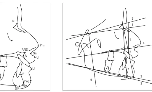
From then on, the following landmarks were identified to obtain angular and linear measurements: S (sella turcica), N (nasion), A (subspinale), B (supramentale), Pog (pogo-nion), Me (menton), Go (go(pogo-nion), Gn (gnathi-on), Ar (articulare), ANS (anterior nasal spine) and PNS (posterior nasal spine) (Fig 1).
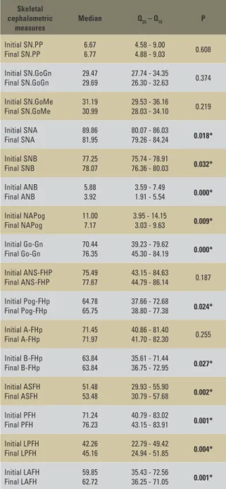
The reference planes used in this study were, as shown in Figure 2: a modified
Frank-fort Horizontal Plane (FHP)29 (1), composed
of a line that forms with the SN line a 7° angle
FIGURE 1 - Cephalometric points (landmarks). FIGURE 2 - Reference planes and lines.
down through point S; Mandibular planes Go-Me (2) and Go-Gn (3); Palatal Plane (PP) (4), formed by points ANS and PNS; lines SN (5), NA (6), NB (7), APog (8) and S-HFp (9).
Angular variables included, as shown in Fig-ure 3: SN.PP (10), SN.GoGn (11), SN.GoMe (12), SNA (13), SNB (14), ANB (15) and NAPog (16); and the linear variables were, as shown in Figure 4: Go-Gn (17), ANS-FHP (18), Pog-FHP (19), A-FHP (20), B-FHP (21), ASFH (22), PFH (23), LPFH (24) and LAFH (25).
REsULTs
This study used a sample of 56 lateral ceph-alograms of 28 young Brazilian of both sexes comprising 16 women (57.1%) and 12 men (42.9%) (Table 1).
Mean age was 13.06 years, with a stan-dard deviation of 1.3 years, with a minimum of 10.33 years and a maximum of 16.58 years, respectively.
As regards facial pattern, 39.3% (11 patients) were dolichofacial while 60.7% (17 patients)
N
Prn
Sn Ul
Ll
Me Gn Pog B Go
Ar S
PNS
ANS
12 11 10
13
14
18
20 23 24
17 19
21 25 22 15
16
were mesofacial. As explained before, during sample selection the brachyfacial pattern was excluded as only one case had this facial type, which might yield unreliable statistical results.
Similarly, under variable MPA model, MPA type 1 was associated with MPA type 2, and type 3 with type 4, since only one patient (3.6%) had been treated with MPA 1 and 5 cases (17.9%) with MPA 3. The remaining per-centages corresponded to 35.7% (10 cases) and 42.9% (12 cases) of MPAs 2 and 4, respectively.
For the variable archwire, the following types were noted: 0.019x0.025-in stainless steel (57.1% or 16 patients), 0.021x0.025-in sta0.021x0.025-inless steel (10.7% or 3 patients) and 0.018x0.025-in stainless steel (32.1% or 9 patients). The latter two archwires were also grouped into a total of 12 cases.
The variable technique showed a frequency of 12 cases (42.9%) for the Straight Wire tech-nique and a total of 16 cases (57.1%) for the Standard Edgewise technique.
The result achieved for the variable total MPA use time was 14.43 months, with a mini-mum of 3 months and maximini-mum of 33 months, and a standard deviation of 9.33 months.
Table 2 shows the means for initial and fi-nal cephalometric measurements of patients of both sexes, their medians, quartiles 25 and 75, and statistical significance value (p), obtained with the Wilcoxon Signed Ranks Test. As can be observed, of all the skeletal cephalometric measures employed in this study, only SNA, SNB, ANB, NAPog, Go-Gn, Pog-FHP, FHP-B, ASFH, PFH, LPFH and LAFH were influenced by treatment with MPA, i.e., showed statisti-cally significant values (p<0.05).
Among the seven independent variables, statistically significant results were found only for age, sex, facial pattern and MPA mod-el. Tables 3 and 4 show differences between cephalometric measurements before and after treatment with MPA related to such variables, including their medians, quartiles 25 and 75,
TABLE 2 - Medians and 25/75 quartiles of initial and final skeletal cepha-lometric measurements and value of statistical significance. (Natal, Rio Grande do Norte state, Brazil, 2005).
TABLE 1 - Relationship between variables and sample distribution. Na-tal, Rio Grande do Norte State, Brazil, 2005.
*Significant difference (p<0.05) based on Wilcoxon test.
and significance value (p) for each individu-al measure. For sex, only Go-Gn and LAFH showed a statistically significant results, and for age, only ANB. As for facial pattern, the only quantities that showed significant differ-ences were PFH and LPFH. Regarding MPA type, statistical differences were found for Go-Gn, ANS-FHP, Pog-FHP, A-FHP, B-FHP, ASFH, PFH and LAFH.
Tables 5 and 6 show the variables associ-ated with the skeletal cephalometric measures that exhibited changes after treatment. The variables were related to these measures prior to treatment. This revealed the influence that they exerted on these measures and whether differences existed in relation to these vari-ables even before starting therapy with MPA. To obtain these results, the Mann-Whitney Test was employed.
As can be seen in Tables 5 and 6, only Go-Gn and LAFH showed statistical relevance even before starting treatment, when related
to variable sex. All other measures, which were influenced by treatment with MPA, exhibited no statistically significant values in this pre-treatment phase.
Variables
Frequency
n %
Age ≤ 13.06 years≥ 13.06 years 1414 5050
Sex Female Male 16 12 57.1 42.9 Facial Pattern Dolicho Meso 11 17 39.3 60.7
MPA Type 1+2 3+4
11 17
39.3 60.7
Archwire 0.019x0.025-in SS
0.021x0.025-in + 0.018x0.025-in SS 16 12
57.1 42.9
Technique Straight WireStandard Edgewise 1216 42.957.1
Skeletal cephalometric
measures
Median Q25 – Q75 P
Initial SN.PP Final SN.PP
6.67 6.77
4.58 - 9.00
4.88 - 9.03 0.608
Initial SN.GoGn Final SN.GoGn
29.47 29.69
27.74 - 34.35
26.30 - 32.63 0.374
Initial SN.GoMe Final SN.GoMe
31.19 30.99
29.53 - 36.16
28.03 - 34.10 0.219
Initial SNA Final SNA
89.86 81.95
80.07 - 86.03
79.26 - 84.24 0.018*
Initial SNB Final SNB
77.25 78.07
75.74 - 78.91
76.36 - 80.03 0.032*
Initial ANB Final ANB
5.88 3.92
3.59 - 7.49
1.91 - 5.54 0.000*
Initial NAPog Final NAPog
11.00 7.17
3.95 - 14.15
3.03 - 9.63 0.009*
Initial Go-Gn Final Go-Gn
70.44 76.35
39.23 - 79.62
45.30 - 84.19 0.000*
Initial ANS-FHP Final ANS-FHP
75.49 77.67
43.15 - 84.63
44.79 - 86.14 0.187
Initial Pog-FHp Final Pog-FHp
64.78 65.75
37.66 - 72.68
38.80 - 77.38 0.024*
Initial A-FHp Final A-FHp
71.45 71.97
40.86 - 81.40
41.70 - 82.30 0.255
Initial B-FHp Final B-FHp
63.84 63.84
35.61 - 71.44
36.75 - 72.95 0.027*
Initial ASFH Final ASFH
51.48 53.48
29.93 - 55.90
30.79 - 57.68 0.002*
Initial PFH Final PFH
71.24 76.23
40.79 - 83.02
43.15 - 83.91 0.001*
Initial LPFH Final LPFH
42.26 45.16
22.79 - 49.42
24.94 - 51.85 0.004*
Initial LAFH Final LAFH
59.85 62.72
35.43 - 72.56
TABLE 3, 4 - Medians, 25/75 quartiles and significance of cephalometric measurements related to independent variables. (Natal, RN, Brazil, 2005).
*Significant difference (p<0.05).
*Significant difference (p<0,05).
Difference between T1 and T2 cephalometric
measurements
ANB Go-Gn ANS-FHP Pog-FHP A-FHP
Median Q25/Q75 p Median Q25/Q75 p Median Q25/Q75 p Median Q25/Q75 p Median Q25/Q75 p
Sex n
Female (16) 2.24 1.12/2.96
0.246
-1.19 -3.76/-0.82
0.029
0.04 -1.76/1.27
0.194
-1.15 -6.13/1.67
0.194
-0.29 -1.02/1.19
0.114
Male (12) 1.18 0.04/3.00 -4.20
-11.44/-2.31 -1.11 -6.48/0.75 -6.13 -10.26/0.51 -1.48 -4.75/0.44
Age (n)
≤ 13.06 (14) 2.65 1.07/3.45 0.035*
-3.72 -13.11/-1.14
0.183
-0.42 -2.64/0.38
0.748
-4.18 -9.66/1.65
0.383
-0.40 -2.37/1.67
1.000
>13.06 (14) 1.29 0.04/2.40 -1.66 -4.16/-0.80 -0.13 -4.89/1.02 0.79 -5.85/0.87 -0.34 -3.21/1.09
Facial Pattern (n)
Dolichofacial (11) 1.13 0.53/2.98
0.410
-2.83 -17.47/-1.23
0.312
-0.82 -4.66/0.78
0.335
-5.09 -9.35/1.84
0.621
-1.07 -3.58/1.24
0.556
Mesofacial (17) 2.37 0.78/3.03 -2.08 -6.31/0.83 0.06 -3.33/1.04 -1.20 -7.28/0.47 -0.37 -1.80/0.80
MPA type (n)
1 + 2 (11) 2.98 0.31/3.61
0.335
-4.83 -17.47/-3.65
0.003*
-1.64 -5.02/-0.03
0.018*
-7.19 -10.58/-1.95
0.001*
-1.26 -3.74/-0.43
0.006*
3 + 4 (17) 1.74 0.75/2.44 -1.23 -3.13/-0.61 0.67 -1.89/1.21 0.39 -3.25/3.30 0.56 -1.27/2.15
Difference between T1 and T2 cephalometric measurements
B-FHP ASFH PFH LPFH LAFH
Mediana Q25/Q75 p Median Q25/Q75 p Median Q25/Q75 p Median Q25/Q75 p Median Q25/Q75 p
Sex (n)
Female (16) -0.84 -5.49/1.14
0.265
-0.89 -1.66/-0.14
0.057
-2.33 -5.50/-0.24
0.210
-2.27 -4.49/-0.27
0.430
-1.44 -2.98/-0.52
0.010*
Male (12) -5.44 -10.39/0.81 -2.38 -5.75/-0.53 -4.95 -7.65/-1.37 -4.39 -5.90/0.80 -5.13 -8.56/-3.11
Age (n)
≤ 13.06 (14) -3.74 -7.91/1.14 0.312
-1.30 -3.74/0.76
0.963
-5.53 -8.64/-1.30
0.081
-4.43 -6.60/-0.68
0.154
-2.83 -5.85/-1.07
0.566
>13.06 (14) -0.43 -5.15/1.01 -1.32 -2.50/-0.23 -2.33 -4.74/-0.01 -1.29 -4.35/-0.56 -2.57 -5.33/-0.47
Facial Pattern (n)
Dolichofacial (11) -4.59 -6.93/1.29
0.724
-1.65 -4.84/-0.24
0.384
-5.54 -11.09/-2.99
0.041*
-4.34 -8.00/-0.99
0.046*
-3.41 -5.71/-0.93
0.371
Mesofacial (17) -0.96 -6.37/0.70 -1.10 -2.38/0.16 -2.31 -5.38/0.28 -2.04 -4.53/-1.97 -2.62 -5.03/-0.58
APM type (n)
1 + 2 (11) -6.29 -10.85/-1.74
0.002*
-2.32 -6.09/-0.94
0.048*
-5.67 -11.09/-3.37
0.015*
-4.52 -8.00/-1.60
0.063
-5.21 -7.57/-2.00
0.041*
TABLE 5, 6 - Values of cephalometric measurements that showed statistically significant changes after treatment - related to independent variables - before starting treatment with MPA. (Natal, RN, Brazil, 2005).
*Significant difference (p<0.05).
*Significant difference (p<0.05).
ANB Go-Gn ANS-FHP Pog-FHP A-FHP
Median Q25/Q75 p Median Q25/Q75 p Median Q25/Q75 p Median Q25/Q75 p Median Q25/Q75 p
Sex (n)
Female (16) 44.14 27.51/66.57
0.003*
Male (12) 75.39 71.04/91.29
Age (n)
≤ 13.06 (14) 5.57 3.26/7.23 0.730
>13.06 (14) 6.04 3.95/7.84
Facial Pattern (n)
Dolichofacial (11)
Mesofacial (17)
MPA type (n)
1 + 2 (11) 70.44 28.51/94.09
1.000
78.39 28.55/102.20
0.410
68.84 22.79/78.16
0.655
74.68 27.14/97.74
0.359
3 + 4 (17) 70.44 44.14/75.43 72.95 43.47/80.19 62.48 37.72/70.58 69.37 41.17/77.70
B-FHP ASFH PFH LPFH LAFH
Median Q25/Q75 p Median Q25/Q75 p Median Q25/Q75 p Median Q25/Q75 p Median Q25/Q75 p
Sex (n)
Female (16) 36.38 26.94/58.82
0.001*
Male (12) 69.29 62.43/79.77
Age (n)
≤ 13.06 (14)
>13.06 (14)
Facial Pattern (n)
Dolichofacial (11) 78.12 39.86/89.06
0.525
45.62 23.68/55.61
0.269
Mesofacial (17) 70.66 40.83/80.62 40.50 22.42/47.09
MPA type (n)
1 + 2 (11) 66.90 23.38/76.33
0.466
52.58 21.36/63.91
0.438
77.53 27.53/89.06
0.466
63.16 26.87/81.10
0.384
DIsCUssION
Angle Class II, division 1 malocclusion is a frequent problem since for its interception and/ or correction a wide range of appliances have been proposed. Moreover, the literature is still scarce in studies that pinpoint which changes re-sult from MPA use, be they skeletal, dental or cutaneous changes. Thus, this study sought to evaluate the skeletal changes triggered by the use of mandibular protraction appliances in patients with Class II, division 1 malocclusion associated with a corrective orthodontic appliance.
As for the sagittal maxillomandibular rela-tionship, only measures ANB and NAPog were verified. The following measures were used to observe vertical changes: SN.PP, SN.GoGn, SN.GoMe, ASFH, PFH, LPFH and LAFH.
According to the results shown in Table 2, only SNA showed statistical significance for the maxillary component, suggesting that MPA acted by hindering anterior maxillary displace-ment, causing a reduction of 1.91°. It is known that during growth the maxilla moves forward and downward. In patients with Class II growth pattern it is common for point A to be posi-tioned more anteriorly. Thus, when associated with the growth tendency observed in the max-illa of untreated patients, SNA often
experienc-es an increase.14 In this study, reduction in this
skeletal cephalometric measurement can there-fore be attributed to the use of the appliance favored by the growth factor, since the treated group had a mean age of 13.06 years.
Concerning mandibular changes, all measures showed significant differences and increased pro-trusion when MPA was used, but this fact does not warrant one to assert that protrusion was solely due to the MPA as this age group shows a predominance of mandibular growth. Cepha-lometric measurements correspond to SNB, Go-Gn, B-FHp and Pog-FHp (Table 2).
The measures used to verify the sagittal max-illomandibular relationship yielded statistically
significant results. After using the appliance, an improved relationship was noted between the maxilla and mandible in the anteroposterior direction, with a more posterior positioning of the maxilla and more anterior positioning of the mandible. There was a decrease in maxillo-mandibular relationship values (ANB, NAPog) which resulted in the correction of the skeletal Class II (Table 2).
In observing the vertical changes result-ing from therapy with MPA, it was found that the angular measures SN.PP, SN.GoGn and SN.GoMe showed no statistically significant differences after MPA use. The former two measures remained fairly constant and the lat-ter experienced a slight downturn. Linear mea-sures ASFH, PFH, LPFH and LAFH showed significant increases (Table 2). Once again the results reinforce Coelho Filho’s finding that de-spite increases in anteroinferior and posterior facial height, the mandibular plane angle is not negatively affected when treatment induces the mandible to move to a more anterior position.
Tables 3 and 4 show the difference ratio be-tween cephalometric initial and final measures, and independent variables. Only variables sex, age, facial pattern and MPA model influ-enced the final cephalometric measures. The other variables used in this research — total use time, archwire and technique used during treatment — showed no statistically significant results and do not seem to exert any influence on the skeletal cephalometric measures target-ed in this study.
As regards age, the only measurement that showed significant alteration was ANB, and the difference between T1 and T2 was higher in
the group aged ≤13.06 years and lower in the
group aged >13.06 years, whose values corre-spond to 2.65 and 1.29, in the order given, due to greater growth potential with more signifi-cant skeletal changes at younger ages (Tables 3 and 4). However, this measure was not initial-ly altered, suggesting that age influenced the treatment (Tables 5 and 6).
According to Enlow,14 during puberty the
growth velocity curve rises to a peak and then begins to fall rapidly. Peak velocity is called maximum height growth speed. Pubertal growth spurt occurs on average two years earli-er in girls than in boys. Spurt initiation, i.e., the age at which the curve shows a steady increase,
represents an age of 10.04
±
1,26 years for girlsand 12.08
±
1.20 years for boys. As for spurtdu-ration there seems to be no significant differ-ence between the sexes (4.73 and 4.91 years for girls and boys, respectively). In this study, the group that exhibited the most significant
changes had a mean age ≤13.06 years, with a
decrease in ANB, as can be seen in Table 4. This can be explained both by the growth factor, since the group in question was experiencing maximum spurt, but also by the mechanics produced by the MPA, confirming once again studies by Coelho Filho which show satisfac-tory results from the use MPAs for facial con-vexity reduction and correction of the maxil-lomandibular relationship.
In analyzing facial pattern, only PFH and LPFH showed enhanced values, with dolicho-facial patients showing slightly more changes than mesofacial patients (Tables 3 and 4). However, these measures showed no signifi-cant changes before starting therapy with the MPA, which may lead one to reason that facial pattern exerted some influence on the treat-ment (Tables 5 and 6).
Facial growth plays a significant role in the prognosis of patients treated orthodontically. A major goal in treating young patients dur-ing the active growth phase is to control facial
growth direction. According to Björk,3
Fran-kel and FranFran-kel15 and Vasconcelos30 increases in the vertical facial factor are deleterious for patients with skeletal Class II malocclusion as the mandible rotates posteriorly, further wors-ening the sagittal malocclusion. In dolichofacial types, treatment of Class II should check an-teroinferior facial height growth and posterior facial height growth. In this study, measures PFH and LPFH — after treatment with MPA — showed increases that were higher for the dolichofacial than for the mesofacial group. The other measures were correlated with the facial pattern and displayed no statistically significant results. This finding has major clinical bearing as it shows an improved profile, control over verti-cal facial increase and mandibular plane angle, or the latter’s anterior rotation, improving the sagittal maxillomandibular relationship.
Regarding MPA type, in all that experienced changes (Go-Gn, ANS-FHP, Pog-FHP, A-FHP, B-FHP, ASFH, PFH, and LAFH) greater changes in measurements were observed in the group using MPA type 1 and 2 than in the second group using MPAs 3 and 4 (Tables 3 and 4). But this difference was not present prior to treat-ment (Tables 5 and 6). Such changes may be associated with the fact that MPAs type 1 and 2 showed more limited mouth opening, greater rigidity, longer-acting time and therefore greater effectiveness.
1. Allen-Noble, P. Clinical management of the MARA. Orthodontic CyberJournal, Auburn, ME, p.1-17, Feb. 1999. 2. Almeida-Pedrin RR, Pinzan A, Almeida RR, Almeida MR,
Henriques JFC. Efeitos do AEB conjugado e do Bionator no tratamento da Classe II, 1ª divisão. Rev Dental Press Ortodon Ortop Facial. 2005;10(5):37-54.
3. Björk A. Prediction of mandibular growth rotation. Am J
Orthod. 1969;55(6):39-53.
4. Calvez X. The universal bite jumper. J Clin Orthod. 1998;32(8):493-500.
5. Castanon R, Waldez M, White LW. Clinical use of the Churro jumper. J Clin Orthod. 1998;32(12):731-45.
6. Coelho Filho CM. Mandibular Protraction Appliance for Class II treatment. J Clin Orthod. 1995;29(5):319-36.
7. Coelho Filho CM. Emprego do Aparelho de Protração Mandibular. In: Grupo Brasileiro de Professores de Ortodontia e Odontopediatria. 9° Livro Anual do Grupo Brasileiro de Professores de Ortodontia e Odontopediatria. 1ª ed. São
Paulo: IMC- Image Maker Comunicações; 2000. p. 122-9. REfERENCEs
CONCLUsIONs
Given the methods employed and the results obtained in this study, it can be concluded that treatment with MPA:
1. Worked by restricting anterior maxillary displacement, with decreased SNA.
2. Influenced the anterior-most mandibular position (SNB, Go-Gn, B-FHp and Pog-FHp).
3. Was effective in reducing facial convexity and correcting the maxillomandibular relationship. 4. Did not influence mandibular vertical growth
since the angular variables showed no signifi-cant posttreatment behavior (SN.PP, SN.GoGn, SN.GoMe). However, anterior and posterior fa-cial heights increased significantly, despite the fact that the mandibular plane angle remained stable. The following influences were noteworthy: (a) variable age (the sample was experiencing pu-bertal growth spurt), (b) variable facial pattern (dolichofacial patients benefited most), and (c) variable MPA type (probably due to the greater stiffness of types 1 and 2).
8. Coelho Filho CM. Clinical application of the Mandibular Protraction Appliance. J Clin Orthod. 1997;31(2):92-102. 9. Coelho Filho CM. The Mandibular Protraction Appliance n 3. J
Clin Orthod. 1998;32(6):379-84.
10. Coelho Filho CM. Emprego clínico do aparelho para projeção da mandíbula. Rev Dental Press Ortodon Ortop Facial, 1998;3(5):69-130.
11. Coelho Filho CM. O Aparelho de Protração Mandibular IV. Rev Dental Press Ortodon Ortop Facial. 2002;7(2):49-60.
12. Coelho Filho CM. O Aparelho de Protração Mandibular (APM)
no tratamento de pacientes adultos. In: Sakai E. et al. Nova
visão em Ortodontia-Ortopedia Facial. 1ª ed. São Paulo: Ed. Santos; 2002. p.457-63.
13. De Vicenzo J. The Eureka Spring: a new interarch force delivery
system. J Clin Orthod. 1997;31(7):454-67.
14. Enlow DH. Crescimento facial. 3ª ed. São Paulo: Artes Médicas;
1993.
15. Fränkel R, Fränkel C. Ortopedia orofacial com o regulador de
Contact address
Emmanuelle Medeiros de Araújo
Av. Lima e Silva, 1611, sala 206 - Lagoa Nova CEP: 59.075-710 - Natal / RN, Brazil E-mail: [email protected]
25. Pancherz H, Ruf S, Kohlhas P. Effective condylar growth and
chin position changes in Herbst treatment: a cephalometric long-term study. Am J Orthod Dentofacial Orthop. 1998;114(4):437-46.
26. Sakima MT, Pinto AS, Raveli DB, Martins LP, Ramos AL. Estudo
do ângulo nasolabial em indivíduos Classe II 1ª divisão com
diferentes padrões faciais. Rev Dental Press Ortodon Ortop
Facial. 2001;6(5):11-5.
27. Silva Filho OG, Freitas SF, Cavassan AO. Prevalência de oclusão normal e má oclusão em escolares na cidade de Bauru (São Paulo). Parte I: relação sagital. Rev Odont USP. 1990;4(2 Pt 1):130-7.
28. Silva Filho OG, Ozawa TO, Ferrari Júnior FM, Aiello CA. Aparelho de Herbst: variação para uso na dentição mista. Rev Dental Press Ortodon Ortop Facial. 2000;5(2):119-28. 29. Siqueira DF. Estudo comparativo, por meio de análise
cefalométrica em norma lateral, dos efeitos dentoesqueléticos e tegumentares produzidos pelo aparelho extrabucal cervical
e pelo aparelho de protração mandibular, associados ao
aparelho ixo, no tratamento da Classe II, 1ª divisão de Angle
[tese]. Bauru: Universidade de São Paulo; 2004.
30. Vasconcelos JCQ. Avaliação das alterações verticais da
face proporcionadas pelo tratamento com o Bionator de Balters [monografia] Goiânia: Associação Brasileira de Odontologia; 2004.
Submitted: September 2007 Revised and accepted: February 2009
16. Gandini Junior LG, Martins JCR, Gandini MREAS. Avaliação
cefalométrica do tratamento da Classe II, divisão 1ª, com aparelho extrabucal de Kloehn e aparelho ixo: alterações esqueléticas (Parte I). Rev Dental Press Ortodon Ortop Maxilar.
1997;2(6 Pt 1):75-87.
17. Garcia C. Jasper Jumper: alternativa para a correção da Classe II. Ortodontia. 1998;3(2):93-100.
18. Jasper JJ. The correction of interarch malocclusions using
a ixed force module. Am J Orthod Dentofacial Orthop.
1995;108(6):641-50.
19. Klapper L. The Superspring II: a new appliance for
non-compliant patients. J Clin Orthod. 1999;33(1):50-4.
20. Konik M, Pancherz H, Hansen K. The mechanism of Class II
correction in late Herbst treatment. Am J Orthod Dentofacial Orthop. 1997;112(1):87-91.
21. Lai M. Molar distalization with the Herbst appliance. Semin
Orthod. 2000;6(5):119-28.
22. Loiola AV, Ramos E, Sakima MT, Sakima T. Aparelho para a projeção da mandíbula modiicado. Rev Clín Ortod Dental
Press. 2002;1(4):31-7.
23. Manfredi C, Cimino R, Trani A, Pancherz H. Skeletal changes of Herbst appliance therapy investigated with more conventional
cephalometrics an European norms. Angle Orthod. 2001;71(3):170-6.