ABSTRACT
self-etching adhesive systems: a 3-D FE analysis
Ana Paula MARTINI1, Rodolfo Bruniera ANCHIETA2, Eduardo Passos ROCHA3, Amilcar Chagas FREITAS JUNIOR4, ALMEIDA4, Renato Herman SUNDFELD5, Marco Antonio LUERSEN6
1- Undergraduate student, Dental School of Araçatuba, São Paulo State University-UNESP, Faculty of Dentistry of Araçatuba, SP, Brazil.
2- DDS, MSc student, Department of Dental Materials and Prosthodontics, Faculty of Dentistry of Araçatuba, São Paulo State University-UNESP, Araçatuba, SP, Brazil.
3- DDS, MSc, PhD, Assistant Professor, Department of Dental Materials and Prosthodontics, Faculty of Dentistry of Araçatuba, São Paulo State University-UNESP, Araçatuba, SP, Brazil.
4-DDS, MSc, PhD student, Department of Dental Materials and Prosthodontics, Faculty of Dentistry of Araçatuba, São Paulo State University-UNESP, Araçatuba, SP, Brazil.
5- PhD, Department of Restorative Dentistry, Faculty of Dentistry of Araçatuba, São Paulo State University-UNESP, Araçatuba, SP, Brazil. 6- PhD, Department of Mechanical Engineering, Universidade Tecnológica Federal do Paraná-UTFPR, Curitiba, Brazil.
Corresponding address: Dr. Eduardo P. Rocha - [email protected] - Departamento de Materiais Odontológicos e Protese - Rua Jose Bonifácio 1193, - 16015-050 - Araçatuba, SP, Brazil - Phone/Fax: +55 18 3636-3290/3636-3245
!"##$%&#
T
he presence of porosities at the dentin/adhesive interface has been observed with the use of new generation dentin bonding systems. These porosities tend to contradict the concept that etching and hybridization processes occur equally and simultaneously. The-refore, the aim of this study was to evaluate the micromechanical behavior of the hybrid ! (FE) analysis. Material and Methods: Three FE models (Mr) were built: Mr, dentin speci-men (41x41x82 Pm) with a regular and perfect (i.e. pore-free) HL based on a self-etchingadhesive system, restored with composite resin; Mp, similar to M, but containing 25% (v/v) voids in the HL; Mpp, similar to Mr, but containing 50% (v/v) voids in the HL. A tensile "#"$&& & & #'! using Ansys Workbench 10.0. The nodes of the base of the specimen were constrained in the x, y and z axes. The maximum principal stress (Vmax) was obtained for all structures at the dentin/adhesive interface. Results: The Mpp showed the highest peak of Vmax in the HL (32.2 MPa), followed by Mp (30 MPa) and Mr (28.4 MPa). The stress concentration in the peritubular dentin was high in all models (120 MPa). All other structures positioned far from voids showed similar increase of stress. Conclusion: Voids incorporated into the HL raised the Vmax in this region by 13.5%. This behavior might be responsible for lower
bond strengths of self-etching and single-bottle adhesives, as reported in the literature.
Key words: Hybrid layer. Voids. Dentin. Finite element analysis.
INTRODUCTION
The structural behavior of the adhesive layer plays an important role in maintaining the integrity of the dentin-resin bond over time1. Self-etching adhesive systems have
been introduced to allow dry bonding based on shallower demineralization with the formation of thinner hybridization of dentin. Consequently, a more homogenous dentin/adhesive interface is
expected to be recreated5. Self-etching adhesives
also reduce the steps necessary for bonding in comparison with etch-and-rinse adhesives. Because many self-etching adhesives leave the bottom of smear plugs intact, they tend to create resin-dentin bonds that exhibit less dentin sensitivity5,6.
Although self-etching adhesives are used in order to form a stable and strong biopolymer18,
in wet enviroments14#* +!
of saving time by using self-etching adhesives may impair the quality of resin–dentin bonds (e.g. incomplete sealing)6.
The complex environment of hybrid layers (HL) created by self-etching adhesives may explain their reduced performance30. For instance, nano
and micro analysis of one-bottle adhesives show the presence of high concentrations of hydrophilic acid monomers as being responsible for the incorporation of voids in these hybrid layers that, in turn, increase their permeability24,29. In
these zones, water is incompletely removed, resulting in regions of imperfect polymerization and/or hydrogel formationbetween the remaining water and the HEMA present in the adhesive systems7,25.Therefore, partial hybridization of
dentin may occur with more aggressive self-etching adhesives (lower pH). The presence of these defects may act as stress raisers19 in
resin-dentin interfaces, reducing adhesion over time. A previous study provided data about bond strength and the characteristics of the adhesive interface obtained using scanning electron microscope (SEM) and nanoleakage9-.
However, little information is available about the mechanical behavior of the dentin/adhesive interface based on hybrid layer quality2,15,19.
The aim of this study was to evaluate the
mechanic behavior of the hybrid layer containing + ! # ' voids were incorporated in different proportions (25% and 50% by volume). The null hypothesis is that voids have no effect on stresses within the hybrid layer.
MATERIAL AND METHODS
In order to perform the micromechanical analysis of dentin/adhesive interfaces, a virtual dentin specimen restored with composite resin18
(41 x 41 x 82 μm) was built using SolidWorks software (SolidWorks Corporation, Concord, MA, USA)2. The dimensions of each structure
of the model and their mechanical properties were based on previous data, assuming a linear, isotropic and linearly elastic study (Table 1).
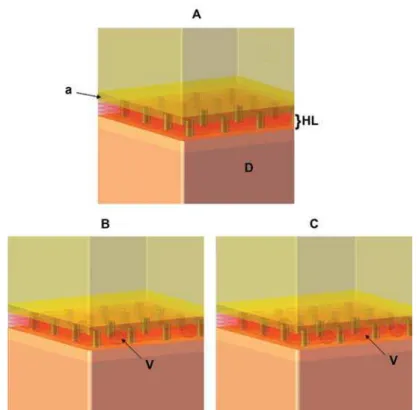
Considering the dimensions reported in Table 18,15,17,18,26, three models were built by varying
their void content in the HL by 0%, 25% or 50% (Mr, Mp, Mpp, respectively) (Table 2, Figure 1). Mr represents a perfect HL, without voids and &!#<&<&&& HL containing 25% and 50% of the volume with voids, respectively.
It was assumed that the adhesive that ! ! models bonded to adjacent and subjacent
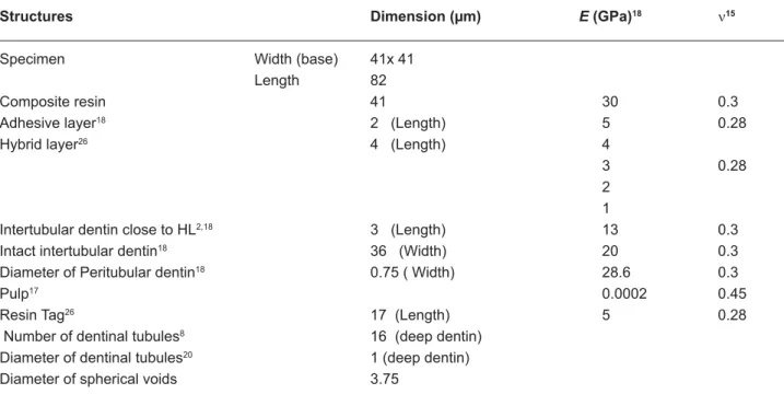
Structures Dimension (μm) E (GPa)18 Q15
Specimen Width (base) 41x 41 Length 82
Composite resin 41 30 0.3
Adhesive layer18 2 (Length) 5 0.28
Hybrid layer26 4 (Length) 4
3 0.28
2
1
Intertubular dentin close to HL2,18 3 (Length) 13 0.3 Intact intertubular dentin18 36 (Width) 20 0.3 Diameter of Peritubular dentin18 0.75 ( Width) 28.6 0.3 Pulp17 0.0002 0.45 Resin Tag26 17 (Length) 5 0.28 Number of dentinal tubules8 16 (deep dentin)
Diameter of dentinal tubules20 1 (deep dentin) Diameter of spherical voids 3.75
structures. Regarding the rigidity of the bonded structures, Misra, et al.18 (2004) established
that the elastic modulus of peritubular dentin and intertubular dentin show values of 28.6 GPa and 20 GPa respectively. However, Katz, et al.12
(2001) reported that the elastic modulus of the intertubular dentin, adjacent to the HL, has a value of 13 GPa, due to effect of previous etching required by the conventional adhesive system. Unfortunately, these data have not been reported for aggressive self-etching adhesive systems.
In their study, Misra, et al.18 (2004) varied
the HL thickness between 2 and 10 μm. These authors considered the HL to be constituted of several sublayers of equal thickness. Thus, the elastic modulus was graded from 4 GPa for &! the adhesive layer, to 1 GPa for the deepest
sublayer in contact with the mineralized dentin base. According to these authors, due to the ! deep demineralized dentin, hybrid layers may be built with different elastic modulus in order to reproduce in vivo conditions. In the present
+=>?! @> thick layers. A 4 GPa elastic modulus was used for the layer closest to the adhesive material; 3 GPa for the second layer, 2 GPa for the third layer and 1 GPa for the deepest layer in contact with the adjacent mineralized intertubular dentin18.
The convergence criterion was applied to reach optimal mesh quality. All models showed up to 38.497 tetragonal elements and 102.580 nodes. The nodes at the base of the specimen !J J+NJJOONO" set up the border line.
In order to determine the loading value, it was observed that in a dentin macro-specimen (5x2x2 mm) restored with composite resin, in an hourglass shape (sectional area of 1.1 mm2)
there was an equivalent tensile force of 18 MPa after 20 N of tensile loading at the top of the resin surface, exactly as previously described22.
Models % of voids in the HL
Mr 0 (ideal bond)
Mp 25
Mpp 50
Table 2- Percentage of voids in the hybrid layer
The cross-sectional area of the micro-specimen model in the present study was equal to 1681 μm2. To apply an 18 MPa tensile force
to the adhesive interface, a tensile load equal to 0.03N was perpendicularly applied to the composite resin surface (Figure 2).
As developed by Misra, et al.18 (2004), the
maximum principal stress (Vmax) was utilized for
identifying failures that could start out of small +X structures, such as dentin. All structures at the interface, i.e. peritubular dentin, intertubular dentin, adhesive layer and HL, were individually analyzed.
The numerical analysis was performed using ANSYS Workbench 10.0 (Swanson Analysis
Figure 2 - A - Hourglass-shaped specimen with 1.1mm2 of sectional area; B - Visualization of the model inside the ! load, perpendicular to the top of the composite resin, with 0.03 N)
System, Canonsburg, PA, USA).
RESULTS
The peak of Vmax was observed in the peritubular dentin (Figure 3) for all models (Figure 4).This behavior was similar in all other structures of the dentin/adhesive interface that showed an increase of Vmax in Mp when compared with Mr and in Mpp when compared with Mp (Figure 4 and 5). Figure 4 shows the Vmax for the
adhesive layer, peritubular dentin, resin tags and
intertubular dentin.
The Vmax in the peritubular dentin was observed in its upper border, exactly where the resin tags start (Figure 3). The Vmax in the tags occurred in their upper border (top of tags), in contact with the dentinal tubule walls (Figure 6).
The increase in the percentage of porosities within the HL, like in other structures, negatively + stress concentration (Figure 5). The increase in stress in the HL was of 6% in Mp when compared with Mr and of 6.5% in Mpp when compared
Figure 4- Maximum principal stress (MPa) in the peritubular dentin, adhesive layer, resin tags and intertubular dentin according to the percentage of voids (0, 25% and 50%) in the hybrid layer
with Mp.
The Vmax in the HL occurred near the porosities and between the second and third micrometers deep in the Mp and Mpp (Figure 6).
DISCUSSION
While a durable seal between several current bonding systems and enamel has been achieved, it is still a challenge to seal the resin-dentin interface, due to the heterogeneous characteristic of the dentin structure, surface morphology10,
and/or intrinsic shortcomings of the design of these modern adhesives21.
Conventional thought is that a perfect seal along the resin-dentin interface can be achieved within the demineralized collagen matrix when &! permanent and primary teeth11. This concept is
based on the assumption that the polymerized resins used for bonding are nonporous and impermeable to fluids23. However, adhesive
phase separation into hybrid and adhesive layers do not create an impervious collagen/polymer network but instead produce a porous web, and
at the same time, affecting the chemical and mechanical properties of the adhesive layer27.
Consequently, in order to evaluate the at dentin/adhesive interfaces in this study, the bond established between the adhesive system and dentin was either considered ideal or incorporated with voids in different contents (25% and 50%).
It was observed that the peritubular dentin showed the highest stresses in all models, in accordance with previous studies2,3. The next
structures to bear high stress were the resin tags, HL, adhesive layer and intertubular dentin.
The increased stress concentration observed in the top of tags can be related to the dentinal tubule diameter (1.0 μm) and closeness to the peritubular dentin which shows the highest elasticity modulus among the structures simulated at the dentin/adhesive interface2,18.
indicates that the bottom of the adhesive layer might not be the site to start up failure when the bond between dentin and HL is porous. The peak of Vmax in the Mr was in the HL close to the peritubular dentin, very similar to that previously found in a perfect bond scenario2,3.
Mollica, et al.19 found that the presence of
voids raised the Vmax by 3.7 times. In our study,
Vmax
only 13.5% (Figure 5). The study of Mollica, et al.19 (2004) showed a generic model with
! ## & and intertubular dentin, adhesive layer based on multiple layers, and resin tags) and mesh (only 5,000 elements and 10 nodes). This might Z of voids.
When voids were incorporated into the HL (Mp and Mpp), the peak of Vmax moved from
the contact with the peritubular dentin to the vicinity of the voids, 2 μm above the base of the HL (Figure 6). This behavior is very similar to that found in studies with micro-tensile loading analyzing the fracture pattern in specimens of dentin sticks24. In these, the failure was
commonly observed at the base of the HL, close to areas with poor hybridization.
Some authors13,27 showed that V
max in the
HL with poor hybridization was higher than the ultimate tensile strength of the self-etching adhesive system, even with a homogenous HL in contact with the dentin base. This issue is very important for the integrity of resin-dentin bonds over time. Lohbauer, et al.16 (2008) showed that
resin tags do not contribute to dentin adhesion in self-etching adhesive systems, in conditions of low bond strength values. In such circumstances, this means that the adhesion of the self-etching system is solely based on the quality of the HL and on its capacity to remain bonded to adjacent structures.
The present 3-D FE measurements showed that tensile stresses are restricted to two main sites in the dentin/adhesive interface: concentrated inside the HL, near the voids (Figure 6); and concentrated at the top of resin tags, in contact with dentinal tubule walls (Figure 6). Thus, our hypothesis can be accepted, as the
Vmax was higher in the presence of voids. Further studies on the porous dentin/adhesive interface should be carried out, considering different degrees of bonding between the HL and the adhesive layer, as well as the intertubular dentin.
CONCLUSIONS
Within the limitations of the present study, we can conclude that:
- The presence of voids in the hybrid layer raised the maximum principal stress in all structures of the dentin/adhesive interface;
- The increase of void content has some #'["\ able to raise the stress by 13.5% inside the HL;
- In the presence of voids, the maximum stress moved from the peritubular dentin to the HL in contact with the voids.
ACKNOWLEDGEMENTS
The authors would like to thank the FAPESP (The State of São Paulo Research Foundation, process # 2008/08479-5; 2008/00209-9) for the grants.
REFERENCES
1- Abdalla AI, Feilzer AJ. Four-year water degradation of a total-etch and two self-total-etching adhesives bonded to dentin. J Dent. 2008;36:611-7.
2- Anchieta RB, Rocha EP, Ko CC, Sundfeld RH, Martin M Jr, Archangelo CM. Localized mechanics of dentin self-etching adhesive system. J Appl Oral Sci. 2007;15:321-6.
3- Anchieta RB, Rocha EP, Sunfeld RH, Martin M Jr, Giannini M, Reis AF. Micromechanics of dentin/adhesive interface in function &#!#`{# press 2010.
4- Bouillaguet S, Gysi P, Wataha JC, Ciucchi B, Cattani M, Godin C, et al. Bond strength of composite to dentin using conventional, one-step, and self-etching adhesive systems. J Dent. 2001;29:55-61.
5- Carvalho RM, Chersoni S, Frankenberger R, Pashley DH, Prati C, Tay FR. A challenge to the conventional wisdom that simultaneous ! # Biomaterials. 2005;26:1035-42.
| } {~+ ? + + }# pressure application before light-curing on the bond strength of adhesive systems to dentin. Braz Dent J. 2008;19:62-7. 10- Giannini M, Chaves P, Oliveira MT. Effect of tooth age on bond strength to dentin. J Appl Oral Sci. 2003;11:342-7.
11- Kaaden C, Schmalz G, Powers JM. Morphological characterization of the resin-dentin interface in primary teeth. Clin Oral Invest. 2003;7:235-40.
12- Katz JL, Bumrerraj S, Dreyfuss J, Wang Y, Spencer P. Micromechanics of the dentin/adhesive interface. J Biomed Mater Res. 2001;58:366-71.
13- Knobloch LA, Gailey D, Azer S, Johnston WM, Clelland N, Kerby RE. Bond strengths of one- and two-step self-etch adhesive systems. J Prosthet Dent. 2007;97:216-22.
14- Koshiro K, Sidhu SK, Inoue S, Ikeda T, Sano H. New concept of resin-dentin interfacial adhesion: the nanointeraction zone. J Biomed Mater Res Part B Appl Biomater. 2006;77:401-8. 15- Le SY, Chiang HC, Huang HM, Shih YH, Chen HC, Dong DR, et al. Thermo-debonding mechanisms in dentin bonding systems !# #""@@@# 16- Lohbauer U, Nikolaenko SA, Petschelt A, Frankenberger R. Resin tags do not contribute to dentin adhesion in self-etching adhesives. J Adhes Dent. 2008;10:97-103.
17- McGuinness NJ, Wilson AN, Jones ML, Middleton J. A stress analysis of the periodontal ligament under various orthodontic loadings. Eur J Orthod. 1991;13:231-42.
18- Misra A, Spencer P, Marangos O, Wang Y, Katz JL. Micromechanical analysis of dentin/adhesive interface by the ! #` <~&& # 2004;70:56-65.
19- Mollica F, De Santis R, Ambrosio L, Nicolais L, Prisco D, Rengo S. Mechanical and leakage behavior of dentin-adhesive interface. J Mater Sci Mater Med. 2004;15:485-92.
20- Nanci A. Ten Cate’s oral histology. 6th ed. St Louis: Mosby;
2003.
21- Pashley DH, Carvalho RM. Dentine permeability and dentine adhesion. J Dent. 1997;25:355-72.
?? + <+'<`#' sectional shape and surface area on the microtensile bond test. Dent Mater. 1998;4:212-21.
23- Prati C, Chersoni S, Acquaviva GL, Breschi L, Suppa P, Tay FR, et al. Permeability of marginal hybrid layers in composite restorations. Clin Oral Investig. 2005;9:1-7.
24- Reis AF, Arrais CA, Novaes PD, Carvalho RM, De Goes MF, Giannini M. Ultramorphological analysis of resin-dentin interfaces produced with water-based single-step and two-step adhesives: nanoleakage expression. J Biomed Mater Res B Appl Biomater. 2004;71:90-8.
25- Spencer P, Wang Y. Adhesive phase separation at the dentin interface under wet bonding conditions. J Biomed Mater Res. 2002;62:447-56.
26- Sundfeld RH, Valentino TA, Alexandre RS, Briso ALF, Sundefeld MLMM. Hybrid layer thickness and resin tag length of a self-etching adhesive bonded to sound dentin. J Dent. 2005;33:675-81. 27- Susin AH, Vasconcellos WA, Saad JR, Oliveira OB Jr. Tensile bond strength of self-etching versus total-etching adhesive systems under different dentinal substrate conditions. Braz Oral Res. 2007;21:81-6.
28- Takahashi A, Sato Y, Uno S, Pereira PNR, Sano H. Effects of mechanical properties of adhesive resins on bond strength to dentin. Dent Mater. 2002;18:263-8.