Drug delivery across blood-brain barrier by means of
intravenous administration of lipid nanoparticles
Thesis submitted to the Faculty of Pharmacy of the University of Porto
PhD in Pharmaceutical Sciences
Pharmaceutical Technology
Susana Amélia Marques Martins
This work was developed under the supervision of Professor Domingos Ferreira
(Faculty of Pharmacy, University of Porto), Professor Eliana Souto (Faculty of
Health Sciences, University of Fernando Pessoa) and Professor Martin Brandl
(Faculty of Health Sciences, University of Tromsoe)
Porto
February 2012
THE REPRODUCTION OF PART OF THIS THESIS IS ONLY AUTHORISED FOR RESEARCH PURPOSES, PROVIDED BY PROPER COMMITMENT AND WRITTEN DECLARATION OF THE INTERESTED PART.
iii My first words of acknowledgement I express them to my supervisor Professor Dr. Domingos Ferreira for his constant advice, guidance, insight and inestimable support. I aim to express my gratitude for the opportunity he gave me to become a PhD student and work with new drug delivery systems.
I would like to thank my co-supervisor Professor Dr. Eliana Souto for offering me excellent scientific guidance and support through helpful discussions, as well as critical revision of my work.
I would like to thank my co-supervisor Professor Dr. Martin Brandl for his scientific guidance, helpful scientific discussion of many subjects presented in this manuscript and revision of my work during all the periods of my PhD. I aim to express my gratitude for the opportunity he gave me to work in his research group.
This thesis would not have been the same without the contribution of my colleagues at the Laboratory of Pharmaceutical Technology. A special thanks to Professor Dr. José Sousa Lobo for his contribution to turn this work possible and for encouraging scientific discussions. I would like to thank Professor Dr. Paulo Costa, Professor Dr. Paulo Silva, Professor Dr. Helena Amaral, Professor Dr. Delfim Santos, Professor Dr. Isabel Almeida, Professor Dr. Paulo Lobão and Dr. Rosa Pena for all the knowledge shared, support and help and for making me feel at home. Special thanks also to Professor Dr. Bruno Sarmento, for his contribution provided both experimentally in the laboratory and in the writing process. I would also like to thank to my PhD colleagues, Ana Catarina Silva, José das Neves, Fernanda Andrade and Sara Baptista, and to the researchers Filipa Antunes and Ana Patrícia Neto for the good mood in the laboratory, exchange ideas and all their help. I would also like to thank the Master student Tiago Carneiro and the Erasmus student Thierry Wendling for their help in some of the experiments.
I would also like to thank the many people from University of Tromsoe who contributed with their time, expertness, instrumentation, and advice to my project. Special thanks to Professor Dr. Ingunn Tho for the scientific ideas and special support on the multivariate analysis. A special thanks to Mereta Skar for her valuable technical assistance, information and help. I would like to thank to the PhD students Stephan Hupfeld and Johanna Kanzer and to the Master student Sara Fischer for their friendship and for being available for scientific and experimental discussions.
The in vitro studies in brain capillary endothelial cells and part of the in vivo studies were performed at the Institute of Pharmacy and Molecular Biotechnology, University of Heidelberg under the supervision of Professor Dr. Gert Fricker to whom I would like to express my gratitude for the opportunity to work in his research group, expert in the
blood-iv
Parmentier, Sonja Pohl and the Master student Dominik Gartzke for the precious help and shared experience, for receiving me so well, and for providing me with a pleasant stay in Heidelberg.
The proteins formulation studies were performed in the Faculty of Pharmaceutical Sciences, University of Copenhagen under the supervision of Professor Dr. Marco van de Weert and Professor Dr. Lene Jørgensen to whom I would like to express my gratitude. Thanks also to Joana Macedo for designing the layout of the cover of this thesis.
My warmest thoughts go to my family and friends who have provided me endless support over the years as a PhD student.
To my mother, Amélia, you have always been an inspiration to me, and thank you for always being there for me. To my dearest sister, Sara thank you for your endless love, kindness, continuous support and encouragement during all this time
I would like to thank my parents-in-law, Dalva and Mario Azevedo, for their support and encouragement.
I want to thank to my best friends, Ana Isabel Carvalho, Ana Maria Coelho, Ana Catarina Lages and Abília Moreno for supporting me and for being part of my life.
My deepest thanks are extended to my friends Joana Pereira, Mafalda Paiva and Marta Perro for all the good moments we shared in and outside the faculty and for the support and exchange of knowledge.
I am deeply thankful to Carlos Azevedo for supporting me in every way possible. When I was geographically distant, you have always been available, encouraging and have given me the best of support. Without your support the distance and all the difficulties associated with this period would be much harder to overcome.
I am grateful to those who have helped me with advices during the period as PhD student. I believe, honestly, that they have positively contributed to my personal growth.
Finally, I want to thank Fundação para a Ciência e Tecnologia for financial support (SFRH/BD/37364/2007) which was essential to the carry out of this research work and thesis.
v undesired chemical compounds. Consequently, for many diseases the delivery of drugs across the blood-brain barrier is poor, thus therapeutic drug levels are not reached in the brain. Looking at high mortality of malignant gliomas and the lack of efficient treatment, this field is a primary area for new intravenous formulations with blood-brain barrier crossing ability. The focus of this thesis was to use nanotechnology in cancer therapy, based on the use of solid lipid nanoparticles (SLN) to assess the suitability of these carriers for anticancer delivery to the brain via intravenous administration.
Bearing in mind the design of SLN for drug brain delivery, understanding the effect of lipid and surfactant composition on SLN physicochemical properties plays a key role. Thus, the first part of this work consisted in the selection of lipids (cetyl palmitate, Dynasan 114, Dyansan 116, Precirol ATO5 and Witepsol E85) and surfactants (polysorbate 20, 40, 60 and 80 and poloxamer 188 and 407) suitable for intravenous administration and a formulation screening study based on 22 factorial designs with centre point. Responses measured within the design space were the mean size and polydispersity index, content of microparticles, macroscopic appearance, pH and zeta potential on the day of production, and one and two years after production. Physical state and polymorphism were also assessed. Multivariate evaluation and modelling were performed to assess both qualitative and quantitative influence of the investigated factors (lipid and surfactant types and concentrations) in the SLN. According to the literature, the most promising SLN for brain drug delivery by means of intravenous injection have a mean size below 200 nm, homogenous size distribution (polydispersity index <0.25), slightly negative zeta potential, absence of microparticles larger than 5 µm and high storage stability. Among the most suitable excipients for producing SLN with appropriated characteristics, the polysorbates and the cetyl palmitate were selected. The models calculated allowed the prediction of the mean size of SLN that could be achieved with a certain lipid/surfactant combination and concentration. These results can be useful for future design of stable SLN formulations. In the second part of this work, with the purpose of anticancer drugs brain delivery, novel formulations of SLN loaded with camptothecin, a potent anticancer candidate, were designed. Incorporation of camptothecin within the hydrophobic and acidic SLN matrix was chosen to stabilise the lactone ring, essential for camptothecin antitumour activity, and to target camptothecin to the brain avoiding premature loss of drug. A multivariate approach was also used to assess the influence of the qualitative and quantitative composition on the physicochemical properties of camptothecin-loaded SLN compared to unloaded SLN. Particles with a mean particle size below 200 nm, a homogenous size distribution, high encapsulation efficiency (> 90%) and high stability were achieved in the
vi
capillary endothelial cells (BCEC) and gliomas was shown in comparison to macrophages. Cytotoxicity studies revealed that camptothecin-loaded SLN induced glioma cell death with the lowest maximal inhibitory concentration (IC50). To assess the SLN coated with
polysorbate 60 and 80 mechanism of cellular internalisation, flow cell cytometry studies were performed in four human glioma cell lines (A172, U251, U373 and U87) and one human macrophage cell line (THP1). The mechanism of internalisation was found to be mainly through a clathrin-dependent endocytic pathway. Flow cell cytometry studies revealed also that SLN have the ability to be internalised by gliomas in a higher amount than by macrophages. In agreement, the cytotoxicity of SLN was higher for gliomas than for macrophages.
Small and wide angle X-ray scattering (SAXS/WAXS) analyses were performed to evaluate the interactions between the SLN and the DMPC lipid bilayer model. These studies suggested favourable interactions of SLN and the lipid bilayers of cells.
A high-performance liquid chromatography (HPLC) assay for the quantification of camptothecin in several rat organs (brain, heart, kidneys, liver, lung, spleen) and serum after the intravenous administration of camptothecin-loaded SLN was developed and validated. The method was shown to be specific, accurate and precise at the intra-day and inter-day levels in all matrices. Stability studies showed that camptothecin was stable in all matrices after 24 h of incubation at room temperature (RT) or in the autosampler, or after three freeze/thaw cycles. Recoveries were higher than 86.4%. The detection limit (DL) was ≤0.2 ng/mL and the quantification limit (QL) was ≤0.5 ng/mL.
Finally, in vivo fluorescence and biodistribution studies of injected rhodamine 123-loaded SLN and camptothecin-loaded SLN were performed in rats and revealed higher brain accumulation when the molecules were incorporated into SLN. Indeed, the incorporation of camptothecin into SLN coated with polysorbate 80 increased more than 8 times the concentration of camptothecin in brain in comparison with camptothecin in suspension. In contrast, camptothecin accumulation decreased in liver and lungs in comparison with camptothecin in suspension. In this work it was demonstrated not only the brain targeting ability of SLN coated with polysorbate 60 and 80, but also the superiority of the antitumour activity of camptothecin-loaded SLN compared with camptothecin in solution/suspension or in physical mixture with SLN. These studies indicate that the camptothecin-loaded SLN are a promising drug brain delivery system worth to be exploited as a novel formulation for brain tumour therapy.
Keywords:
Solid lipid nanoparticles; Brain drug delivery; Camptothecin; Malignant glioma; Biodistribution.vii moléculas indesejáveis presentes no sangue. A maioria dos fármacos não consegue atravessar essa barreira em concentrações terapêuticas, não sendo portanto eficazes no tratamento da generalidade das doenças cerebrais. Observando a elevada mortalidade associada aos gliomas malignos e a falta de tratamento eficiente, o desenvolvimento de novas formas farmacêuticas, com capacidade de atravessar a barreira hematoencefálica e simultaneamente aumentar a esperança média de vida desses doentes, é uma área de elevado interesse. O presente trabalho teve como objetivo o emprego de nanopartículas de matriz lipídica sólida (SLN) na vetorização para o cérebro de anticancerígenos, após a sua administração intravenosa, para o tratamento de tumores cerebrais.
A primeira parte deste trabalho consistiu no estudo do efeito da composição qualitativa e quantitativa de diferentes lípidos e tensioativos nas propriedades físico-químicas, determinantes para vetorização cerebral, das nanopartículas de lípidos sólidos (SLN). Após a seleção de lípidos (Dynasan 114, Dynasan 116, palmitato de cetilo, Precirol ATO5 e Witepsol E85) e tensioativos (polissorbato 20, 40, 60 e 80 e poloxâmero 188 e 407), compatíveis com a via intravenosa, esses excipientes foram combinados para produzir SLN tendo por base o desenho experimental fatorial do tipo 22 com ponto central. As respostas medidas foram o diâmetro médio e índice de polidispersão, número e tamanho de micropartículas, aspeto macroscópico, pH e potencial zeta no dia da produção, e ao fim de um e dois anos após a produção. O estado físico e polimorfismo das SLN foram também avaliados por calorimetria diferencial de varrimento. Os estudos estatísticos multivariados iniciaram-se com uma Análise dos Componentes Principais seguida por uma Regressão por Mínimos Quadrados Parciais. Estes estudos permitiram avaliar a influência qualitativa e quantitativa dos fatores investigados (tipos e concentrações de lípidos e tensioativos) nas propriedades físico-químicas das SLN. De acordo com a literatura as SLN mais promissoras para a vetorização de fármacos para o cérebro devem possuir um diâmetro médio inferior a 200 nm, reduzido índice de polidispersão (<0.25), um potencial zeta ligeiramente negativo, ausência de micropartículas com dimensões superiores a 5 µm e elevada estabilidade. Entre os excipientes estudados, os mais adequados para a produção de SLN com as características pretendidas foram os polissorbatos e a cera palmitato de cetilo. Os modelos de previsão calculados podem ser utilizados no desenvolvimento futuro de SLN.
Com o objetivo de veicular anticancerígenos para o cérebro, foram desenvolvidas novas formulações de SLN com o fármaco camptotecina encapsulado. Novamente, neste estudo foi efetuada uma abordagem estatística multivariada para comparar a influência da composição qualitativa e quantitativa sobre as propriedades físico-químicas das
viii
eficiência de encapsulação (> 90%) e estáveis. Estudos de libertação in vitro, em plasma humano, revelaram um perfil de libertação prolongado da camptotecina incorporada nas SLN. Estudos in vitro em culturas celulares indicaram que as SLN apresentaram maior afinidade para as células endoteliais de capilares cerebrais e para gliomas do que para macrófagos. Estudos de citotoxicidade demonstraram que a toxicidade da camptotecina está aumentada quando este fármaco se encontra incorporado nas SLN. Para determinar o mecanismo de internalização celular das SLN, marcadas com rodamina 123 e revestidas por polissorbato 60 e 80, foram efetuados estudos in vitro, em quatro linhas celulares de glioma humano (A172, U251, U373 e U87) e uma linha celular de macrófagos humanos (THP1). Utilizando a técnica de citometria de fluxo determinou-se que as SLN foram internalizadas principalmente por endocitose mediada por clatrina e que estas foram mais internalizadas pelos gliomas do que pelos macrófagos.
Estudos de dispersão de raios-X de pequenos ângulos (SAXS) e grandes ângulos (WAXS) foram efetuados para avaliar as interações entre as SLN e o modelo de bicamada lipídica, constituído por DMPC. Estes estudos indicam que possa ocorrer, in vivo, uma interação favorável entre as SLN e as membranas celulares.
Neste trabalho foi desenvolvido e validado um método de cromatografia líquida de alta eficiência (HPLC) para quantificar, por fluorescência, a concentração de camptotecina em vários órgãos de rato (cérebro, coração, rins, fígado, pulmão, baço) e soro após a administração intravenosa de camptotecina incorporada nas SLN. O método mostrou-se específico, exato e preciso em todas as matrizes. Estudos de estabilidade mostraram que o fármaco é estável em todas as matrizes após 24 h à temperatura ambiente e após três ciclos de congelamento/descongelamento. A recuperação foi superior a 86,4%. O limite de deteção foi ≤ 0.2 ng/mL e o limite de quantificação foi ≤ 0.5 ng/mL.
Estudos in vivo de fluorescência e de biodistribuição em ratos confirmaram o aumento de rodamina 123 e camptotecina no cérebro quando veiculada nas SLN. Esses estudos confirmaram a aptidão das SLN em melhorarem a acumulação, retenção e distribuição do anticancerígeno camptotecina no cérebro dos animais. De facto, a concentração de camptotecina no cérebro foi cerca de 8 vezes superior quando este fármaco estava incorporado nas SLN revestidas com polissorbato 80. Em contraste, a acumulação de camptotecina diminuiu no fígado e pulmões. Os estudos efetuados nesta tese indicam as SLN como vetores coloidais promissores na veiculação de anticancerígenos para o cérebro e que devem ser explorados na terapia de tumores cerebrais.
Palavras-chave: Nanopartículas de lípidos sólidos; Vetorização de fármacos para o cérebro; Camptotecina; Gliomas malignos, Biodistribuição.
ix
Acknowledgments ………... iii
Abstract ……….. v
Resumo ………. vii
List of Figures ... xvi
List of Tables ... xxvi
List of Abbreviations and Symbols ... xxx
Chapter I - Aims and Organisation of the Thesis ... 1
1.1. Aims of the Thesis ... 3
1.2. Organisation of the Thesis ... 4
Chapter II - Introduction ... 5
2.1. Blood-brain barrier as a key role for drug brain delivery ………. 7
2.1.1. Blood-brain barrier structure ……… 7
2.1.2. Transport across the blood-brain barrier ………... 8
2.2. State of the art in brain drug delivery ………... 10
2.3. Lipid nanoparticles for parenteral drug delivery ……… 15
2.3.1. Solid lipid nanoparticles ………. 16
2.3.2. Nanostructured lipid nanoparticles ……….. 20
2.3.3. Lipid-drug conjugates ………. 21
2.3.4. Lipid nanoparticles for targeting the central nervous system for cancer treatment ………. 21
2.3.5. In vivo fate of lipid nanoparticles: protein adsorption pattern ……….. 24
2.3.6. The role of surfactants in targeting the central nervous system ………. 26
2.3.7. Lipid nanoparticles toxicological concerns ………. 29
2.4. Malignant gliomas ………... 31
2.5. Camptothecin ………... 36
2.5.1. DNA topoisomerase I poison ……… 37
2.5.2. Camptothecin physicochemical characteristics and their relevance in vivo antitumour activity ………... 38
2.5.3. Camptothecin analogues ………... 42
2.5.4. Camptothecin formulations ……… 42
2.5.5. Considerations on therapeutically relevant camptothecin concentrations …. 45 References ………... 45
x
3. Methods for lipid nanoparticles production and characterisation ………... 65
3.1. Solid lipid nanoparticles production ……….. 65
3.1.1. Hot high pressure homogenisation ……….. 66
3.1.2. High shear homogenisation followed by ultrasonication ……….. 68
3.2. Solid lipid nanoparticles characterisation methods ……… 70
3.2.1. Measurement of particle size and zeta potential ………... 71
3.2.1.1. Photon correlation spectroscopy ………. 71
3.2.1.2. Optical single particle sizing ………. 72
3.2.1.3. Zeta potential ……….. 73
3.2.2. Lipid crystallinity ……….. 75
3.2.2.1. Differential scanning calorimetry ……….. 75
3.2.2.2. X-ray scattering ……….. 77
3.2.3. Association efficiency of drugs in solid lipid nanoparticles ..……….... 78
3.2.3.1. High performance liquid chromatography ……….. 79
3.2.4. Solid lipid nanoparticles morphology ………... 80
3.2.4.1. Scanning electron microscopy ………. 80
References ………... 81
Chapter IV – Physicochemical properties of lipid nanoparticles: Effect of lipid and surfactant composition ……….... 85
Abstract ………. 87
4.1. Introduction ……….. 88
4.2. Materials and methods ………... 91
4.2.1. Materials ………91
4.2.2. Formulations ……….91
4.2.3. Methods ………... 91
4.2.3.1. Production of lipid nanoparticles ……….. 91
4.2.3.2. Assessment of particle size and size distribution ……….. 92
4.2.3.3. Zeta potential ……….. 92
4.2.3.4. Differential scanning calorimetry analysis ……….. 93
4.3. Results ……….. 93
4.3.1. Particle size and size distribution ………. 93
4.3.2. Zeta potential ………... 97
4.3.3. Differential scanning calorimetry ……….. 98
xi
4.4.3. Differential scanning calorimetry ……… 102
4.5. Conclusions ……… 102
References ………. 103
Chapter V - Multivariate design for the evaluation of lipid and surfactant composition effect for optimisation of lipid nanoparticles ……….. 107
Abstract ……….. 109
5.1. Introduction ……… 110
5.2. Materials and methods ………. 112
5.2.1. Materials ………. 112
5.2.2. Experimental design ………. 112
5.2.3. Annotation of formulation ……… 113
5.2.4. Methods ………. 113
5.2.4.1. Production of lipid nanoparticles ……… 113
5.2.4.2. Assessment of particle size and size distribution ……… 114
5.2.4.3. pH ………... 114
5.2.4.4. Zeta potential………. 114
5.2.4.5. Assessment of storage stability ………. 114
5.2.4.6. Multivariate analysis ……… 115
5.3. Results ……… 115
5.3.1. Lipid nanoparticles characteristics at day of production ………. 120
5.3.1.1. Influence of HPH parameters ………. 120
5.3.1.2. Influence of formulation parameters on particle size and size homogeneity (PI) ……… 120
5.3.1.3. Influence of formulation parameters on pH ……….. 128
5.3.2. Lipid nanoparticles storage stability ……….……. 129
5.3.2.1. Coded storage stability parameter ……… 129
5.3.2.2. Influence of formulation parameters on particle size and size homogeneity upon storage ……… 130
5.3.2.3. Influence of formulation parameters on pH changes ………. 132
5.3.2.4. Correlation between the responses ……….. 133
5.4. Discussion ……….. 134
5.5. Conclusion ………. 138
xii
Abstract ……….. 143
6.1. Introduction ……… 144
6.2. Materials and Methods ………. 146
6.2.1. Materials ………. 146
6.2.2. Experimental design ………. 146
6.2.3. Annotation of formulation ……….…146
6.2.4. Methods ………. 147
6.2.4.1. Production of solid lipid nanoparticles ……….. 147
6.2.4.2. Assessment of particle size and size distribution ……… 147
6.2.4.3. pH ………... 147
6.2.4.4. Assessment of storage stability ……….… 147
6.2.4.5. Zeta potential ……… 148
6.2.4.6. Differential scanning calorimetry analysis ……….…... 148
6.2.4.7. Association efficiency of camptothecin with solid lipid nanoparticles ……… 148
6.2.4.8. Camptothecin in vitro release studies ………...……….149
6.2.4.8.1. In PBS pH 7.4 ……….. 149
6.2.4.8.2. Human Plasma ………..…149
6.2.4.9. Isolation of porcine brain capillary endothelial cells and cell culture ... 149
6.2.4.10. Cell culture ………...……... 150
6.2.4.10.1. Endothelial porcine brain capillary endothelial cells and cell culture ………...… 150
6.2.4.10.2. Macrophages cell culture ………...…150
6.2.4.11. Cytotoxicity assay ………...…150
6.2.4.12. Internalisation of solid lipid nanoparticles by BCEC ………..…………151
6.2.4.13. Internalisation of camptothecin-loaded solid lipid nanoparticles by macrophages ………152
6.2.4.14. Animal studies ……….……152
6.2.4.15. Multivariate analysis ………...…153
6.2.4.16. Statistical analysis ………..…153
6.3. Results and Discussion ……… 154
6.3.1. Physicochemical characteristics of camptothecin-loaded solid lipid nanoparticles in comparison to plain solid lipid nanoparticles ………...…154
6.3.2. Camptothecin-loaded solid lipid nanoparticles compared to unloaded solid lipid nanoparticles ……….157
xiii
6.3.5. In vitro release ………165
6.3.6. In vitro cell culture studies of camptothecin-loaded solid lipid nanoparticles ………..………... 168
6.3.7. In vivo camptothecin biodistribution by means of rhodamine 123-loaded solid lipid nanoparticles ………...………. 172
6.4. Conclusion ………. 174
References ……… 174
Chapter VII - Solid lipid nanoparticles as intracellular drug transporters: An investigation of the uptake mechanism and pathway ………... 179
Abstract ……….. 181
7.1. Introduction ……… 182
7.2. Materials and Methods ………. 184
7.2.1. Materials ………. 184
7.2.2. Annotation for formulation ………... 185
7.2.3. Methods ………. 185
7.2.3.1. Production of solid lipid nanoparticles ……….. 185
7.2.3.2. Assessment of particle size and size distribution ……… 185
7.2.3.3. Zeta potential ……… 186
7.2.3.4. Differential scanning calorimetry analysis ……… 186
7.2.3.5. Cell culture ……… 186
7.2.3.6. Fluorescence microscopy ………... 187
7.2.3.7. MTT assay ………. 188
7.2.3.8. Flow cell cytometry ……….. 188
7.2.3.9. Statistical analysis ……… 189
7.3. Results ……… 190
7.3.1. Physicochemical characterisation of solid lipid nanoparticles ………... 190
7.3.2. In vitro studies in cells ……….. 193
7.3.2.1. Relative uptake of solid lipid nanoparticles in human cell lines ……… 193
7.3.2.2. Effect of solid lipid nanoparticles on cell viability and cytotoxicity …... 195
7.3.2.3. Uptake mechanism of solid lipid nanoparticles ……….. 196
7.3. Discussion ………. 202
7.4. Conclusion ………. 206
xiv
for the determination of camptothecin in animal organs following administration in
solid lipid nanoparticles ……… 211
Abstract ……….. 213
8.1. Introduction ……… 214
8.2. Experimental ……….. 216
8.2.1. Chemical and reagents ……… 216
8.2.2. Solid lipid nanoparticles production ………... 216
8.2.3. Physicochemical characterisation of solid lipid nanoparticles ………... 216
8.2.4. Analytical procedure ………. 217
8.2.4.1. Instruments and analysis conditions ………. 217
8.2.4.2. Preparation of stock and standard solutions ……… 218
8.2.4.3. Preparation of biological matrices sample ………... 218
8.2.5. Method validation ………. 218
8.2.5.1. Selectivity ……….. 218
8.2.5.2. Linearity and range ……….. 219
8.2.5.3. Accuracy and precision ………... 219
8.2.5.4. Detection limit and quantification limit ……….. 219
8.2.5.5. Robustness ………... 219
8.2.5.5.1. Stability ……….. 219
8.2.5.5.2. Extraction recovery ……….. 220
8.2.6. Application of the method to pharmacokinetic studies ………... 220
8.2.7. Statistical analysis ……… 221
8.3. Results and discussion ……… 221
8.3.1. Physicochemical characterisation of solid lipid nanoparticles ………... 221
8.3.2. Chromatography ………... 221
8.3.3. Application of the method to pharmacokinetic studies ………... 228
8.4. Conclusion ………. 230
References ………. 230
Chapter IX – Brain targeting effect of camptothecin-loaded solid lipid nanoparticles in rat after intravenous administration ……….. 233
Abstract ………... 235
9.1. Introduction ……… 236
9.2. Materials and Methods ………. 238
xv 9.2.3.1. Production of solid lipid nanoparticles and camptothecin formulations
……… 239
9.2.3.2. Assessment of particle size and size distribution ………..……. 239
9.2.3.3. Zeta potential ……… 240
9.2.3.4. pH ………..………. 240
9.2.3.5. Camptothecin association efficiency ………. 240
9.2.3.6. Assessment of storage stability ………. 241
9.2.3.7. Scanning electron microscopy ………...…… 241
9.2.3.8. Small and wide angle X-ray scattering (SAXS/WAXS) ……….. 241
9.2.3.9. Cell culture ……… 242
9.2.3.10. Cytotoxicity assay ……….. 243
9.2.3.11. Biodistribution in animals ……….. 243
9.2.3.12. Data analysis ………..… 244
9.3. Results and Discussion ……… 245
9.3.1. Physicochemical characterisation of solid lipid nanoparticles ………... 245
9.3.2. Interactions of solid lipid nanoparticles with lipid membranes: SAXS and WAXS results ………... 248
9.3.3. Cytotoxicity of camptothecin-loaded solid lipid nanoparticles ………... 253
9.3.4. Camptothecin-loaded solid lipid nanoparticles biodistribution ……….. 257
9.4. Conclusion ………. 266
References ……… 267
Chapter X – Conclusions and Future Perspectives ………... 273
10. Conclusions and Future Perspectives ……….… 275
10.1. Conclusions ………. 275
10.2. Future perspectives ……… 277
Chapter XI – Appendix ……….….. 279
11.1. Appendix 1 - Improving oral absorption of salmon calcitonin by trimyristin lipid nanoparticles ………... 281
xvi
Chapter II – Introduction ……… 5
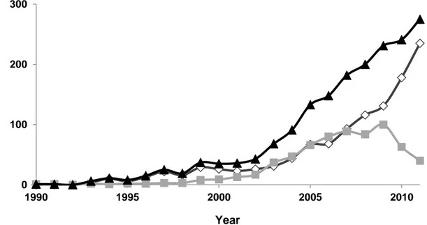
Figure 2.1. Schematic representation of the blood-brain barrier. Adapted from van Rooy et al. [13] ………... 7 Figure 2.2. Transport pathways across the blood-brain barrier. Adapted from van Rooy et al. [13] ……… 9 Figure 2.3. Number of articles (◊), patents (■) and total number of publications (▲)
related to lipid nanoparticles from 1990 to 2011. Data obtained in Scopus searching the words: solid lipid nanoparticles (SLN), nanostructured lipid carriers (NLC), lipid nanospheres and drug-lipid conjugates (LDC) nanoparticles ………... 16 Figure 2.4. Macroscopic aspect of solid lipid nanoparticles (SLN) aqueous dispersions (left), schematic representation of SLN aqueous dispersion (middle) and schematic representation of SLN stabilised with surfactant (right) ………... 18 Figure 2.5. Models of drug incorporation in solid lipid nanoparticles. Adapted from Müller et al. [102]……… 19 Figure 2.6. Cellular effects of topoisomerase I inhibitors in dividing and non-dividing
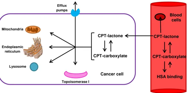
cells. Adapted from Frese et al. [247] ….………... 38 Figure 2.7. Camptothecin pentacyclic structure ………. 39 Figure 2.8. Camptothecin pentacyclic structure with a dynamic pH dependent equilibrium between the lactone (closed E ring) and carboxylate (open ring) form of the drug ……….. 40 Figure 2.9. Schematic representation of the mechanisms involved in camptothecin
distribution. Adapted from Beretta et al. [263] ………... 41 Figure 2.10. Schematic representation of camptothecin-loaded solid lipid nanoparticles coated with surfactant. Adapted from Kuo et al. [135] ………. 44
xvii Figure 3.1. Schematic diagram of the formation of solid lipid nanoparticles. A) Formation of an emulsion after homogenisation of the molten lipid, drug and surfactant mixture added with water heated at the same temperature in an ultra-turrax. B) The droplets are broken down into droplets of nanometre size by high pressure homogenisation or ultrasonication techniques. C) Formation of the solid lipid nanoparticles though the solidification of the nanodroplets below the lipid crystallisation temperature. Adapted from Wang et al [8] ……….. 65 Figure 3.2. Schematic illustration of high pressure homogenisation. Adapted from APV [12, 13] ………. 68 Figure 3.3. Schematic representation of solid lipid nanoparticles (SLN) production by
high pressure homogenisation and high speed stirring followed by ultrasonication …….……… 69 Figure 3.4. Schematic illustration of ultrasonication ……….. 70 Figure 3.5. Schematic representation of zeta potential. Adapted from Malvern [28] ... 74
Chapter IV – Physicochemical properties of lipid nanoparticles: Effect of lipid and
surfactant composition ……….... 85
Figure 4.1. Photon correlation spectroscopy (PCS)-data: mean diameters (mean ± SD) and PI (mean ± SD) on production day of five parallels. Given are the formulations of cetyl palmitate (CP), Dynasan 114 (D14), Dynasan 116 (D16), Precirol ATO5 (POL5) and Witepsol E85 (WE85) with 5% of lipid and 2% of the surfactants polysorbate 20 (P20), 40 (P40), 60 (P60), 80 (P80), poloxamer 188 (PL188) and 407 (PL407) (particle size given as volume-weighted mean) ……… 94 Figure 4.2. Photon correlation spectroscopy (PCS)-data: mean diameter (mean ± SD) and PI (mean ± SD) on production day of five parallels. Given are the formulations of Dynasan 114 (D14) based lipid nanoparticles (LN) with 5, 10 and 15% of lipid and 0.8, 1.2 and 2% of the surfactants polysorbate 20 (P20), 40 (P40), 60 (P60), 80 (P80), poloxamer 188 (PL188) and 407 (PL407) (particle size given as volume-weighted mean) ………. 95
xviii
formulations of Dynasan 114 (D14) based lipid nanoparticles (LN) with 5, 10 and 15% of lipid and 0.8, 1.2 and 2% of the surfactants polysorbate 20 (P20), 40 (P40), 60 (P60), 80 (P80), poloxamer 188 (PL188) and 407 (PL407) (particle size given as volume-weighted mean). Absence of results means that phase separation occurred ……….. 96 Figure 4.4. Typical diameter of relative distribution of lipid nanoparticles (LN) (particle size obtained as a measure of number distribution) ………. 96 Figure 4.5. Optical single particle sizing (OSPS) 95% (■) and 99% (■) data of lipid nanoparticles (LN) based on cetyl palmitate (CP), Dynasan 114 (D14) and Witepsol E85 (WE85) (5%lipid/0.8% surfactant) on day production (particle size obtained as a measure of number distribution) ………. 97 Figure 4.6. DSC thermograms of lipid nanoparticles (LN) CP10P801.2 (A), D145P802 (B),
D165P802 (C), POL55P602 (D) and WE855P600.8 (E) on day production
(black) and 1 year after production (grey) ……….. 99
Chapter V - Multivariate design for the evaluation of lipid and surfactant composition effect for optimisation of lipid nanoparticles ……….. 107 Figure 5.1. Regression coefficients from PLS of mean size (A), polydispersity index (PI) (B) and the number of microparticles larger than 1 μm (C) of lipid nanoparticles (LN) on the production day (90 samples). CP: cetyl palmitate; D14: Dynasan 114; WE85: Witepsol E85; P20, P40, P60 and P80: polysorbate 20, 40, 60 and 80; PL188 and PL407: poloxamer 188 and 407 ………. 121 Figure 5.2. PCA bi-plot of mean particle size, polydispersity index (PI) and number of
microparticles larger than 1 μm per mL ……… 123 Figure 5.3. Surface plot from PLS model of the surfactants polysorbate 20 (P20) (A, B and C), 60 (P60) (D, E and F), 80 (P80) (G, H and I) and poloxamer 407 (PL407) (J, K and L) with the lipids cetyl palmitate (CP) (A, D, G and J), Dynasan 114 (D14) (B, E, H and K) and Witepsol E85 (WE85) (C, F, I and L). CP + P20: R2cal: 0.98, R2pred: 0.54, explained Y-variance 98%, two PC; CP + P60: R2cal: 0.98, R2pred: 0.67, explained Y-variance 98%, two PC; CP + P80: R2cal: 0.96, R2pred: 0.42, explained Y-variance 96%, two
xix PC; D14 + P60: R2cal: 0.96, R2pred: 0.91, explained Y-variance 96%, two PC; D14 + P80: R2cal: 0.95, R2pred: 0.89, explained Y-variance 95%, two PC; D14 + PL407: R2cal: 0.99, R2pred: 0.96, explained Y-variance 99%, two PC; WE85 + P20: R2cal: 0.91, R2pred: 0.58, explained Y-variance 91%, one PC; WE85 + P60: R2cal: 0.99, R2pred:0.61, explained Y-variance 99%, two PC; WE85 + P80: R2cal: 0.97, R2pred: 0.93, explained Y-variance 97%, two PC; WE85 + PL407: R2cal: 0.91, R2pred: 0.61, explained Y-variance 91%, one PC ……….………… 127 Figure 5.A.1. Regression coefficients of the PLS model of pH in lipid nanoparticles (LN) formulations measured on production day ………... 128 Figure 5.4. Regression coefficients of PLS models of the coded storage stability parameter after 1 year. CP: cetyl palmitate; D14: Dynasan 114; WE85: Witepsol E85; P20, P40, P60 and P80: polysorbate 20, 40, 60 and 80; PL188 and PL407: poloxamer 188 and 407 ……… 129 Figure 5.A.2. Regression coefficients of PLS models of the coded storage stability
parameter after two years ……….. 130 Figure 5.5. Regression coefficients from PLS model of lipid nanoparticles (LN) A) mean
particle size after 1 year storage and B) polydispersity index (PI) after 1 year storage. CP: cetyl palmitate; D14: Dynasan 114; WE85: Witepsol E85; P20, P40, P60 and P80: polysorbate 20, 40, 60 and 80; PL188 and PL407: poloxamer 188 and 407 ………...………..…. 131 Figure 5.A.3. Regression coefficients of the PLS model of pH in lipid nanoparticles (LN) formulations A) pH change after one year storage and B) pH measured after one year storage ………. 132 Figure 5.6. A) PCA correlation loadings PC1 vs PC2 and B) PCA correlation loadings PC1 vs PC3. PI: polydispersity index ……… 134
xx
Figure 6.1. Physicochemical characteristics of unloaded solid lipid nanoparticles (SLN) versus camptothecin-loaded SLN. A) mean SLN size (nm), B) polydispersity index (PI), C) number of particles larger than 1 µm per mL, D) pH and E) zeta-potential. The target line is indicated ………... 157 Figure 6.2. Correlation plot from PCA of unloaded and camptothecin-loaded and unloaded solid lipid nanoparticles. Parameters correlated were mean particle size, polydispersity index (PI), number of particles larger than 1 µm per mL, pH and zeta potential. Explained variance on first 2 PCs 57% .. 159 Figure 6.3. Regression coefficients from PLS of mean size (A) Polydispersity index (B) and the association efficiency (C) of camptothecin-loaded solid lipid nanoparticles stabilised with polysorbate 80 on the production day (10 samples). CP: cetyl palmitate; D14: Dynasan 114; WE85: Witepsol E85; P80: polysorbate 80. Size: R2cal: 0.99, R2pred:0.93, explained Y-variance 86%, 2 PC; PI: R2cal: 0.31, R2pred:0.21, explained Y-variance 20%, 2 PC; Association efficiency: R2cal: 0.86, R2pred:0.74, explained Y-variance 85%, 2 PC ……….….. 160 Figure 6.4. PCA bi-plot of mean particle size, polydispersity index (PI), number of
microparticles larger than 1 µm per mL, storage stability and association efficiency (AE). Explained variance on first two PCs 65% ……… 163 Figure 6.5. DSC thermograms of A) solid lipid nanoparticles (SLN) based on 5% cetyl
palmitate (CP) stabilised with 2% polysorbate 20 (black), 40 (dark grey), 60 (grey) and 80 (light grey) with (__) or without (….) camptothecin (CPT) on day production; B) to D) lipid bulk 1st (black, __) and 2nd (black, ….) heating, CPT-loaded SLN on day production (light grey, __) and one year after production stored at 4 ºC (light grey, ---) or room temperature (light grey, ….) and unloaded SLN on day production (dark grey, __) and one year
after production stored at room temperature (dark grey, ….) …………... 165 Figure 6.6. Camptothecin in vitro release curve in human plasma (__) and PBS 7.4 (---) from cetyl palmitate- (CPT-CP5P802, black (◊)), Dynasan114-
(CPT-D145P802, dark grey (•)) and Witepsol E85-based solid lipid nanoparticles
xxi Figure 6.7. In vitro MTT viability of BCEC exposed for 24 h to CPT solution (- . -) CP5P802 (..◊..), CPT-CP5P802 (_◊_),CPT + CP5P802 (--◊--), D145P802 (..•..),
CPT-D145P802 (_•_), CPT + D145P802 (--•--), WE855P602 (..■..),
CPT-WE5P602 (_■_) and CPT + WE855P802 (--■--) at the CPT concentrations
between 0.25 and 10 µM. * P<0.05 (Dunnett-test: < control (CPT in solution)) ……… 168 Figure 6.8. Confocal images of rhodamine B-loaded solid lipid nanoparticles internalisation by BCEC. BCEC were exposed during two hours to A) rhodamine B-CP5P802 (red); B) rhodamine B-D145P802 (red); C) rhodamine
B-WE855P600.8 (red) ……… 170
Figure 6.9. Fluorescence images of camptothecin-loaded solid lipid nanoparticles internalisation by macrophages. Macrophages were exposed during two hours to A) CPT-CP5P802 (blue); B) CPT-D145P802 (blue); C)
CPT-WE855P600.8 (blue); (D) FluoSpheres® carboxylated nanospheres (green).
Macrophages membrane was stained with Alexa Fluor® 594 (red) …… 171 Figure 6.10. Cerebral distribution of rhodamine 123-loaded solid lipid nanoparticles (SLN) in rat brain after i.v. administration via the tail vein: 0.5 mL nanoparticle suspension (10 mg/mL) in 0.9% saline was administered. A) Free rhodamine 123 (control), B) polysorbate 60 stabilised SLN (rhodamine 123-CP5P602) C) polysorbate 80 stabilised SLN (rhodamine 123-CP5P802).
2 hours after i.v administration of SLN coated with polysorbates have reach the brain and revealed a spread distribution across the brain tissue ………..………..……. 173
Chapter VII - Solid lipid nanoparticles as intracellular drug transporters: An investigation of the uptake mechanism and pathway ………... 179 Figure 7.1. Physicochemical characteristics of cetyl palmitate-based solid lipid nanoparticles (SLN) stabilised with polysorbate 60 (CP60) and 80 (CP80). Mean diameter, polydispersity index (PI)(up) and zeta potential (down) of the SLN CP60 (left) and CP80 (right) stored at 4 ºC (■), 22 ºC (■) and 40 ºC
xxii
Figure 7.2. DSC thermograms of bulk cetyl palmitate (CP), solid lipid nanoparticles (SLN) stabilised with polysorbate 60 (CP60) and 80 (CP80). (A): DSC thermograms of the bulk CP ( - . -) and the SLN CP80 (black) and CP60 (grey) stored at 4 ºC (. .), 22 ºC (- -), and 40 ºC (_). (B): DSC thermograms of CP80 (black) and CP60 (grey) stored at 4 ºC (. .), 22 ºC (- -), and 40 ºC (_) ……… 192 Figure 7.3. Rhodamine 123 (R123)-loaded solid lipid nanoparticles (SLN) cell uptake.
Fluorescence microscope images of R123-loaded SLN uptake (2 h) by THP1, A172, U251, U373 and U87 cell lines. Nucleus are stained with DAPI (blue) and cell membrane with Alexa (red). R123-loaded SLN shown green fluorescence. Representative images are shown. (Magnification 400x) ……….. 194 Figure 7.4. Cell viability of the cells exposed at rhodamine 123 (R123)-loaded solid lipid
nanoparticles (SLN) stabilised with polysorbate 60 (R123-CP60) and 80 (R123-CP80). Viability of different cells (THP1, A172, U251, U373 and U87) exposed for 24 h at R123-CP60 and R123-CP80 at the concentrations of 50 and 500 µg/mL of solid content ……… 195 Figure 7.5. Cell viability of different cells after incubation with cetyl palmitate-based solid
lipid nanoparticles (SLN) stabilised with polysorbate 60 (CP60) and 80 (CP80). Cell viability of THP1, A172, U251, U373 and U87 incubated for 24 h with CP60 (♦), CP80 (■) and water (▲) ……… 196 Figure 7.6. Uptake of rhodamine 123 (R123)-loaded solid lipid nanoparticles (SLN)
stabilised with polysorbate 60 (R123-CP60) and 80 (R123-CP80). Flow cytometry histograms data for THP1 (A), A172 (B), U251 (C), U373 (D) and U87 (E) in medium (control (black)) and that were incubated with R123-CP60 (---) or R123-CP80 ( ) ………. 197 Figure 7.7. Uptake mean of rhodamine 123 (R123)-loaded solid lipid nanoparticles (SLN) stabilised with polysorbate 60 (R123-CP60) and 80 (R123-CP80). Flow cytometry data for THP1, A172, U251, U373 and U87 in medium (control (■)) and that were incubated with R123-CP60 (■) or R123-CP80 (■). Control (cells) was always significant different than cells incubated with R123-loaded SLN ……….… 198
xxiii Macropinocytosis, engulfing nanoparticles, forming macropinosomes (B1) which can be exocytosed or fused with lysosomes (L). C) Clathrin-mediated endocytosis, leading to primary endosomes (C1) and late endosomes (C2) with multivesicular bodies (C3). D) Clathrin- and caveolae-independent endocytotic pathways. E) Caveolae-mediated endocytosis, leading to caveosomes (E1) which fuse with the endoplasmic reticulum (E2) or translocate through the cell (E3). F) Particle diffusion or transport through the apical plasma membrane, leading to particles situated in the cytosol. The figure and descriptions are adapted from [25, 26] ……….. 199 Figure 7.9. Effect of temperature on rhodamine 123 (R123)-loaded solid lipid
nanoparticles (SLN) internalisation. Cells were treated with 50 µg/mL of R123-loaded SLN stabilised with polysorbate 60 (R123-CP60, ■) and 80 (R123-CP80, ■) at 4 °C. Subsequent analysis by flow cytometry showed a significant decrease in fluorescence for all cells treated at 4 °C compared with 37 °C (100% uptake) ………... 200 Figure 7.10. Endocytosis mechanism on rhodamine 123 (R123)-loaded solid lipid nanoparticles (SLN) internalisation. Effect of clathrin-(A) and caveolae-(B) mediated endocytosis inhibition on R123-loaded SLN internalisation. Cells were pretreated with sucrose 0.45 M (A) or filipin (5 µg/mL) (B) during 30 min and then treated with 50 µg/mL of R123-loaded SLN stabilised with polysorbate 60 (R123-CP60, ■) and 80 (R123-CP80, ■). Subsequent analysis by flow cytometry showed a significant decrease in fluorescence for all cells treated with sucrose 0.45 M (A) but not for cells treated with filipin (5 µg/mL) (B) compared with 37 °C (100% uptake) ……...……….. 201 Figure 7.11. Effect of macropinocytosis/phagocytosis inhibition on rhodamine 123
(R123)-loaded solid lipid nanoparticles (SLN) internalisation. Cells were pretreated with cytochalasin B (5µg/mL) during 30 min and then treated with 50 µg/mL of R123-loaded SLN stabilised with polysorbate 60 (R123-CP60, ■) and 80 (R123-CP80, ■). Subsequent analysis by flow cytometry showed a significant decrease in fluorescence only for A172 and U87 incubated with R123-CP80 (■) treated with cytochalasin B compared with 37 °C without inhibition (100% uptake) ………. 202
xxiv
solid lipid nanoparticles ……… 211
Figure 8.1. CPT pentacyclic structure with a dynamic pH dependent equilibrium between the lactone (closed ring, left) and carboxylate (open ring, right) form of the drug ……… 214 Figure 8.2. Chromatograms of blank brain (A), heart (B), kidneys (C), liver (D), lungs
(E), spleen (F), serum (G) and CPT in PB3 (10 ng/mL) (H) ……….. 222 Figure 8.3. Chromatogram of CPT in PB3 (10 (A), 50 (B) and 100 (C) ng/mL) ……. 223 Figure 8.4. Linearity studies for the proposed HPLC method. Response factors versus
CPT standard solutions concentration ……….. 224
Chapter IX - Camptothecin-loaded solid lipid nanoparticles biodistribution in rat after intravenous administration and targeting effect on brain ……….. 233 Figure 9.1. Representative SEM micrographs of unloaded (left) and
camptothecin-loaded (right) cetyl palmitate-based solid lipid nanoparticles ……..……. 247 Figure 9.2. X-ray diffraction patterns (SAXS and WAXS) at 37 ºC for: A) physical
mixture of excipient (CP+P60); B) solid lipid nanoparticles (SLN, CP60); C) physical mixture of excipient (CP+P80) and D) SLN (CP80) ………..….. 248 Figure 9.3. X-ray diffraction patterns (SAXS and WAXS) for: A) DMPC at 18 ºC (Lβ' phase); B) DMPC mixture with camptothecin 18 ºC (Lβ' phase); C) DMPC at 40 ºC (Lα phase) and D) DMPC mixture with camptothecin 40 ºC (Lα phase) ……… 250 Figure 9.4. X-ray diffraction patterns (SAXS and WAXS) at 18 ºC (Lβ' phase) for: A)
DMPC mixture with SLN (CP60); B) DMPC mixture with SLN (CPT-CP60); C) DMPC mixture with SLN (CP80); D) DMPC mixture with SLN (CPT-CP80) and at 40 ºC (Lα phase) for: E) DMPC mixture with SLN (CP60); F) DMPC mixture with SLN (CPT-CP60); G) DMPC mixture with SLN (CP80); H) DMPC mixture with SLN (CPT-CP80).……..……….………. 251
xxv CPTCP60 ( ), CPTCP80 ( ■ ), CPT+CP60 (- -), CPT+CP80 (- ■ -) and CPT susp. (..▲..). *means that formulation induces significantly higher cytotoxicity (P<0.05) than camptothecin in suspension in some concentrations. **means that formulation induces significantly higher cytotoxicity (P<0.05) than camptothecin in suspension in all concentrations range (n=3) ……… 255 Figure 9.A.1. Representative images of organs (brain, heart, kidneys, liver, lungs and spleen) extracted from tested animals ……….. 258 Figure 9.6. Camptothecin (CPT) concentration in serum, brain, heart, kidneys, liver, lungs and spleen after intravenous administration of CPTCP60 (_____), CPTCP80 (__■__), CPT+CP60 (- -), CPT+CP80 (- ■ -) and CPT susp.
(..▲..) (n=6) ……… 260
Figure 9.7. The F values of CPT+CP60 (□), CPTCP60 (■), CPT+CP80 (■) and CPTCP80 (■) after intravenous administration of 0.5 mg CPT/kg ……... 264
xxvi
Chapter II – Introduction ……… 5
Table 2.1. Anticancer drugs incorporated into solid lipid nanoparticles for brain tumour targeting ………... 23 Table 2.2. WHO grading scheme and features of common astrocytic tumours (98) ……… 33 Table 2.3. CNS drug physicochemical features for brain penetration according to (260, 261) and camptothecin physicochemical features ……… 39 Table 2.4. Examples of camptothecin and its analogues in the market or clinical development ……… 42 Table 2.5. Examples of camptothecin-loaded colloidal carrier in clinical development ……… 44
Chapter IV – Physicochemical properties of lipid nanoparticles: Effect of lipid and
surfactant composition ……… 85
Table 4.1. Main properties of the lipids and wax used in the preparation of the LN (manufacturer information) ……… 89 Table 4.2. Main properties of the surfactants used in the preparation of the LN (manufacturer information) ……… 90 Table 4.3. Zeta potential (ZP), size (mean ± SD) and polydispersity index (PI) (mean ± SD) of Dynasan 114 (D14) based lipid nanoparticles (LN) with 5% of lipid and 2% of surfactant ……….. 98 Table 4.4. Enthalpy, peak onset and maximum and RI of the lipid bulk and the LN on day production and 1 year after ………... 98
Chapter V - Multivariate design for the evaluation of lipid and surfactant composition effect for optimisation of lipid nanoparticles ……….. 107 Table 5.1. Experimental design: investigated factors and levels for 22 factorial designs with centre point ………113
xxvii storage stability) of lipid nanoparticles (LN) formulations measured production day and after one year and two years storage ……… 116 Table 5.2. PLS models of lipids separately; effect on size of lipid nanoparticles (LN) on the production day (bold numbers indicate significant regression coefficients) ………... 125 Table 5.3. PLS models of surfactants separately; effect on size of lipid nanoparticles (LN) on the production day (bold numbers indicate significant regression coefficients) ………... 126
Chapter VI - Brain delivery of camptothecin by means of solid lipid nanoparticles: Formulation design, in vitro and in vivo studies ……… 141
Table 6.1. Physicochemical characteristics (mean particle size, polydispersity index (PI), microparticles larger than 1 µm/mL, pH, zeta potential (ZP) and storage stability) of camptothecin-loaded solid lipid nanoparticles formulations measured on production day and after one year storage ………... 155 Table 6.2. 50% Inhibition concentration (IC50) of unloaded solid lipid nanoparticles
(SLN), camptothecin in solution, camptothecin in physical mixture with unloaded SLN and camptothecin-loaded SLN against porcine BCEC ………... 169
Chapter VII - Solid lipid nanoparticles as intracellular drug transporters: An investigation of the uptake mechanism and pathway ………... 179 Table 7.1. Uptake pathways and inhibition conditions ……… 189 Table 7.2. Physicochemical characteristics of solid lipid nanoparticles (SLN). Mean size, polydispersity index (PI) and zeta potential (ZP) of SLN on day production and dispersed in culture medium ……… 191 Table 7.3. DSC parameters of solid lipid nanoparticles (SLN). Onset and melting
temperatures, melting enthalpies and recrystallisation index (RI) of cetyl palmitate-based SLN stabilised with polysorbate 60 (CP60) and 80 (CP80) stored at 4 ºC, 22 ºC and 40 ºC ………. 192
xxviii
solid lipid nanoparticles ……… 211
Table 8.1. Regression analysis of calibration curves, detection limit (DL) and quantification limit (QL) for CPT in PB3, serum, brain, heart kidneys, liver, lungs, spleen, over the specified concentration rate ……….. 223 Table 8.2. Accuracy and precision for the analysis of CPT in PB3, serum, brain, heart kidneys, liver, lungs, spleen (n = 6) ………... 225 Table 8.3. Stability of CPT at different experimental conditions in PB3, serum, brain, kidneys, liver, heart, kidneys, liver, lungs and spleen (n = 6) ……… 226 Table 8.4. Extraction efficiency of CPT from rat organs at different concentration levels (n = 6) ………. 227 Table 8.5. Parameters comparison between the proposed method and other methods for the detection of camptothecin ……….. 228 Table 8.6. Concentration of CPT (ng/g organ) in rat 1 h after i.v. administration of CPT
suspension, CPT-loaded SLN and physical mixture of CPT and unloaded SLN (n = 6) ……… 229
Chapter IX - Camptothecin-loaded solid lipid nanoparticles biodistribution in rat after intravenous administration and targeting effect on brain ……….. 233 Table 9.1 Physicochemical characteristics of solid lipid nanoparticles (SLN). Mean size, polydispersity index (PI), zeta potential (ZP), association efficiency (AE) and pH of SLN on day of production and one year after production (storage at 4 ºC)……….... 245 Table 9.2. Long and short range distances determined from SAXS and WAXS patterns ………. 249 Table 9.3. Long range distances determined from SAXS patterns and correlation length (ξ) at the Lα and Lβ' for the different formulations mixture with DMPC ... 252 Table 9.4. 50% Inhibition concentration (IC50) of unloaded solid lipid nanoparticles
(SLN, CP60 and CP80), camptothecin in suspension (CPT susp.), camptothecin in physical mixture with unloaded SLN (CP60+CPT and CP80+CPT ) and camptothecin-loaded SLN (CP60CPT and CP80CPT)
xxix Table 9.5. Camptothecin (CPT) concentration in serum, brain, heart, kidneys, liver, lungs and spleen after intravenous administration of CPT formulations ………. 259 Table 9.6. Pharmacokinetic parameters of camptothecin (CPT) formulations after
xxx
2-D PAGE Two-dimensional polyacrylamide gel electrophoresis ABC transporter ATP-binding cassette transporter
AE Association efficiency
AFM Atomic force microscopy
Alexa Alexa Fluor® 594 conjugated of wheat germ agglutinin
ANOVA One-way analysis of variance
Apo Apolipoprotein
Apo ER-2 Apolipoprotein E receptor-2 ATCC American Type Culture Collection
AUC Area under the curve
AUMC Area under the first moment curve
BBB Blood-brain barrier
BCEC Brain capillary endothelium cells
B0W B-coefficient zero weighted
BSA Bovine serum albumin
CNS Central nervous system
CP Cetyl palmitate
CPT Camptothecin
Cryo-SEM Cryo scanning electron microscopy Cryo-TEM Cryo transmission electron microscopy CV Coefficient of variation
D14 Dynasan® 114 (trimirystin)
D16 Dynasan® 116 (tripalmitin)
Da Dalton
DAPI 4',6-diamidino-2-phenylindole dihydrochloride
DGV Direcção Geral de Veterinária
DL Detection limit
DLS Dynamic light scattering
DMEM Dulbecco’s Modified Eagle’s Medium
DMPC 1,2-Dimyristoyl-sn-glycero-3-phosphocholine
DMSO Dimethyl sulfoxide
DNA Deoxyribonucleic acid
DoE Design of experiments
DO-FUdR 3′,5′-dioctanoyl-5-fluoro-2′-deoxyuridine DSC Differential Scanning Calorimetry
xxxi EGFR Epithelial growth factor receptor
EPR effect Enhanced permeation and retention effect
FBS Fetal bovine serum
FD Fluorescence detector
FDA Food and Drug Administration
FFEM Freeze fracture electron microscopy
GeV Gigaelectron volt
GRAS Generally recognised as safe
h Hour
HBSS Hanks’ Balanced Salt solution HLB Hydrophilic-lipophilic balance
HPH High pressure homogenisation
HPLC High performance liquid chromatography
HSA Human serum albumin
HTMAB Hexadecyltrimethylammonium
IC50 50% Inhibition concentration
ICH International Conference on Harmonisation
i.e. id est
i.v. Intravenous
J/g Joule per gram
kHz Kilohertz
KRB Krebs–Ringer buffer
LD Laser diffraction
LDC Drug-lipid conjugates
LDL Low density lipoproteins
LN Lipid nanoparticles
LRP-1 Low density lipoprotein receptor-related protein 1
Min Minute(s)
MGMT Methylated-DNA-protein-cysteine methyltransferase
MPa Mega Pascal
MPS Mononuclear phagocyte system
MTT 3-[4,5-dimethylthiazol-2-yl]-2,5-diphenyltetrazolium bromide
MW Molecular weight
NLC Nanostructured lipid carriers
xxxii
P20 Polysorbate 20
P40 Polysorbate 40
P60 Polysorbate 60
P80 Polysorbate 80
PALS Phase analysis light scattering
PB Phosphate buffer
PB3 Phosphate buffer pH 3
PB10.5 Phosphate buffer pH 10.5
PBCA Poly(butyl cyanoacrylate)
PBS Phosphate buffered saline
PC Principal component
PCA Principal component analysis
PCL Poly ɛ-caprolactone
PCS Photon correlation spectroscopy
PEG Polyethylene glycol
PET Polyethyleneterephthalat
PI Polydispersity index
PHDCA Poly(hexadecylcyanoacrylate)
PL188 Poloxamer 188
PL407 Poloxamer 407
PLA Polylactic acid
PLGA Poly(lactic-co-glycolic acid)
PLS Partial least square regression analysis PMA Phorbol 12-myristate 13-acetate
POL5 Precirol ATO5
ppt Parts per trillion
PVA Poly(vinyl alcohol)
QbD Quality by Design
QC Quality control
QL Quantification limit
R123 Rhodamine 123
R Correlation coefficient
R2 Squared correlation coefficient
R2cal Squared correlation coefficient of calibration R2pred Squared correlation coefficient of prediction
xxxiii RMSEP Root mean square error of prediction
RP-HPLC Reversed-phase high performance liquid chromatography
rpm Rotation per minute
RT Room temperature
RTT Rat-tail tendon
SAXS Small-angle X-ray scattering
SD Standard deviation
SDS Sodium dodecylsulfate
SEM Scanning electron microscopy
SLN Solid lipid nanoparticles SR-BI Scavenger receptor B class I
STM Scanning tunnelling microscopy
T.C. Theoretical concentration
TEA Triethylamine
TEER Transendothelial electric resistance TEM Transmission electron microscopy
US Ultrasonication
UV Ultraviolet
VEGF Vascular endothelial growth factor VLDL Very low density lipoprotein
vs Versus
v/v Volume/volume
WGA Wheat germ agglutinin
w Weight
WAXS Wide-angle X-ray scattering
WE85 Witepsol E85
WHO World Health Organisation
w/o/w Water-in oil-in water
w/v Weight/volume
w/w Weight/weight
ZP Zeta potential
ºC Degrees
ºC/min Degrees per minute
3 1.1. Aims of the thesis
During the last decades, dosage form design in general has been gaining increasing importance and with the advent of nanotechnology new pharmaceutical dosage forms and drug delivery systems based on nanoparticles are under development to deliver different drugs to the intended site of action.
The present thesis is focussing on the potential use of solid lipid nanoparticles (SLN) as a strategy to overcome the blood-brain barrier upon intravenous injection, more specifically enable the delivery of anticancer drugs to the brain parenchyma intending the improvement of malignant gliomas treatment.
In the present thesis, several lipids and surfactants compatible with intravenous administration were selected and the influence of these excipients on the physicochemical properties of SLN was studied, since such properties are important for brain drug delivery. The drug camptothecin was selected as a model drug for the study of brain delivery abilities of SLN. It was aimed to overcome the poor solubility and stability of camptothecin in aqueous solution, which so far has hindered a therapeutic application of this potent anticancer drug, by incorporating camptothecin into SLN.
In vitro and in vivo studies in rats were performed to assess the suitability of the developed camptothecin-loaded for brain delivery.
Specific aims:
I. To study the qualitative and quantitative influence of lipids and surfactants on SLN physicochemical properties by both univariate and multivariate analysis and to identify formulation design parameters within the HPH technology to be applied during future formulation development studies.
II. To investigate the influence of the qualitative and quantitative composition on physicochemical properties of camptothecin-loaded SLN compared to unloaded SLN, and to study the camptothecin-incorporation capacity of SLN of different lipid composition and its in vitro release from SLN.
III. To study the cytotoxicity of camptothecin-loaded SLN in brain capillary endothelial cells (BCEC), in four human glioma cell lines (A172, U251, U373 and U87) and in one human macrophage cell line (THP1).
IV. To assess the cellular mechanism and pathway of SLN internalisation to evaluate if these systems have the ability to be internalised by four human glioma cell lines (A172, U251, U373 and U87) and by one human macrophage cell line (THP1) and to understand the mechanisms behind this internalisation.
4
V. To investigate the interactions of camptothecin and camptothecin-loaded SLN with lipid membranes by synchrotron small and wide angle X-ray scattering (SAXS/WAXS) analysis.
VI. To develop and validate a simple, sensitive and specific high-performance liquid chromatography (HPLC) assay for the quantification of camptothecin incorporated into SLN in several rat organs (brain, heart, kidneys, liver, lung, spleen) and serum.
VII. To perform in vivo fluorescent and biodistribution studies to assess the suitability of SLN as drug delivery system for brain targeting.
1.2. Organisation of the thesis
The thesis is organised in 11 chapters.
Chapter 1 includes the aims and organisation of the thesis and also includes a brief description of the subsequent chapters.
Chapter 2 comprises a general introduction to contextualise the state of art of key topics within the thesis, such as the blood-brain barrier as a key role for drug brain delivery, lipid nanoparticles for parenteral drug delivery, lipid nanoparticles for targeting to the central nervous system for cancer treatment, malignant gliomas and the anticancer camptothecin. Chapter 3 contains a theoretical review of the methodologies of SLN production and characterisation used in the present thesis.
Chapter 4 to 9 contain the main studies performed, incl. materials, methods, results and discussion which are presented as manuscripts or published articles. Supplementary figures or tables of published or submitted papers were embedded in the manuscript as supplementary to the original manuscript. The number of figures of each chapter starts by the Arabic number of the chapter followed by the original number in the manuscript.
Chapter 10 includes conclusions from the present studies and future research perspectives.
Chapter 11 comprises appendix 1 that include one article that was published during the PhD but are out of the scope of the thesis. It was included only as appendix and no further discussion will be carried out.
The thesis was written in British English. Despite that some of the chapters are in American English since the original articles were published on that style.
7
2.1. Blood-brain barrier as a key role for drug brain delivery 2.1.1. Blood-brain barrier structure
The blood–brain barrier is a metabolic and cellular structure that acts as a central nervous system (CNS) firewall by limiting the passage of toxins, bacteria, viruses and various chemical substances from the systemic circulation to the neural tissue, but at the same time allowing the passage of substances essential to CNS metabolic function [1, 2].
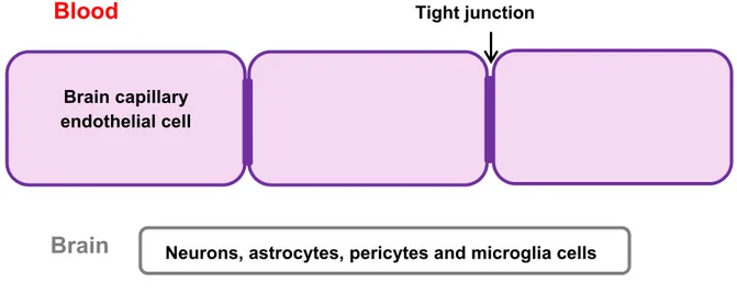
Paul Ehrlich in 1885 [3] and later his student Edwin Goldman [4, 5] were the first to notice about the restricted water-soluble dyes exchange, existing between the blood and the brain. In 1900 Lewandowsky was the first to use the term blood-brain barrier while studying the limited permeation of potassium ferrocyanate into the brain [6]. The anatomy of the blood-brain barrier was elucidated in the 1960s by electron microscopic studies with brain slices [7, 8]. Since blood-brain barrier discovery until today several studies have been performed to characterise the structure and properties of such barrier (e.g. [9-12]). The blood-brain barrier is formed by brain capillary endothelial cells (BCEC) lining and tightly sealing the cerebral capillaries, together with perivascular elements such as astrocytes, neurons, pericytes and microglia cells (Figure 2.1). This set forms the so-called neurovascular unit [1, 2, 13].
Blood
Tight junction
Figure 2.1. Schematic representation of the blood-brain barrier. Adapted from van Rooy et al. [13]. Despite not participate in the physiological activity of the blood-brain barrier, perivascular elements release various soluble factors which help in the differentiation and maintenance of blood-brain barrier properties [1, 14, 15].
The BCEC exhibit important morphological characteristics, closely related to their physiological activity, such as the presence of tight junctions, the absence of fenestrations, and a low pinocytic activity [16].
Neurons, astrocytes, pericytes and microglia cells Brain capillary
endothelial cell


![Figure 3.2. Schematic illustration of high pressure homogenisation. Adapted from APV [12, 13]](https://thumb-eu.123doks.com/thumbv2/123dok_br/15840289.1084233/102.892.94.746.142.522/figure-schematic-illustration-high-pressure-homogenisation-adapted-apv.webp)
