Neuropsychological abnormalities in children with the Panayiotopoulos
syndrome point to parietal lobe dysfunction
Ricardo Lopes
a,⁎
, Mário R. Simões
a, Alberto J.R. Leal
b,c,da
Faculty of Psychology and Educational Sciences, University of Coimbra, Coimbra, Portugal
bInstituto Universitário de Lisboa (ISCTE-IUL), Cis-IUL, Lisboa, Portugal c
Department of Neurophysiology, Centro Hospitalar Psiquiatrico de Lisboa, Lisbon, Portugal
d
Department of Pediatric Neurology, Centro Hospitalar Lisboa Central, Portugal
a b s t r a c t
a r t i c l e i n f o
Article history: Received 23 July 2013 Revised 8 November 2013 Accepted 12 November 2013 Available online 15 December 2013 Keywords: Epilepsy Panayiotopoulos syndrome EEG Neuropsychology Parietal lobePanayiotopoulos syndrome (PS) is a common epilepsy syndrome associated with rare clinical seizures and un-known localization of the epileptogenic area. Despitefindings of normal development in patients with PS, recent neuropsychological studies point to subtle and diverse cognitive impairments. No well-outlined hypothesis about the localization of the brain dysfunction responsible for these impairments has been proposed. We further explored the cognitive dysfunctions in PS and made inferences on the most likely anatomical localization of brain impairment. A group of 19 patients (aged 6–12) with PS was rated according to spike activity and lateralization. The patients were submitted to a neuropsychological evaluation to assess general intelligence, memory, lan-guage, visual–perceptual abilities, attention, and executive functions. Using 35-channel scalp EEG recordings, the N170 face-evoked event-related potential (ERP) was obtained to assess the functional integrity of the ventral pathway. All patients with PS showed normal IQ but subtle and consistent neurocognitive impairments. Namely, we found abnormalities in the copy task of the Rey–Osterrieth Complex Figure and in the Narrative Memory Test. There was no correlation between neuropsychological impairments with spike activity and hemispheric spike lateralization. The N170 ERP was normal in all patients except for one. Our neuropsychologicalfindings demon-strate impairments in visual–perceptual abilities and in semantic processing. These findings, paired with the ab-sence of occipital lobe dysfunction in all neuropsychological studies of PS performed to this date, support the existence of parietal lobe dysfunction.
© 2013 Elsevier Inc. All rights reserved.
1. Introduction
Panayiotopoulos syndrome (PS) is a frequent childhood epilepsy syndrome with consistent and easily identifiable clinical manifestations during seizure events[1,2]but with unknown brain area of onset of the epileptic activity. As in other benign childhood epilepsies, there is no sig-nificant cognitive impairment relevant to daily activities, and the neuro-logical status is normal between seizure events. Because the EEG scalp paroxysms are complex and variable between patients[3], no consistent clues to the localization of the epileptogenic area are obtainable besides the general idea that the posterior cortex is mainly involved[4,5].
Several researchers have attempted to characterize the neuropsy-chological functions in patients with PS in detail, which could be a way to gain insight into the dysfunctional brain areas originating the epileptic activity. Subtle and diffuse neuropsychological impairments have been described, ranging from attention, memory, or intellectual dysfunction[6] involvement of visual-perceptive attention and[7]
language (reading and writing), arithmetic, and perceptive abilities
[8]. Nevertheless, no consistent picture of cognitive dysfunction could be drawn from these previous studies.
The interpretations given to the reported dysfunctions have also been heterogeneous. Bedoin et al.[7]suggest that visual–perceptive at-tention abnormalities might be due to a top-down deficit resulting from the propagation of the interictal activity to the frontal lobes. Germanò et al.[8]postulate the integrity of higher-order processes in proposing an alternative bottom-up impairment due to defective acquisition of vi-sual stimuli. The last interpretation does notfind support in the study of De Rose et al.[9], in which no visual or perceptual abnormalities were found in an extensive battery of tests for occipital lobe function.
Cases of PS associated with structural lesions have been described
[2,10], but despite the general suggestion that posterior brain areas are mainly involved, no consistent hypothesis as to the localization of the ep-ileptic area has been put forward. An exception is a recent case report of a patient with PS with a parietal lobe lesion[11]whose electroclinical data led the authors to propose the inferior parietal lobe (IPL) as a possible lo-calization of the epileptogenic area in PS. This hypothesis was proposed to explain both the neurophysiological and clinical manifestations of the syndrome. The available evidence from the neuropsychological studies,
⁎ Corresponding author at: Rua do Colégio Novo, Apartado 6153, 3001-802 Coimbra, Portugal. Fax: +351 217819809.
E-mail address:[email protected](R. Lopes).
1525-5050/$– see front matter © 2013 Elsevier Inc. All rights reserved.
http://dx.doi.org/10.1016/j.yebeh.2013.11.013
Contents lists available atScienceDirect
Epilepsy & Behavior
revealing mainly visual–spatial impairments, does not clearly support pa-rietal lobe dysfunction but is certainly compatible with this hypothesis.
The main goal of the present study was to obtain a profile of the neu-ropsychological dysfunctions in a representative sample of patients with PS that could lead to an anatomical hypothesis for the localization of the epileptogenic area.
2. Methods and subjects 2.1. Patients
Twenty-one children (Table 1: demographic information) referred for a clinical EEG sleep study in the Pediatric Clinical Neurophysiology Laboratory of Hospital Dona Estefânia in Lisbon, in the 2009–2012 peri-od, were selected for this study after a clinical diagnosis of PS was established based on the following criteria: 1) age range between 6 and 12 at the time of assessment; 2) normal cognitive development; 3) history of prolonged (longer than 10 min) seizures, including impaired consciousness and/or eye deviation and autonomic manifestations; 4) normal EEG background; and 5) EEG with focal or multifocal spike activ-ity compatible with PS, including the posterior cortex. The age range was selected in order to ensure that the same neuropsychological tests were used across every patient. After the identification of cases using the pre-vious criteria, informed consent to participate in the study was obtained from the children's parents, and a further session was booked, where additional neurophysiology and neuropsychology evaluations were performed. One patient was under methylphenidate drug trial by his neurologist because of bad school performance tentatively attributed to attentional deficit. Two patients were excluded after the neuropsy-chological assessment revealed an IQ below 80.
2.2. Neurophysiological data
Patients were submitted to a clinical 1-h sleep EEG, using the 19 electrodes of the 10–20 system, and to a separate 1-h wake recording using a 35-channel montage (Fp1/2, F3/4, C3/4, P3/4, O1/2, F7/8, T7/8, P7/8,
F11/12, Fz, Cz, Pz, FT9/10, FC5/6, FC1/2, TP9/10, CP5/6, CP1/2, P11/12) in a cap
with ring sintered-AgCl electrodes (EasyCap Inc.). Impedances were kept under 5 kΩ, the high and low pass filters were set at 0.5 and 70 Hz, respectively, and the sampling rate was set at 1000 Hz.
Both sleep and wake EEGs were visually inspected by an experi-enced clinical neurophysiologist (AL), and the hemispheric distribution
of posterior interictal spikes was determined (Table 3). The abundance of spike activity was subjectively quantified using a qualitative 5-grade system, where grade 1 had few isolated spikes; grade 2 bursts of contin-uous spiking (less than 3 s between spikes) in less than 20% of the re-cording duration; grade 3 had 20–50%; grade 4 had 50–80%; and grade 5 had more than 80%.
During the wake EEG, visual-evoked potentials with the presenta-tion of faces was performed, as well as the N170 potential quantified for both hemispheres, as described in Lopes et al.[12]. Briefly, black and white photos of Portuguese faces taken from the Coimbra's Neuro-psychological Assessment Battery[13]were randomly presented for 200 ms in a LCD screen at a distance of 1 m, with an intertrial period of 750 ms. The 35-channel EEG recording was converted to the activity of twofixed regional sources placed in the fusiform gyrus position of a predefined standard BEM model[14]in such a way that each source expressed the contribution of a hemisphere to the N170 potential asso-ciated with faces[12]. The ratios between the left and right hemispheres were calculated for each patient (Table 3).
2.3. Neuropsychological evaluation
The neuropsychological evaluation consisted of three different parts: clinical interview, general intelligence assessment, and the evaluation of four specific cognitive domains (Memory, Language, Attention, and Executive Functions).
2.3.1. Clinical interview
A clinical interview was performed with the patients and their par-ents in order to update the clinical records on seizure frequency, school achievements, and general health complaints.
2.3.2. General intelligence measure
We used, as a measure of general intelligence, the Portuguese version of the Wechsler Intelligence Scale for Children, III Edition (WISC-III)[15], including all six verbal subtests (information, similari-ties, arithmetic, vocabulary, comprehension, and digit span) and the seven performance subtests (picture completion, coding, picture ar-rangement, block design, object assembly, symbol search, and mazes). 2.3.3. Cognitive function assessment
Coimbra's Neuropsychological Assessment Battery (CNAB,Table 2), a set of 16 tests, was used to evaluate specific cognitive functions
Table 1
Characteristics of patients: demographic and clinical.
Pts Age
(year)
Sex Age at seizure onset (years)
Seizure semiologya
Total number of seizures Seizure drugs Others
PS1 6 F 5 IC, Vo 2 None
PS2 6 M 3 IC, H 3 None
PS3 10 M 7 Vo, H 3 None
PS4 11 M 6 IC, Vo, Hypert 3 CBZ + VPA
PS5 6 M 4 IC, DH 2 None PS6 10 M 5 IC, Cy, TCS 3 CBZ PS7 6 M 4 IC, ED, DH 5 CBZ PS8 11 M 5 IC, DH 3 LMT PS9 12 F 6 IC, FC 3 None PS10 9 F 7 IC, Vo 4 None PS11 9 M 7 IC, DH, FC, Vo 3 VPA PS12 10 M 6 IC 2 None PS13 7 M 5 IC 3 None Photosensitivity
PS14 11 F 7 IC, Hypert 2 None
PS15 10 F 6 IC, Pa, Hypert 2 None
PS16 6 M 4 IC, Vo 8 LVT
PS17 9 M 4 IC, Hypert 2 CBZ
PS18 12 M 6 IC, Hypert 2 LVT
PS19 12 M 5 IC, Vo 4 None Rubifen 20 + 20
aIC (impaired consciousness); ED (eye deviation); DH (diffuse hypotonia); FC (focal clonus); Vo (vomiting); Pa (pallor); TCS (tonic–clonic seizure; Hypert (hypertonia); H (headache);
(Memory, Language, Attention, and Executive Functions). This compre-hensive battery was previously subject to normalization in a represen-tative sample of 1104 Portuguese children and adolescents from 5 to 15 years old[13]. Several validation studies were made with different clinical groups, including epilepsy[16], oppositional defiant disorder
[17], or specific learning difficulties[18,19]. From the normalization sample of the CNAB, 19 healthy children protocols were chosen (matching age, sex, and parents' abilities with the patient group) in order to compare results.
3. Results
3.1. Clinical and neurophysiological evaluation
Overall clinical features of the group of patients with respect to age, sex, age at onset, total number of seizures, ictal semiology, and medica-tion are presented inTable 1.
The large majority of the patients (13 out of 16,Table 3) had mild grades 2 (n = 5) and 3 (n = 8) spike activity in the sleep recordings,
Table 2
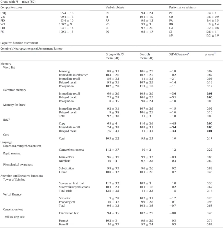
Neuropsychological assessment. General intelligence measure
Wechsler Intelligence Scale for Children, III Edition (WISC-III, Portuguese version) Group with PS— mean (SD)
Composite scores Verbal subtests Performance subtests
FSIQ 95.4 ± 16 IN 9.4 ± 2.4 PC 9.4 ± 1 VIQ 99.4 ± 16 SI 10.1 ± 1.9 CD 9.6 ± 0.9 PIQ 93.4 ± 10 AR 9.4 ± 1.3 PA 9.4 ± 1.5 VCI 100.2 ± 9 VO 9.9 ± 3 BD 9 ± 1.4 POI 94.1 ± 14 CO 9.7 ± 2.6 OA 9.2 ± 0.8 PSI 100.3 ± 13 DS 9.5 ± 1.7 SS 10.8 ± 1.1 MA 10.2 ± 1.6
Cognitive function assessment
Coimbra's Neuropsychological Assessment Battery
Group with PS mean (SD) Controls mean (SD) SSP differencesX p valueX Memory Word list Learning 8.8 ± 3.1 10.6 ± 2.9 −1.8 0.07 Immediate interference 10.4 ± 2.6 10.2 ± 2.5 0.2 0.87 Immediate recall 8.9 ± 3.3 11 ± 3.1 −2.1 0.05 Delayed recall 9.3 ± 3.1 10.7 ± 2.9 −1.4 0.16 Recognition 10.2 ± 2.8 11.3 ± 1.8 −1.1 0.12 Narrative memory Immediate recall 6.9 ± 2.9 10.5 ± 2.9 −3.6 0.01 Delayed recall 7.5 ± 2.8 10.6 ± 2.9 −3.1 0.00 Recognition 8 ± 3.5 9.8 ± 2.4 −1.8 0.06
Memory for faces
Immediate recall 9.2 ± 3.1 10.7 ± 2.6 −1.5 0.09 Delayed recall 9 ± 3.8 10.6 ± 2.9 −1.6 0.15 Total 9.2 ± 3.8 11 ± 3 −1.8 0.08 ROCF Copy 6.8 ± 4 11.6 ± 2.6 −4.8 0.00 Immediate recall 7.4 ± 3.8 10.8 ± 3.1 −3.4 0.00 Delayed recall 7.6 ± 4.1 11 ± 3.1 −3.4 0.01 Corsi Corsi 10.5 ± 2.2 9.5 ± 2.3 1.0 0.17 Language
Directions comprehension test
Comprehension test 11.2 ± 3.7 10 ± 2 1.2 0.29 Rapid naming Form colors 9.6 ± 3.9 9.9 ± 3.2 −0.3 0.80 Numbers 10 ± 4 9.7 ± 2.8 0.3 0.80 Phonological awareness Substitution 9.8 ± 3.9 9.6 ± 2.6 0.2 0.89 Elision 10.8 ± 3.2 10.1 ± 2.6 0.7 0.45
Attention and Executive Functions Tower of Coimbra Success onfirst trial 11.7 ± 3.2 10.7 ± 3 1.0 0.30 Successful reproductions 10.3 ± 2.3 10.1 ± 1.6 0.2 0.67 Total trials 12.5 ± 3.5 11 ± 2.8 1.5 0.14 Verbal Fluency Semantic 9 ± 2.8 10.2 ± 3.1 −1.2 0.20 Phonological 10 ± 3.7 9.9 ± 2.8 0.1 0.96 Total 9.6 ± 3.2 10.3 ± 3.6 −0.7 0.66 Cancelation test Cancelation test 9.4 ± 3.5 10.2 ± 2.9 −0.8 0.43
Trail Making Test
Form A 10.2 ± 3 9.9 ± 2.9 0.3 0.74
Form B 10 ± 3.7 9.7 ± 2.4 0.3 0.84
while six did not show spiking while awake. Sleep not only put in evidence spikes in patients with normal awake EEGs but also induced the appearance of independent foci in some with focal spikes. Eleven patients had spikes over the left hemisphere and an equal number over the right hemisphere, demonstrating an overall symmetry of our group in spike lateralization (Table 3).
Eight patients had spikes in both hemispheres, but whereas they occurred independently in four cases, in the remaining four, there was evidence for secondary propagation from a leading hemisphere to the contralateral one.
A comparison of the spike grade of the eight patients medicated with antiepileptic drugs (Table 1) and nonmedicated ones revealed similar average values (2.43 and 2.56, respectively) and statistically did not dif-fer from each other (t(14) = 0.29, pb 0.05).
The N170 right to left hemisphere ratio was within the normal range defined in Lopes et al.[12]in all cases except in patient 1 where it was increased (1.82). The average N170 ratios for the patients with left hemisphere spikes (1.00), right spikes (0.96), or independent bilateral spikes (0.98) were also within the normal range and overlapping. No functional difference between occipital lobe function, measured by the N170, could be found between the patient subgroups.
3.2. Neuropsychological evaluation
All patients had Full Scale IQ N80 (M = 95, SD = 16) in the Wechsler Intelligence Scale for Children. The Verbal IQ (M = 99) and Performance IQ (M = 93) were normal, as well as all the remaining subtests. Complete neuropsychological results are presented inTable 2. When comparing the mean CNAB test scores between patients with epilepsy and the normative group, only two tests showed a significant difference (more than 3 standard score points, range: 1–19), with the clinical group scoring worse than the controls: the copy task of the Rey–Osterrieth Complex Figure (ROCF) with a difference of 4.8 standard score points and Narrative Memory Test (NMT) with a difference of 3.6 standard score points. The overall mean score difference of all 16 CNAB tests between groups was 0.9. Moreover, ROCF was abnormal in 11/19 patients, while the Narrative Memory Test (NMT) was abnormal in 7/19. For the ROCF test, in the copy task, most of the items were copied but misconstructed (misplaced or rotated) leading to below average results in the copy. Consequently, immediate and delayed memory retrieval tasks also showed below average standardized values, but no significant (mean: 0.6 standard score) differences were found between the standardized results of the two memory tasks and the copy task.
For the NMT test, below average results were due to the immediate and delayed memory task but not to the recognition one. Most of the er-rors were omitted elements of the“stories”, with no significant intrusions. 3.3. Comparison of neuropsychological and neurophysiological results
A comparison of the spike abundance score for the patients with ab-normal and ab-normal results in either the ROCF or NMT tasks (Table 3) did not reveal relevant differences, with average scores of 2.64/2.20 and 2.14/2.30, respectively, demonstrating a medium/rare spike activity for all subgroups.
An analysis of the hemispheric spike lateralization for the patients with low scores in the NMT supported a slight asymmetry because left independent spikes were present in 5 out of 6 patients, while only 3 out of 6 had independent right spikes. For the ROCF, there was also a slight left hemisphere dominance as 5 out of 9 patients had independent left spikes, while only 3 out of 9 had independent right spikes.
The N170 ratio (Table 3) did not demonstrate differences between patients with abnormalfindings on the ROCF and NMT (average ratios of 0.98 and 0.97) and the ones who scored in the normal range for each test (average ratios of 1.03 and 1.03). These results do not support a role for the fusiform gyrus area, responsible for the N170 generation, in the neuropsychological abnormalities.
4. Discussion
Overall, our work demonstrates very subtle neuropsychological ab-normalities in patients with PS, with more consistent difficulties in the visual–spatial domain (ROCF) and narrative memory recall (NMT). The normal results of the N170 visual-evoked potentials support normal lower occipital lobe function in the neighborhood of the fusiform gyrus
[12], a result consistent with the normal function of the ventral visual pathway. This is in line with previous studies which have demonstrated a normal level of performance of these patients in a wide range of neuropsychological tests, namely, those evaluating occipital lobe function[9,20]. The more consistent abnormalities across these and our study converge in the area of visual–spatial organization, most likely representing dysfunction of the parietal–occipital visual dorsal pathway, while the more basic occipital lobe functions are remarkably normal. The clinical features of our group are comparable with other PS series[8,9,20]. All 19 patients had normal cognitive development and MRIs, mainly rare (b5) and prolonged (N10mn) autonomic seizures and EEG spike activity over the posterior brain regions.
Table 3
Neurophysiology and neuropsychology data.
Pts Spike lateralization Spike propagation N170 ratio Memory for
historiesX
Rey–Osterrieth Complex FigureX
Wake EEG (35 ch) Sleep EEG Spike grade
p1 Right Right 3 1.82
p2 Left Right and left 4 Left/right independent 0.77
p3 No spikes Right and left 3 Left/right independent 1.08
p4 Right and left Left/right independent 1.10 X
p5 Left Left 3 1.16
p6 Left Left 1 0.91 X
p7 Left Right and left 2 Left/right independent 0.97 X X
p8 Left Left 3 Left to right hemisphere 1.00 X
p9 Left Left 2 0.92 X
p10 No spikes Left 3 0.93 X
p11 Right Right 3 1.05 X
p12 No spikes Left 1 1.04 X X
p13 Right Right 2 Right to left hemisphere 1.10
p14 No spikes Right 2 0.78
p15 Right 0.83
p16 No spikes Left 3 1.07 X
p17 Right Right 2 Right to left hemisphere 0.82 X
p18 No spikes Right 3 Occipital to frontal 0.86 X
p19 Right Right Right to left hemisphere 0.86 X
X
The ROCF is a well-known test that had initially been proposed to assess perceptive organizational and visual memory[21]. Later, this test was also used to assess other cognitive functions such as planning, inhibition, or perseverative deficits[22]. Several scoring systems have been proposed[23–25], most of them having two main features in com-mon: presence and quality of the element reproduction and the relative element's position in thefigure. Different types of errors have been cor-related with different cognitive impairments: patients with frontal lobe lesions typically demonstrate repetitions and perseverations and parie-tal–occipital lesion patients demonstrate difficulty with spatial organi-zation of thefigure, (for a review see Lezak et al.)[26]. In our study, ROCF copy errors made by our patient group with PS are similar with the ones found in patients with parietal–occipital lesions[27]: most of thefigure elements are present; they are neither replicated nor their units multiplied but (slightly to mildly) rotated or misaligned between each other. Solms et al.[28]described a group of 16 patients who showed gross rotation on the copy task of ROCF. In 9 of those patients, an accurate localization of lesions could be made. Interestingly, all 9 pa-tients had lesions of the frontal lobe, but only 4/9 showed involvement of the parietal lobe dorsal stream. According to the authors, such results could be due to strong functional links between the dorsolateral frontal lobes and the parietal dorsal stream.
A deficit in “higher-order” functions such as planning or visual attention could explain this kind of poor ROCF copy results[7,29,30]. However, we have not found Attention (e.g. Cancelation tests and Trail Making Test Form A) or Executive Function (e.g. Tower of London and Verbal Fluency) deficits. Furthermore, the WISC-III perfor-mance IQ was normal. Ourfindings agree well with Germanò et al.[8]
results, showing also deficits in perceptive visual–spatial tasks, but not in any higher-order process, in a group of PS. Overall, these results suggest that top-down processes are spared.
On the other hand, the possibility of an early impairment in visual information processing at the occipital lobe as a possible cause of the observed ROCFfindings does not find support either in the existing neu-ropsychology literature[9]which failed to demonstrate specific occipital lobe dysfunction in patients with PS or in our own results demonstrating normal N170 visual-evoked responses. In sum, we suggest that attribut-ing dysfunction to the frontal lobe (attention and plannattribut-ing) or the occip-ital lobe (N170 and basic visual–perceptual functions) are not satisfactory explanations for the visual–organizational deficits found. A parietal lobe dysfunction seems to us a more consistent possibility.
Concerning the NMT results, we hypothesize a semantic processing impairment more than a specific memory one. Semantic processing refers to the capacity to access knowledge about the world, central to access word meaning, (verbal) reasoning, planning, and conceptual organization[31]. Against an episodic memory deficit is the fact that re-sults of all other memory tasks are normal: memory for faces, ROCF memory tasks, and, most importantly, the second verbal memory CNAB task, the wordlist test. We found that our patients with PS did not use the logical association between different sequences of the“stories” of NMT, only retrieving isolated items or short sequences and, therefore, are unable to integrate the complex information that composes the story. That might explain why the results of the word list tasks are normal, since these tasks only use isolated words as retrieval items, not requiring any integration of the complex information. Along this line of reasoning, a meta-analysis of 120 fMRI studies[31]found that the left posterior parietal lobe plays a fundamental role on concept retrieval and integration. Our argument that the NMT difficulties we found are the result of a poor conceptual organization and integration seems to be in accordance with a PET study conducted by Ruby et al.[32]showing that the parietal lobe is involved in the processing of scripts. Scripts are a set of expectations about what will happen next in a well-understood situation[33]; they are composed of goal-oriented sequences of events that typically occur in a specific and systematic order[34].
From the 12 patients with PS who showed poor results on ROCF or NMT, only 2 showed deficits on both tests simultaneously. Because
there is a preferential hemispheric contribution either to semantic pro-cessing (left) or to visual–organizational functions (right), these find-ings suggest that a selective involvement of one of the hemispheres could be a contributing factor to the small overlap of the two types of test impairment. Nevertheless, our neurophysiological data do not allow us to consistently determine which hemisphere is more strongly affected since there is no correlation between spike frequency, topo-graphic distribution, and neuropsychological deficits.
Overall, our results pointing towards parietal lobe dysfunction in our group of patients agree well with the recent evidence obtained from a symptomatic case of PS in which a parietal lobe localization of the epi-leptogenic area could reproduce the main clinical and neurophysiologi-cal features of PS[11].
Acknowledgments
The authors are grateful to Daniel Carvalho and Adília Seabra for their technical support and to Professor Rita Jerónimo for the paper re-view and suggestions.
Ricardo Lopes has been supported by the Grant SFRH/BD/65617/ 2009 from the Portuguese Foundation for Science and Technology (FCT). References
[1]Panayiotopoulos C. Vomiting as an ictal manifestation of epileptic seizures and syn-dromes. J Neurol Neurosurg Psychiatry 1988;51:1448–51.
[2]Panayiotopoulos C. Panayiotopoulos syndrome: a common and benign childhood epileptic syndrome. London: John Libbey & Company; 2002.
[3]Kokkinos V, Koutroumanidis M, Tsatsou K, Koupparis A, Tsiptsios D, Panayiotopoulos C. Multifocal spatiotemporal distribution of interictal spikes in Panayiotopoulos syn-drome. Clin Neurophysiol 2010;121:859–69.
[4]Leal A, Nunes S, Dias A, Vieira J, Moreira A, Calado E. Analysis of the generators of ep-ileptic activity in early-onset childhood benign occipital lobe epilepsy. Clin Neurophysiol 2007;118:1341–7.
[5]Leal A, Ferreira J, Dias A, Calado E. Origin of frontal lobe spikes in the early onset be-nign occipital lobe epilepsy (Panayiotopoulos syndrome). Clin Neurophysiol 2008;119:1985–91.
[6]Gülgönen S, Demirbilek V, Korkmaz B, Dervent A, Townes B. Neuropsychological functions in idiopathic occipital lobe epilepsy. Epilepsia 2000;41(4):405–11.
[7]Bedoin N, Ciumas C, Lopez C, Redsand G, Herbillon V, Laurent A, et al. Disengagement and inhibition of visual–spatial attention are differently impaired in children with rolandic epilepsy and Panayiotopoulos syndrome. Epilepsy Behav 2012;25:81–91.
[8]Germanò E, Gagliano A, Magazù A, Sferro C, Calarese T, Mannarino E, et al. Benign childhood epilepsy with occipital paroxysms: neuropsychologicalfindings. Epilepsy Res 2005;64:137–50.
[9]De Rose P, Perrino F, Lettori D, Alfieri P, Cesarini P, Battaglia D, et al. Visual and visuoperceptual function in children with Panayiotopoulos syndrome. Epilepsia 2010;51(7):1205–11.
[10]Yalçin A, Toydemir H, Çelebi L, Forta H. Panayiotopoulos syndrome with coincidental brain lesions. Epileptic Disord 2009;11:270–6.
[11]Leal A, Lopes R, Ferreira J. Origin and dynamics of epileptic activity in a symptomatic case of Panayiotopoulos syndrome: correlation with clinical manifestations. Clin Neurophysiol 2013;124:20–6.
[12]Lopes R, Cabral P, Canas N, Breia P, Foreid J, Calado E, et al. N170 asymmetry as an index of inferior occipital lobe dysfunction in patients with symptomatic occipital lobe epilepsy. Clin Neurophysiol 2011;122(1):9–15.
[13]Simões MR, Albuquerque C, Pinho M, Pereira M, Seabra-Santos M, Alberto I, et al. Co-imbra Neuropsychological Assessment Battery— CNAB (Bateria de Avaliação Neuropsicológica de Coimbra (BANC)). Lisboa: Cegoc; 2013 [in press, Portuguese].
[14]Fuchs M, Kastner J, Hawes S, Ebersole J. A standardized boundary element method volume conductor model. Clin Neurophysiol 2002;113(5):702–12.
[15]Wechsler D. Escala de Inteligência de Wechsler para Crianças– Terceira Edição (WISC-III). Cegoc: Lisboa; 2003.
[16]Lopes A, Simões MR, Robalo C, Fineza I, Gonçalves O. Neuropsychological assessment of children with epilepsy: attention and executive functions in temporal lobe epilep-sy (Evaluación neuropsicológica en niños con epilepsia: Atención y funciones ejecutivas en epilepsia del lóbulo temporal). Rev Neurol 2010;5:265–72 [Spanish].
[17]Sá D, Albuquerque C, Simões MR. Neuropsychological assessment of oppositional defiant disorder (Avaliação neuropsicológica da perturbação de oposição e desafio). Psicol Saude Doencas 2008;9(2):299–317 [Portuguese].
[18]Albuquerque C, Simões MR. Rapid naming tests: developmental course and relations with neuropsychological measures. Span J Psychol 2010;13(1):88–100.
[19]Albuquerque C, Simões MR, Martins C. Phonological awareness tests of Coimbra's Neu-ropsychological Assessment Battery: reliability and validity studies (Testes de Consciência Fonológica da Bateria de Avaliação Neuropsicológica de Coimbra: Estudos de precisão e validade). Rev Iberoam Diagn Eval Psicol 2011;29(1):55–76 [Portuguese].
[20]Specchio N, Trivisano M, Di Ciommo V, Cappelletti S, Masciarelli G, Volkov J, et al. Panayiotopoulos syndrome: a clinical, EEG, and neuropsychological study of 93 con-secutive patients. Epilepsia 2010;51(10):2098–107.
[21]Rey A. L'examen psychologique dans les cas d’encephalopathie traumatique. Arch Psychol 1941;28:286–340.
[22]Ogino T, Watanabe K, Nakano K, Kado Y, Morooka T, Takeuchi A, et al. Predicting executive function task scores with the Rey–Osterrieth Complex Figure. Brain Dev 2009;31:52–7.
[23]Osterrieth P. Le test de copie d'unefigure complex: l'etude de la perception et de la memoire. Arch Psychol 1944;28:1021–34.
[24]Meyers J, Meyers K. The Meyers scoring system for the Rey Complex Figure and the recognition trial: professional manual. Odessa, FL: Psychological Assessment Resources; 1995.
[25]Stern R, Javorsky D, Singer E, Singer N, Somerville J, Duke L, et al. The Boston quali-tative scoring system for the Rey–Osterrieth Complex Figure. Professional Manual Psychological Assessment Resources, Inc.; 1999
[26]Lezak M, Howieson D, Loring D. Neuropsychological assessment. 4th ed. Oxford: Oxford University Press; 2004.
[27]Pillon B. Troubles visuo-construtifs et méthodes de compensation: résultats de 85 patientes atteints de lésion cérébrales. Neuropsychologia 1981;19(3):375–83 [French].
[28]Solms M, Turnbull O, Kaplan-Solms K, Miller P. Rotated drawing: the range of perfor-mance and anatomical correlates in a series of 16 patients. Brain Cogn 1998;38:358–68.
[29]Watanabe K, Ogino T, Nakano K, Hattori J, Kado Y, Sanada S, et al. The Rey–Osterrieth Complex Figure as a measure of executive function in childhood. Brain Dev 2005;27:564–9.
[30]Weber R, Riccio C, Cohen M. Does Rey Complex Figure copy performance measure executive function in children? Appl Neuropsychol Child 2012;1:1–7.
[31]Binder J, Desai R, Graves W, Conant L. Where Is the semantic system? A critical review and meta-analysis of 120 functional neuroimaging studies. Cereb Cortex 2009;19:2767–96.
[32]Ruby P, Sirigu A, Decety J. Distinct areas in parietal cortex involved in long-term and short-term action planning: a pet investigation. Cortex 2002;38:321–39.
[33]Schank R, Abelson R. Knowledge and memory: the real story. In: Wyer Jr Robert S, editor. Knowledge and memory: the real story. Hillsdale, NJ: Lawrence Erlbaum Associates; 1995. p. 1–85.
[34]Schank R, Abelson R. Scripts, plans, goals and understanding. Hillsdale, NJ: Lawrence Erlbaum Associates; 1977.