(will be inserted by the editor)
Probing Deep Tissues with Laser-Induced Thermotherapy
Using Near Infrared Light
Received: date / Accepted: date
Abstract Optically tunable gold nanoparticles have been widely used in research with near-infrared light as a means to enhance laser-induced thermal therapy since it capitalises on nanoparticles’ plasmonic heating properties. There have been published several studies on numerical models replicating this therapy in such conditions. However, there are several limitations on some of the models which can render the model unfaith-ful to therapy simulations. In this paper, two techniques of simulating laser induced thermal therapy with a high absorbing localised region of interest inside a phantom are compared. To validate these models we conducted an experiment of an agar-agar phantom with an inclu-sion reproducing it with both models. The phantom was optically characterized by absorption and total attenu-ation. The first model is based on the macro perspec-tive solution of the radiaperspec-tive transfer equation given by the diffusion equation, which is then coupled with the Pennes bioheat equation to obtain temperature. The second is a Monte Carlo model that considers a stochas-tic solution of the same equation and is also consid-ered as input to the Pennes bioheat transfer equation which is then computed. The Monte Carlo is in good agreement with the experimental data having an av-erage percentage difference of 4.5% and a correlation factor of 0.98, while the diffusion method comparison with experimental data is 61% and 0.95 respectively. The optical characterisation of the phantom and its in-clusion were also validated indirectly since the Monte Carlo, which used those parameters, was also validated. While knowing the temperature in all points inside a body during photothermal therapy is important, one has to be mindful of the model which fit the conditions and properties. There are several reasons to justify the
discrepancy of the diffusion method: low scattering con-ditions, absorption and reduced scattering are compa-rable. The error bars that are normally associated when characterizing an optical phantom can justify also a part of that uncertainty. For low size tumours in depth, one may have to increase the light dosage in photother-mal therapies to have a more effective treatment. Keywords Phototherapy · Photothermal therapy · Monte Carlo · Diffusion Approximation · Near Infrared Light
Introduction
Breast cancer remains the leading cancer killer in mid-dle aged women around the world [15]. Despite recent advances in therapy and diagnosis, cancer continues to be a difficult disease to treat. Depending on its loca-tion, stage or whether it became resistant to the on-going treatment, the therapies can also change. There-fore, it is imperative that the resources the oncologist has to tackle this disease, be numerous and diverse. In this work, we study near infrared photothermal ther-apy (PTT), which allow non-ionizing radiation to dif-fuse inside tissue and cause low temperature elevation [11, 14]. When compared to the most conventional ther-apies like surgery and chemotherapy, it is non-invasive and produces minimal damage to healthy tissue. A frac-tion of photon energy is absorbed and converted into heat, depending on tissue properties, which can destroy malignant tissue through processes like coagulation or hyperthermia if the achieved temperature and time of exposure at that temperature is sufficient [2]. A limit-ing factor in this process is large optical scatterlimit-ing in biological tissue which imposes restrictions on how deep one can heat a predetermined region of interest. With
the advent of gold nanoparticles (GNP) in medicine, there is a promising opportunity to bypass such limita-tions.
Gold nanoparticles have tunable high absorption pro-perties in the NIR regions that allow local higher ab-sorbed energy. Several authors [25, 5, 4, 19], report a tem-perature change at up to 3 cm depth when using these particles while no temperature change was observed without them. This increase in temperature caused by exogenous contrast agents changes the rate of deposited heat which in turn is influenced by irradiation con-ditions and tissue properties that ultimately influence therapy’s success [24].
A myriad of cell damaging effects can be observed starting from ∼ 39oC where protein denaturation hap-pens. Depending on the time of exposure these effects can do reversible or irreversible damage to cells[20, 14, 11]. Hence, a precise temperature control is crucial since it is impossible to have probes to measure the tempera-ture in all space. Consequently, it is important to have models that can mimic the illumination conditions and tissue properties, and allow an accurate temperature representation.
Among the many light models already developed two stand out for being the most used in the commu-nity: diffusion model and Monte Carlo. Monte Carlo simulations give a stochastic representation of the so-lution to the radiative transfer equation (RTE), which is the equation that governs how light propagates in-side biological tissue [26]. The diffusion equation can be obtained from the RTE considering some of its pa-rameters as first-order spherical harmonic expansions which allows only isotropic and first-order anisotropic terms. In practice, this imposes restrictions on the con-ditions in which this theory is valid, i.e. on tissues with low absorption and high scattering properties, among others [24].
The studies that are published using nanoparticles can be separated into two categories. There are some approaches when using gold nanoparticles in the liter-ature that consider an homogeneous mixture of a so-lution and gold nanoparticles and replicate the tem-perature or irradiance with simulations [23, 25]. Other approaches consider two layers of tissue stacked with and without the presence of gold nanoparticles [4, 5]. Depending on the number of gold nanoparticles used, the optical properties of the tissue vary significantly to a point where absorption is comparable to scattering [18], which might render the diffusion theory ineffec-tive. In this work, we will address this problem using an agar phantom with an inclusion that mimics this con-dition. First, it is presented the conventional method of measuring some of the phantoms’ optical properties.
After, an experiment is conducted in which the phan-tom is irradiated and the temperature is measured with a thermocouple inside the phantom.
The numerical methods consider the light distribu-tion using diffusion theory and Monte Carlo, which is considered the gold standard to model light [26]. To model the heat transfer mechanism we choose the Pennes bioheat equation [17] that reproduces well the temperature inside a phantom [24].
In this paper, we present a study with a nanopar-ticle based inclusion in a phantom far from its surface. To the best of the author’s knowledge this study is not presented elsewhere and is important; the nanoparticles aggregate around a tumour of finite size and the other studies present homogeneous mediums with nanoparti-cles[5]. We also characterize the optical properties of the mediums considered and the irradiation experiment.
Mathematical formulation
The models of optical and thermal propagations of light and heat through the tissue are presented in this sec-tion. In the first part, we consider the diffusion the-ory and Monte Carlo simulations to solve the radia-tive transfer equation, while the Pennes bioheat transfer equation is considered to model the heat propagation mechanism.
The diffusion equation with a constant wave source at its border is defined as [1]
−∇ · κ(r)∇φ(r) + µa(r)φ(r) = 0, r ∈ Ω, (1)
where κ = 1/(3(µ0s+ µa)) is the diffusion coefficient, µa
the absorption coefficient, µ0sis the reduced scattering coefficient, φ the fluence rate, Ω its domain and position vector r. The fluence rate boundary conditions between phantom and exterior are defined by
φ(m) + 2ξ(c)κ(m)∂φ(m)
∂ν = q(m), m ∈ ∂Ω, (2) where the term ξ(c) represents the refractive bound-ary mismatch between different regions, ∂ν represents the outward boundary normal, q is the source distri-bution at the boundary ∂Ω, and m the position vec-tor restricted to ∂Ω. This model is derived under two premises, it is valid in high diffusive versus absorbing tissues and it is not valid near source points, where the gradient of fluence rate is not linear [24].
In a Monte Carlo simulation, each photon is simu-lated individually with a predetermined starting posi-tion, direction and interaction mechanisms. In the near
infrared region, light either scatter or gets absorbed in-side an homogeneous tissue. The scattering phase func-tion of the radiative transfer equafunc-tion in soft biologi-cal tissues can be modelled by the Henyey-Greenstein phase function [24]. At the boundaries, refraction and reflection are considered with Fresnel and Snell equa-tions [7].
The laser induced heating source term is described by Q(r, t) ≡ µa(r) × φ(r, t) [24]. Since this term is
ex-ternal to the heat generated in the body, we add it to the Pennes bioheat transfer equation as follows
ρc∂T (r, t)
∂t =∇ · (k(r)∇T (r, t)) + Qm(r) + Q(r, t) + ωbρbcb(T (r, t) − Ta), r ∈ Ω,
(3)
where ρ is the tissue density, c is the specific heat, T (r, t) the temperature, k(r) the thermal conductivity, Qmmetabolic heat rate, ωb rate of blood perfusion, ρb
density of blood, cb blood’s specific heat and Tabody’s
temperature.
The convection heat transfer mechanism is also con-sidered and is determined by Newton’s law:
Qc= h(T∞− T (m, t)), m ∈ ∂Ω, (4)
where h is the convective heat transfer coefficient and T∞is the room temperature. Radiation, thermal
evap-oration and/or phase change of tissue during the heat transfer process are not considered in the simulations.
Materials and Methods
An agar-agar phantom was produced in two steps. First, a mixture of 1.0% agarose powder (Agar-Agar, Vahin´e, France) in distilled water. The water was heated along with the agarose powder and mixed (at a mixing tem-perature of ≈ 90oC) three times to ensure an
homoge-neous optical density of the gel. It was then let cool off inside a 60×60×60 mm3 cube using a cylinder of 15.5
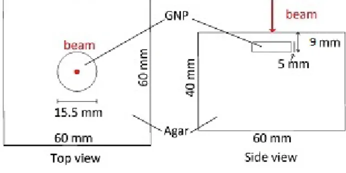
mm in diameter to create a 5mm deep hole on top of the phantom. Once it cooled down to ambient temper-ature, the hole created by the cylinder was filled with two equal parts of agar and gold nanoparticles prepared according to [22]. The two were mixed at 35o C since the gold nanoparticles had a bio-coating in them. This proportion was used to ensure the gold nanoparticles were in a fixed place, but also to have a good balance between scattering and absorption. A 4 mm layer of agar-agar solution was added to the phantom. The fi-nal phantom is depicted in figure 1.
Fig. 1 Gel phantom setup. The laser beam entry position and direction is depicted in red. The main component (agar) is made of 1% agar and water, while the smaller component (GNP) is made of 1% agar, 10% GNP solution and 89% water.
Determination of Materials’ Properties
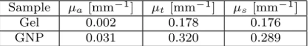
The optical properties of the gel and inclusion were measured using an integrating sphere (International Light, INS 250), a laser diode source (Roithner, RLTMDL-808-5W-5) 808 nm wavelength and a spec-trometer (Avaspec 2048, Avantes). The measured co-efficients were the absorption coefficient and the total attenuation coefficient. Other optical properties such as anisotropy and refractive index were taken from litera-ture [5, 10].
Similarly to the methods described in [16], the ab-sorption coefficient (µa) was measured placing the
cu-vette with a sample of agar or nanoparticles and agar in the integrating sphere’s centre, and compared with a cuvette with distilled water. Coupled with the inte-grating sphere was the spectrometer whose signal was then deconvoluted from the laser wavelength distribu-tion peaking at 808nm. The absorpdistribu-tion coefficient was determined using the equation µa = I0I−I0 , where I0 is
the measurement with distilled water, and I the signal measured with the sample solution.
To measure the attenuation coefficient (µt) a
proce-dure similar to the one described in [16] was followed. Additionally, a 1mm-pinhole was placed in front of the detector and both were positioned 40cm away from the sample. This procedure is reported to limit multi-ple scattering events to some extent[12, 13]. The Beer-Lambert equation was used to determine the attenua-tion coefficient, µt= − logII
0, where I is the measured
signal of the cuvette with the sample and I0 the
mea-sured signal of the cuvette with distilled water. Each measurement was repeated 5 times to ensure consis-tency. The scattering coefficient was determined indi-rectly by the equation µs= µt− µa. Table 1 shows the
Sample µa[mm−1] µt[mm−1] µs[mm−1]
Gel 0.002 0.178 0.176 GNP 0.031 0.320 0.289
Table 1 Measured optical properties of the agar gel phantom and a mixture of gold nanoparticles (GNP) and agar gel.
Experimental setup
The phantom was irradiated in the top surface of the box with a continuous wave diode laser of 1.1 W power and a full width at half maximum of 7 mm, as repre-sented in figure 1.
Temperatures were registered using a type K ther-mocouple (Labfacility, Z2-T-1M) and a therther-mocouple converter (Seneca K109TC) connected to a LabVIEW R
interface and with a tunable data acquisition rate of 1s. The thermocouple was placed on top of the cylinder, at 4 mm depth. The constant wave laser was on for 706 s, then turned off. The radiation absorbed by ther-mocouple was taken into account and was subtracted from experimental results as suggested in [3]. The am-bient temperature was measured at 22.9 oC and the
temperature measured in the thermocouple the instant before the laser irradiation phase was 23.88oC, which is considered the reference temperature when determining temperature change.
Simulations
Several software applications were used to compute the solution to the temperature determination. Along with the commercial software COMSOL Multiphysics R and
MATLAB R, which were used to perform the data
anal-ysis. Iso2mesh [8] and GMSH [9] were used to build and characterize the meshed phantom. This mesh was used as an input in both TOAST++ [21] and the Monte Carlo software Mesh-based Monte Carlo [6]. Both of these codes are considered to be the state of the art in their own domain to determine the irradiance within biological tissue. One only needs to provide the laser parameters and the tissue’s optical properties since the appropriate functions are already built-in. All codes were run in a 8x Intel Core i7 4790 (4.0 GHz) CPU with 4x 4GB DDR3 1600MHz.
The diffusion approximation software provides the selection of a gaussian profiled source with σ = 3.07 mm, modelled as a Neumann source which considers it as a diffuse boundary current. The same laser profile and the standard deviation were selected in the Monte Carlo simulator. 10 million events were simulated. The output fluence rate of both programs was then used as 3D heat source in COMSOL.
The temperature for both cases was computed by COMSOL Multiphysics R. The top surface transferred
convective heat using the Newton convection heat trans-fer equation with the respective coefficient at 5 W K−1 m−2 [24] while the other surfaces were considered to be
in thermal isolation. Although this consideration could be considered erroneous, its effect is considerable only after the laser-on phase when the phantom is cooling down. The blood density and the metabolic heat coef-ficient of the Pennes bioheat equation were set to zero. This consideration converts the Pennes bioheat equa-tion into the classical heat transfer equaequa-tion. The den-sity, heat capacity and thermal conductivity coefficients of water were considered in this test.
The used optical parameters for the phantom are the following: absorption coefficient: µa: 0.002 mm−1,
reduced scattering coefficient: µ0s= µs(1 − g) = 0.0176
mm−1an anisotropy (g) of 0.9 is assumed [5], refractive index = 1.33 [10]. The inclusion’s optical properties dif-fer from the previous ones on the absorption coefficient, which is µa = 0.031 mm−1and reduced scattering
coef-ficient µ0s= 0.029 mm−1, while the others are the same. Inside the inclusion the absorption coefficient and the reduced scattering coefficient are comparable and can make the diffusion approximation ineffective.
Results and Discussion
To check that both models work within reasonable agre-ement a simulation was conducted. Consider a case in which both Monte Carlo and the diffusion approxima-tion are effective; an homogeneous 2 cm wide cube with the following optical properties: µa = 0.002 mm−1,
µ0s= 1.22 mm−1; g = 0.9 and refractive index of 1.33. A continuous wave gaussian near infrared laser beam of power = 1.5 W and FWHM = 5 mm aims at one of the cube’s side at the centre for 180 s. Figure 2 shows a comparison between temperature increase of both simulators 1mm deep of the irradiated side. One can observe the diffusion approximation is almost 1 K higher at 180 s between simulated temperatures which is caused by the different approaches of dealing with scattering anisotropy by the two models.
Figure 3 presents the comparison between experi-ment and simulation results given from Monte Carlo and diffusion theory. Both models are in good agree-ment with the experiagree-ment measureagree-ments. There are some variations in the measured temperature along time which is explained by non-linear photon absorption and heat diffusion from the thermocouple probe unto its sur-roundings. The instant the irradiation phase stopped this behaviour was not observed. These variations de-crease in amplitude with smaller thermocouple probes.
Fig. 2 Temperature increase comparison between Monte Carlo (blue) and the diffusion theory (green) simulators as a function of time, at a point located 1mm below the irradi-ated spot.
The average percentage difference between the Monte Carlo model is 4.5% and for the diffusion approxima-tion 61%, while the correlaapproxima-tion coefficient was of 0.98 and 0.95, respectively.
These results can be compared to e.g., Elliot et al. [5] where this problem is explored in a similar exper-iment. Joined in a single phantom cylinder are two smaller cylinders with different optical properties and lengths. The diffusion approximation was used to model the experiment. The physical parameters for the finite element calculations were adjusted within the experi-mental error (± 10%) to optimize results. The average percentage difference and correlation coefficient were 4.5% and 0.99 respectively, for the similar conditions as this experiment. Changing the physical parameters in the diffusion approximation model in this study by 20%, decreases the average percentage difference from 61% to 23%. Although this variation is far from the 4.5% reported in [5] this result highlights the model sensitivity on these parameters.
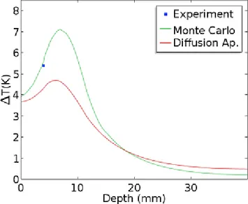
Figure 4 shows the temperature increase in depth along the axis that is aligned with the laser beam axis the second before the laser was shut down. The distri-bution of both models is slightly different. Using more thermocouple probes would allow a more comprehen-sive study of this distribution. When comparing it with similar temperature change distributions in other stud-ies, such as [5, 4], increases in temperature greater than 10 K can be observed. This result suggests that the size of the inclusion with nanoparticles is a relevant compo-nent to temperature increase, along with irradiance as
Fig. 3 Comparison of simulation and experimentally mea-sured increase in temperature, 4 mm below surface. It shows temperature change as a function of time. Both models are shown in green and red and are compared with the experi-mental results shown in blue.
well as the inclusions’ optical properties, density and location inside tissue.
Fig. 4 Temperature increase at t = 706 s right before the laser was turned off. The x-axis represents an axis that is aligned with the laser beam axis, which is located at the cen-tre of two faces of the cube. The Monte Carlo simulation and diffusion approximation results are presented in green and red, respectively. The experimental point is shown in blue.
Conclusions
It is of crucial importance to know the temperature in every point inside the tissue when performing pho-tothermal therapy to better control the light dosage delivered. We present two ways of simulating laser in-duced thermal therapy in a specific experimental setup with a high absorbing region of interest located at 4 mm of depth, and compared to experimental data. The two models are in reasonable agreement with the exper-iment results. The size of inclusion affects the maximum temperature one can achieve, as well as concentration and depth.
Conflict of interests The authors declare that they have no conflict of interest.
Role of funding source This work was partially sup-ported by national funding by the Portuguese FCT - Funda¸c˜ao para a Ciˆencia e Tecnologia through the projects PD/BD/105920/2014, UID/FIS/04559/2013 (LIBPhys) and UID/BIO/00645/2013.
Ethics approval Not applicable.
References
1. Arridge, S.R.: Optical tomography in medical imaging. Inverse Problems 15(2), R41 (1999). DOI 10.1088/0266-5611/15/2/022
2. Brunetaud, J.M., Mordon, S., Maunoury, V., Beacco, C.: Non-PDT Uses of lasers in oncology. Lasers In Medical Science 10(1), 3–8 (1995). DOI 10.1007/BF02133156 3. Cain, C.P., Welch, A.J.: Thin-Film Temperature
Sen-sors for Biological Measurements. IEEE Transactions on Biomedical Engineering BME-21(5), 421–423 (1974). DOI 10.1109/TBME.1974.324415
4. Cheong, S.S.K., Krishnan, S., Cho, S.H.: Modeling of Plasmonic Heating From Individual Gold Nanoshells for Near-Infrared Laser-Induced Thermal Therapy. Medical Physics 36(6), 4664–4671 (2008). DOI 10.1118/1.2961677 5. Elliott, A.M., Stafford, R.J., Schwartz, J., Wang, J., Shetty, A.M., Bourgoyne, C., O’Neal, P., Hazle, J.D.: Laser-induced thermal response and characterization of nanoparticles for cancer treatment using magnetic res-onance thermal imaging. Medical Physics 34(7), 3102– 3108 (2007). DOI 10.1118/1.2733801
6. Fang, Q.: Mesh-based Monte Carlo method using fast ray-tracing in Pl¨ucker coordinates. Biomedical optics ex-press 1(1), 165–175 (2010). DOI 10.1364/BOE.1.000165 7. Fang, Q., Boas, D.a.: Monte Carlo simulation of pho-ton migration in 3D turbid media accelerated by graph-ics processing units. Optgraph-ics express 17(22), 20178–20190 (2009). DOI 10.1364/OE.17.020178
8. Fang, Q., Boas, D.A.: Tetrahedral mesh generation from volumetric binary and grayscale images. Proceedings - 2009 IEEE International Symposium on Biomedical
Imaging: From Nano to Macro, ISBI 2009 pp. 1142–1145 (2009). DOI 10.1109/ISBI.2009.5193259
9. Geuzaine, C., Remacle, J.F.: Gmsh: a three-dimensional finite element mesh generator with built-in pre-and post-processing facilities. International Journal for Numeri-cal Methods in Engineering 79(11) 0, 1309–1331 (2009). DOI 10.1002/nme.2579
10. Jacques, S.L.: Optical Properties of Biological Tissues: A Review. Physics in Medicine and Biology 58(11), R37 (2013). DOI 10.1088/0031-9155/58/11/R37
11. Jaque, D., Mart´ınez Maestro, L., del Rosal, B., Haro-Gonzalez, P., Benayas, A., Plaza, J.L., Mart´ın Rodr´ıguez, E., Garc´ıa Sol´e, J.: Nanoparticles for photothermal ther-apies. Nanoscale 6(16), 9494–9530 (2014). DOI 10.1039/c4nr00708e
12. Kim, A., Wilson, B.C.: Measurement of ex vivo and in vivo tissue optical properties: Methods and theories. In: Optical-Thermal Response of Laser-Irradiated Tissue, pp. 267–319. Springer Netherlands, Dordrecht (2010). DOI 10.1007/978-90-481-8831-4 8
13. Marchesini, R., Bertoni, A., Andreola, S., Melloni, E., Sichirollo, a.E.: Extinction and absorption co-efficients and scattering phase functions of human tissues in vitro. Applied optics 28(12), 2318– 24 (1989). DOI 10.1364/AO.28.002318. URL http://www.ncbi.nlm.nih.gov/pubmed/20555518 14. Niemz, M.H.: Laser-Tissue Interactions.
Springer Berlin Heidelberg, Berlin, Heidelberg (1996). DOI 10.1007/978-3-662-03193-3. URL http://link.springer.com/10.1007/978-3-662-03193-3 15. Organization, W.H.: Women’s health (2013). URL
http://www.who.int/mediacentre/factsheets/fs334/en/ 16. Patterson, M.S., Wilson, B.C., Wyman, D.R.: The
agation of optical radiation in tissue. II: Optical prop-erties of tissues and resulting fluence distributions. Lasers in Medical Science 6(4), 379–390 (1991). DOI 10.1007/BF02042460
17. Pennes, H.H.: Analysis of tissue and arterial blood tem-peratures in the resting human forearm. 1948. Journal of applied physiology (Bethesda, Md. : 1985) 85, 5–34 (1998). DOI 9714612
18. Phadnis, A., Kumar, S., Srivastava, A.: Numerical in-vestigation of thermal response of laser-irradiated bio-logical tissue phantoms embedded with gold nanoshells. Journal of Thermal Biology 61, 16–28 (2016). DOI 10.1016/j.jtherbio.2016.08.002
19. Reynoso, F.J., Lee, C.d., Cheong, S.k., Cho, S.H.: Imple-mentation of a multisource model for gold nanoparticle-mediated plasmonic heating with near-infrared laser by the finite element method. Medical Physics 40(7), 073301 (2013). DOI 10.1118/1.4808361
20. Sapareto, S.A., Dewey, W.C.: Thermal dose determina-tion in cancer therapy. Internadetermina-tional journal of radiadetermina-tion oncology, biology, physics 10(6), 787–800 (1984). DOI 10.1016/0360-3016(84)90379-1
21. Schweiger, M., Arridge, S.: The Toast++ software suite for forward and inverse modeling in optical tomography. Journal of biomedical optics 19(4), 040801 (2014). DOI 10.1117/1.JBO.19.4.040801
22. Silva, C.O., Rijo, P., Molpeceres, J., Ascens˜ao, L., Roberto, A., Fernandes, A.S., Gomes, R., Pinto Coelho, J.M., Gabriel, A., Vieira, P., Reis, C.P.: Bioproduction of gold nanoparticles for photothermal therapy. Thera-peutic delivery 7(5), 287–304 (2016). DOI 10.4155/tde-2015-0011
23. Vera, J., Bayazitoglu, Y.: A note on laser penetration in nanoshell deposited tissue. International Journal of Heat
and Mass Transfer 52(13-14), 3402–3406 (2009). DOI 10.1016/j.ijheatmasstransfer.2009.02.014
24. Welch, A.J., Van Gemert, M.J.C.: Optical-thermal re-sponse of laser-irradiated tissue, vol. 2. Springer, Dor-drecht (2011). DOI 10.1007/978-90-481-8831-4
25. West, J.L., Hirsch, L.R., Stafford, R.J., Bankson, J.A., Sershen, S.R., Rivera, B., Price, R.E., Hazle, J.D., Ha-las, N.J., West, J.L.: Nanoshell-mediated near-infrared thermal therapy of tumors under magnetic resonance guidance. Proceedings of the National Academy of Sciences 100(23), 13549–13554 (2003). DOI 10.1073/pnas.2232479100
26. Yao, R., Intes, X., Fang, Q.: Generalized mesh-based Monte Carlo for wide-field illumination and detection via mesh retessellation. Biomedical Optics Express 7(1), 171 (2016). DOI 10.1364/BOE.7.000171