Universidade de Aveiro 2012
Departamento de Biologia
Cátia Sofia Andrade dos
Santos
Biomonitorização de aves em recuperação:
um estudo de longo termo
Biomonitorization of birds under recovery: a
long term study
Universidade de Aveiro 2012
Departamento de Biologia
Cátia Sofia Andrade dos
Santos
Biomonitorização de Aves em Recuperação: um
estudo de longo termo
Biomonitorization of Birds under recovery: a long
term study
Dissertação apresentada à Universidade de Aveiro para obtenção do grau de Mestre em Biologia Aplicada – Ramo de Ecologia, Biodiversidade e Gestão dos Ecossistemas, realizada sob a orientação científica da Doutora Susana Patrícia Mendes Loureiro, Investigadora auxiliar do Departamento de Biologia e CESAM (Centro de Estudos do Ambiente e do Mar) da universidade de Aveiro, e co-orientação da Doutora Marta Sofia Soares Craveiro Alves Monteiro dos Santos, Investigadora de Pós-Doutoramento do Departamento de Biologia e CESAM da Universidade de Aveiro.
Dedico este trabalho à minha família, em especial à minha mãe, ao meu pai e aos meus avós, sempre incansáveis no carinho e apoio prestado não só durante estes dois anos mas durante uma vida.
o júri
Presidente Prof. Doutor João António de Almeida Serôdio
professor auxiliar, Departamento de Biologia, Universidade de Aveiro
Prof. Doutora Lúcia Maria das Candeias Guilhermino
professora catedrática, Instituto de Ciências Biomédicas Abel Salazar, Universidade do Porto
Doutora Susana Patrícia Mendes Loureiro
investigadora auxiliar, Departamento de Biologia e CESAM, Universidade de Aveiro
Doutora Marta Sofia Soares Craveiro Alves Monteiro dos Santos
investigadora de Pós-Doutoramento, Departamento de Biologia e CESAM, Universidade de Aveiro
agradecimentos Ao longo dos últimos dois anos foram muitos os desafios que encontrei no meu percurso. Felizmente, durante esses momentos tive a sorte de poder contar com o apoio e contributo de pessoas absolutamente extraordinárias. Assim sendo, gostaria de agradecer:
Às minhas orientadoras, professora Susana Loureiro e Marta Monteiro, por toda a aprendizagem científica e principalmente pela sua infinita paciência, apoio e disponibilidade em me aturar. Não poderia ter tido a sorte de encontrar melhores orientadoras neste meu percurso.
Ao Dr. Ricardo Brandão do CERVAS, à Dr.ª Vanessa Soeiro do Parque Biológico de Gaia e à Fábia Azevedo do RIAS, por desde o início se terem disponibilizado em participar no meu trabalho e gentilmente ceder amostras.
Ao Abel Ferreira, pelo indispensável apoio técnico no laboratório.
A todos os meus colegas de laboratório pela ajuda, apoio e incentivo, em especial ao Gonçalo Ferreira, ao Diogo Cardoso e ao Rui Morgado.
À Ana Catarina Bastos, pela sua imensa ajuda na minha análise estatística, sugestões e correções.
A todos os meus amigos, em especial à Vera Silva e à Sónia Pinho por me aturarem e me acompanharem nesta longa jornada que foi escrever uma tese. À BBVI -Música Nova de Ílhavo, ao maestro, contra mestre, direção e músicos que me apoiaram e viram crescer profissionalmente.
E por último, mas não menos importante, agradeço à minha família. Em especial, aos meus pais e avós, por todo o seu amor, carinho e suporte ao longo de todo o meu percurso académico. Sem eles eu nunca chegaria aqui.
palavras-chave aves, ecotoxicologia, biomarcadores, genotoxicidade, neurotoxicidade.
resumo O uso de aves, em particular aves aquáticas, como bioindicadores de qualidade ambiental tem vindo a ser aplicado em estudos de diversos tipos de ecossistemas. São vários os atributos que tornam as aves espécies de interesse na biomonitorização ambiental, como a sua abundância, facilidade de encontrar no campo e particularmente sensibilidade a contaminantes ambientais, nomeadamente toxinas e contaminantes bioacumuláveis. Nos últimos anos, uma parte significativa dos estudos de biomonitorização realizados em Portugal tem-se focado essencialmente em organismos de níveis tróficos inferiores (ex. larvas, crustáceos e bivalves), mas pouca atenção tem sido dada a organismos de níveis tróficos superiores, tais como mamíferos ou aves.
O presente trabalho teve como principais objetivos: (i) avaliar a exposição ambiental das aves portuguesas a contaminantes ambientais, em particular de aves aceites para reabilitação em centros de recuperação de animais selvagens, (ii) esclarecer se esses fatores podem ou não conduzir à doença das aves e influenciar a sua recuperação, e (iii) entender se o uso de ferramentas ecotoxicológicas pode ou não ser uma mais-valia na monitorização e recuperação dessas mesmas aves. No sentido de esclarecer estas questões foram avaliados marcadores de neurotoxicidade e genotoxicidade em aves aquáticas das ordens Ciconiiformes, Charadriiformes e Pelecaniformes. Na primeira parte deste trabalho (análise de biomarcadores de neurotoxicidade), e em dois estudos independentes, foi feita a caracterização das colinesterases presentes no plasma da cegonha branca (Ciconia ciconia), garça-real (Ardea cinerea) e do ganso-patola (Morus
bassanus) e avaliada a reativação da colinesterase (ChE) presente no
cérebro da gaivota-argêntea (Larus michahellis). A impossibilidade da interligação entre estes estudos e uma exploração mais detalhada deve-se à (in)disponibilidade de amostras/aves. Na segunda parte deste trabalho, para a avaliação de efeitos genotóxicos, foi analisada a frequência de micronúcleos e outras anomalias nucleares em eritrócitos de cegonha branca (Ciconia ciconia), garça-real (Ardea cinerea), garça-vermelha (Ardea
purpurea) e garça-branca-pequena (Egretta garzetta).
A pseudocolinesterase (PChE) foi o principal tipo de colinesterase identificada no plasma de C. ciconia, A. cinerea e M. bassanus. Por sua vez, nos ensaios de reativação observou-se um aumento significativo (superior a 50%) na atividade da colinesterase presente no cérebro de L. michahellis, sugerindo exposição prévia destes indivíduos a anticholinesterásicos .
Estes resultados sugerem que as aves portuguesas poderão estar expostas a diferentes graus de contaminação ambiental, podendo esta contaminação deteriorar a saúde das aves. O uso de ferramentas ecotoxicológicas na monitorização de aves em reabilitação afigura-se-nos, por isso, como sendo uma mais-valia pois permitirá identificar de forma mais precoce sinais fisiológicos de toxicidade e assim executar uma avaliação mais criteriosa do estado físico das aves. Para além disso, a monitorização através da utilização deste tipo de biomarcadores poderá permitir seguir a potencial recuperação dessas aves.
keywords birds, ecotoxicology, biomarkers, genotoxicity, neurotoxicity.
abstract Birds, including waterbirds, have been used as bioindicators of environmental quality in a broad range of ecosystems. Amongst other attributes, their abundance, conspicuousness and sensibility to environmental contaminants, including bioaccumulative chemicals and toxins, are some of the characteristics that make them key species in environmental biomonitorization. Over the past years a significant part of the Portuguese biomonitoring studies has focused on organisms at lower trophic levels (e.g. larvae, crustacean and mollusks), but failed to address contaminants’ effects upon organisms at higher trophic levels such as mammals or birds.
The present study aims were to: (i) assess the exposure of Portuguese birds to environmental contaminants, in particular birds accepted for rehabilitation in wildlife recovery centres, (ii) clarify if these factors could lead to birds illness and influence their recovery, and (iii) understand if ecotoxicological tools can help and be useful tools in the future to monitor and aid bird’s recovery. In order to address these issues, it was assessed markers of neurotoxic and genotoxic exposure in aquatic birds from the orders Ciconiiformes, Charadriiformes and Pelecaniformes. In the first part of this study (the analysis of neurotoxic markers), it was characterized the cholinesterase form present in plasma of the white stork (Ciconia ciconia), grey heron (Ardea cinerea) and northern gannet (Morus bassanus) and the cholinesterase (ChE) reactivation in brain of the yellow-legged gull (Larus
michahellis) was assessed. The impossibility of interconnection between
these studies and a more detailed exploration was due to the (un)availability of samples/birds. In the second part of this work, for the assessment of genotoxic effects, the frequency of micronucleus and other nuclear abnormalities was analysed in erythrocytes of the white stork (Ciconia
ciconia), grey heron (Ardea cinerea), purple heron (Ardea purpurea) and the
little egret (Egretta garzetta).
Pseudocholinesterase (PChE) was the main cholinesterase present in plasma of C. ciconia, A. cinerea and M. bassanus. Moreover, cholinesterase activity in brain of L. michahellis was found to get reactivated at a significant extent (activity increase in 50%), suggesting a previous exposure of these individuals to anticholinesterase agents. High levels of genotoxic damage were also observed in the species of Ciconiiformes studied, with these values varying significantly between different years and geographical origins (P < 0.05).
These results suggest that Portuguese birds might be exposed to different levels of environmental contamination and that this contamination may impair birds’ health. The use of ecotoxicological tools seems to be a very promising way to help monitor and aid bird’s recovery as it will probably allow screening for early physiological signs of toxicity, therefore enabling a more insightful evaluation of birds’ health condition. Moreover, the use of these types of biomarkers may allow to monitor the potential rehabilitation of these birds.
i
Index
Page List of figures iv List of tables vi Chapter 1General introduction, Objectives and Relevance of the Work 1
1.1. General introduction 2
1.1.1. Wildlife toxicology: a field within ecotoxicology 2 1.1.2. Perturbations in birds’ populations: contaminants as a cause 4 1.1.3. Waterbirds as indicators of environmental quality 6
1.2. Objectives and species studied 10
1.3. Relevance of the study 12
1.4. Organization of the thesis 13
1.5. References 14
Chapter 2
Characterization of Cholinesterases in Plasma of Three Portuguese Native
Bird Species: Application to Biomonitoring 19
2.1 Abstract 20
2.2 Introduction 21
2.3 Materials and methods 22
2.3.1. Sample collection 22
2.3.2 Sample preparation and ChE determinations 22
2.3.3 Cholinesterase characterization 23
2.3.4 Basal level for the activity of the dominant ChE 23
2.3.5 Chemicals 23 2.3.6 Data analysis 24 2.3.7 Ethics statement 24 2.4 Results 24 2.5 Discussion 26 2.6 Acknowledgements 31
ii
2.7 References 32
Chapter 3
Brain Cholinesterase Reactivation as a Marker of Pesticide Exposure in the Yellow-legged Gull Larus michahellis (Naumann, 1840): A Case Study. 35
3.1Abstract 36
3.2 Introduction 37
3.3 Materials and methods 39
3.3.1. Samples collection and preparation 39
3.3.2 Chemicals 39
3.3.3 Protein quantification 39
3.3.4 ChE determination 40
3.3.5 Reactivation assays 40
2.3.6 Set up, data treatment and analysis 40
3.4 Results 41
3.5 Discussion 44
3.6 Acknowledgements 47
3.7 References 48
Chapter 4
Ciconiiformes as Sentinels of Chemicals’ Genotoxic Effects: A 5-year Study
on Portuguese Birds 50
4.1Abstract 51
4.2 Introduction 52
4.3 Materials and methods 54
4.3.1. Birds sampled and blood collection 54
4.3.2 Slide preparation and genotoxic damage scoring 54
4.3.3 Statistical analysis 54
4.4 Results 56
4.4.1 Micronucleus (MN) and total nuclear abnormalities (TNA) frequencies
in Ciconiiformes 56
iii Ciconiiformes
4.4.3. Genotoxic recovery following bird rehabilitation 62
4.5 Discussion 63
4.6 Acknowledgements 67
4.7 References 68
Chapter 5
General Discussion and Conclusions 70
5.1 General Discussion and Conclusions 71
iv
List of Figures
Page Fig. 2.1. Plasma ChE activity at increasing concentrations of the substrates
acetylthiocholine iodide (AcSCh), iodide propionylthiocholine iodide (PrSCh) and S-butyrylthiocholine iodide (BuSCh) in: (A) M. bassanus, (B) C. ciconia and (C) A. cinerea. Results are expressed as the mean ± SE of three birds. 25
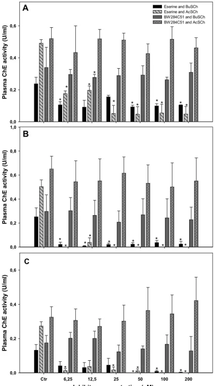
Fig. 2.2. Effects of the inhibitors eserine and BW284C51 on the plasma ChE activity of: (A) M. bassanus, (B) C. ciconia and (C) A. cinerea using AcSCh or BuSCh as substrates. Results are expressed as the mean ± SE of three birds; *significantly
different from control (P<0.05). 27
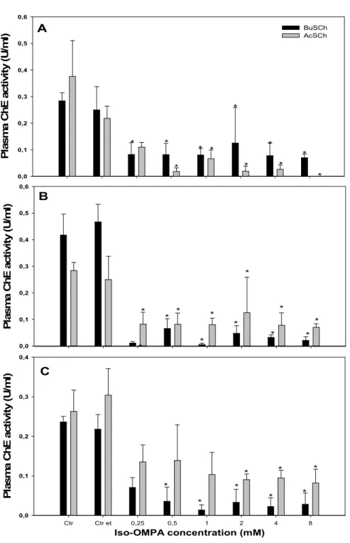
Fig. 2.3. Effects of the inhibitor iso-OMPA on the plasma ChE activity of: (A) M. bassanus, (B) C. ciconia and (C) A. cinerea using AcSCh or BuSCh as substrates. Results are expressed as the mean ± SE of three birds; *significantly different from
control (P<0.05). 30
Fig. 3.1. Chemical reactivation of a phosphorilated ChE with 2-PAM. (A) OP inhibited enzyme, (B)- 2-PAM attaches itself at the site the OP and its negatively charged atom of oxygen binds to the positively charged phosphorus atom of the OP, (C) The bond between the phosphorus atom and the oxygen of the serine is broken, and the OP molecule is removed from the ChE, leaving the “regenerated” enzyme
ready to work normally again. 38
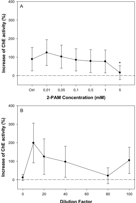
Fig. 3.2. Performance of chemical (A) and spontaneous (B) reactivation assays in brain ChE of Larus michahellis. Results are expressed as the mean ± SE of eleven birds, Ctrl= ultrapure water control, *= significantly different from control (Dunn’s
test, p<0.05). 42
Fig. 4.1. Erythrocyte nuclear abnormalities (NA) in Ciconia ciconia: A) normal nuclei; B-C) lobed; D-E) Kidney shaped; F-G) Segmented; H) notched; I)
Micronuclei. 55
Fig. 4.2. Variability of micronuclei (MN) frequency in Portuguese populations’ of Ciconiiformes and its deviation from the minimum (0) and maximum (2.14) normal values of MN (depicted by the dashed lines) reported in the literature for healthy
v
Fig. 4.3. Principal Component Analysis (PCA) of micronuclei (MN) and total nuclear abnormalities (TNA) in Ciconia ciconia with: (A) district and year, (B) survival and age, as explaining variables. N=52 individuals; Percentage of variability explained by each principal component is indicated in brackets for each axis; Dotted blue lines correspond to clusters identified graphically; Doted green lines within each cluster are only used to group points for identification porpoises; Chi= live chick, L-Juv=live juvenile, L-Ad=live adult, D-Ch=deceased chick, D-Juv=deceased juvenile,
D-Ad= deceased adult. 59
Fig. 4.4. Temporal variation of the TNA frequency in Portuguese populations of Ciconiiformes. (A)- Ciconia ciconia and Ardea cinerea variation in continental territory; (B)- District variation of TNA frequency in Ciconia ciconia. Significant differences (Holm-Sidak, P<0.05): a= different from 2007, b= different from 2008, c= different from 2009, d= different from 2010; *= sample size equal to 1. 60 Fig. 4.5. Temporal variation of the MN frequency in Portuguese populations of Ciconiiformes. (A)- Ciconia ciconia and Ardea cinerea variation in continental territory; (B)- District variation of TNA frequency in C. ciconia. a= significantly different from 2008 (Dunn’s test, p<0.05); *= sample size equal to 1. 60 Fig. 4.6. Principal Component Analysis (PCA) of total nuclear abnormalities (TNA) and micronuclei (MN) in Ardea cinerea with: (A, B) district and year, (C, D) age and survival as explaining variables. N=13 individuals; Percentage of variability explained by each principal component is indicated in brackets for each axis; PCA analysis of TNA and MN frequencies are presented in separate because Ardea cinerea retained the highest variability; Dotted blue lines correspond to clusters identified graphically; L-Chi= live chick, L-Juv=live juvenile, L-Ad=live adult, D-Ch=deceased chick, D-Juv=deceased juvenile, D-Ad= deceased adult. 61 Fig. 4.7. Number of micronuclei (MN) and total nuclear abnormalities (TNA) variation over time per sampled individual of: (A) Ciconia ciconia, (B) Ardea cinerea and (C) Ardea purpurea. Dashed black lines correspond to the maximum normal level of MN for healthy individuals (2.14 MN/1000 erythrocytes) reported for Ciconiiformes and other avian species in the literature [6,11,12]. 62
vi
List of Tables
Page Table 1.1. Overview of the species studied, with summarized information of the
Order and Family characteristics. Adapted from [53-57]. 11
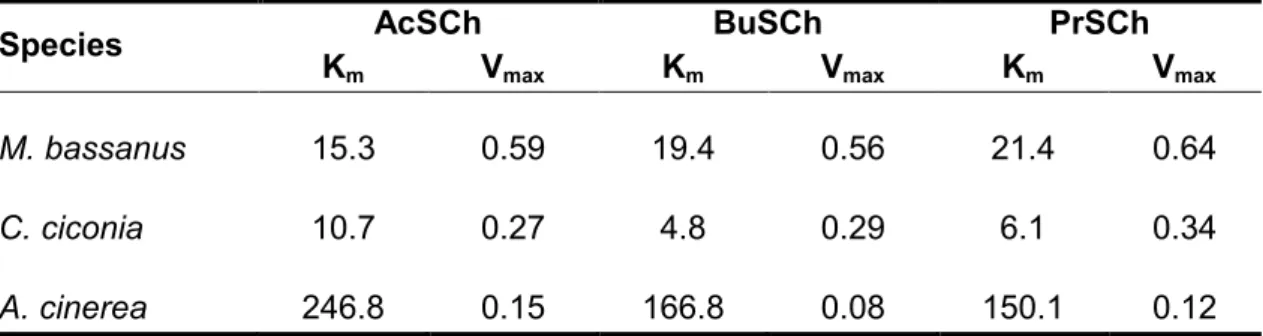
Table 2.1. Apparent values of Km(µM) and Vmax(µmol/min/min) as estimated by Michaelis-Menten equation for the substrates AcSCh, BuSCh and PrSCh. Values for this study are expressed as the mean value of 3 individuals. 26 Table 2.2. Normal range of ChE activity in non-exposed individuals of the Northern Gannet, the White stork and the Grey heron respectively, including sample size of individuals (n), minimum (min), maximum (max), mean and standard deviation (SD)
values. 28
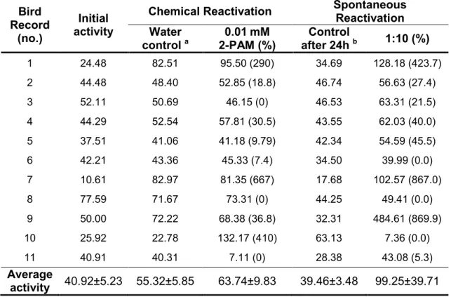
Table 3.1. Brain ChE activity of Larus michahellis before (initial activity) and after reactivation assays. Activities are expressed as U/mg of protein and values in brackets correspond to the percentage of reactivation. Averages of activities are presented by mean ± (SE). a= enzymatic activity obtained when incubating the samples with ultrapure water (1 hour, 25ºC), b=enzymatic activity obtained after
incubating the original (not diluted) samples for 24h, 4ºC. 43
Table 4.1. Total nuclear abnormalities (TNA) and micronuclei (MN) frequency per 1000 erythrocytes in Ciconiiformes. When n>2, values are presented by the mean and standard error is given in brackets; x and y are used to present depicted values
of TNA and MN frequencies when n=2 57
Table 4.2. Spearman correlation coefficients of different categories of nuclear abnormalities scored and the total nuclear abnormalities (TNA) for each studied species. Values highlighted correspond to statistical significant values (p<0.05). Sample size (n) of Ciconia ciconia, Ardea cinerea and Ardea purpurea was 57, 18 and 7, respectively; L= lobed, S= segmented, K= kidney-shaped, N= notched, MN=
Chapter 1
General Introduction, Objectives and Relevance of the Work
Chapter 1 – General Introduction, Objectives and Relevance of the Work
2
1. 1. General introduction
Since the time of Neanderthal Man, when fire was discovered resulting into the release to the environment of soot and polycyclic aromatic hydrocarbons from combustion processes, pollution has always been present in human civilizations [1]. Environmental pollution as we know today, though, only arisen after industrial revolution. The emergence of industries and consequent consumption of coal and other fossil fuels, allied to the tremendous demographic boost, leaded to an unprecedented increase on volume of both industrial and human wastes, disposed off through flagrant release into the environment [2]. It wasn’t however until the 1950s to 1970s that first concerns over environmental pollution began to draw public attention.
Prompted up by Rachel’s Carson “The Silent Spring”, published in 1962, the crescent evidences of the harmful effects of chemicals upon wildlife spurred the approval of legislation aimed at regulating and limiting the release of chemical wastes into the environment [2]. Impelled also by these same concerns, it was in the late 1960s that the modern environmental toxicology and its subfield of ecotoxicology arisen.
Contemporarily understood as being the science that relates the harmful effects of chemical agents in living beings, the term “ecotoxicology” was firstly introduced in 1969 by René Truhaut as the branch of toxicology that studied the toxic effects caused by natural or synthetic pollutants on the living systems [3]. A multidisciplinary science, ecotoxicology recognizes that the toxicity induced by contaminants is originated at the molecular level, by altering essential cellular mechanisms, which in turn results in a hierarchical cascade of changes from physiological processes to ecosystem function [4]. Being its main aim to understand and anticipate the distribution path of pollutants in the environment (as well as the ecological effect at them associated), ecotoxicology employs ecological parameters to access toxicity, providing a wide range of valuable tools in ecosystems monitoring and therefore playing a crucial role in the management, restoration and conservation of natural systems [3].
1.1.1. Wildlife toxicology: a field within ecotoxicology
Wildlife toxicology is the branch of ecotoxicology focused on the study of the effects of environmental contaminants (e.g. survival, health, reproduction) upon amphibians, reptiles, mammals and birds [5,6]. Unlikely other scientific fields whose knowledge is acquired through deliberate hypothesis testing and discovery, wildlife toxicology has been
Chapter 1 – General Introduction, Objectives and Relevance of the Work
3
driven by chemical use and misuse, catastrophic animal poisonings, ecological mishaps and the research conducted in the field of human toxicology [6].
Recognition of deleterious effects of contaminants upon wildlife can be traced back to the end of industrial revolution, when initial reports of lead poisoning from ingestion and retention of spent shot on pheasant Phasianus colchicus (England, 1876) and waterfowls (USA, 1894) appeared [6]. Lead toxicity was manifested by emaciated birds with paralyzed legs and feet, with feet being held similarly to the “drop-hand” condition observed in lead-poisoned humans. [7]. Increased awareness of environmental problems and endangered species protection was gained during the early twentieth century. With expanded production and use of petroleum right after World War I, thus leading to an increase of the oil transported across the ocean, first oil spills occurred resulting in the death of numerous marine birds [6,8]. As reports of harmful effects of contaminants’ upon birds and other wildlife populations continued to drawn public and scientific attention, prompting the development of a wide range of tools to assess exposure and effects of contaminants on impacted populations, wildlife toxicology made a standing.
The use of improved analytical, molecular and biochemical tools allied to the scientific advances made in ecology and other complementary fields has also greatly contributed to the development of wildlife toxicology. By 1950s, first bioaccumulation studies were developed, aiming to assess organochlorine pesticides burdens in wildlife [6]. On this same decade, first studies that aimed to relate lethality and pathology data of contaminants to tissue concentrations on wildlife were published. Earliest work, by Coburn et al. [9], tested phosphorus tissue concentrations indicative of exposure in both black and mallard duck (Anas rubripes and Anas platyrhynchos, respectively), through a combination of field investigations and dosing trials. Investigation of toxicity thresholds continued, and towards the latter half of 1970s first controlled exposure studies were initiated in game and captive wild bird species to determine sublethal responses (e.g. reproduction, behaviour, biochemical indicators of exposure, physiological and endocrine function) of pesticides, metals and crude oil [6].
As methodologies used in wildlife toxicology were successively improved, in field studies, it was demonstrated for the first time that pesticide use and agricultural practices could also affect indirectly farmland birds’ population by altering habitat vegetation and availability of preferred insects, thus reducing dramatically their food supplies [10]. Back in 1990s, diagnose of contaminant exposure and effects was further improved by the development of new molecular assays (e.g. markers of genetic damage and oxidative stress) and use of nondestructive sampling methodologies [6,11]. In more recent years,
Chapter 1 – General Introduction, Objectives and Relevance of the Work
4
other tools to assess contaminant exposure and effects upon wildlife have been developed recurring to techniques such as DNA fingerprinting, Polymerase chain reaction (PCR) and microarrays, allowing to acquire information more detailed on the effects of contaminants beyond individual and cellular levels [5]
It is a fact that the development of new and improved sophisticated tools to assess exposure, effects of contaminants upon wildlife populations has greatly contributed to the development of wildlife toxicology as a science, as well as in the protection of endangered wildlife species. In spite of this, however, the ability to predict and prevent ecological mishaps remains nowadays still very limited, which can be partly attributable to the logistical difficulties of studying wild animal populations and/or extrapolating data generated in laboratory [6]. The varying sensitivity of animals at the species and even individual level poses a problem, being difficult to assess contaminants’ concentration-effects on wildlife; moreover, the fact that multiple contaminants may coexist within a natural system enhances the probability of occurring combined effects, which may act as a confounding factor in the prediction and/estimation of contaminants’ effects on wildlife and their supporting habitat.
1.1.2. Perturbations in birds’ populations: contaminants as a cause
Similarly to other vertebrates, avian exposure to environmental contaminants is dependent on contaminants’ and habitats’ physiochemical properties, life history characteristics (e.g. age, gender, size), and behavioural traits that are intrinsic to each bird species, among others [12]. Exposure routes in birds may occur through four pathways: ingestion, inhalation, dermal contact and maternal transfer.
Ingestion is the principal pathway of contaminant exposure in birds and may occur through direct intake of free chemicals (e.g. accidental ingestion of liquid formulations following spills, or through intentional poisoning by humans) or more commonly, indirectly, through ingestion of contaminated food (e.g. ingestion of prey items that have previously accumulated contaminants in their tissues), or water [13]. Dermal exposure and maternal transfer are also important routes of exposure in avian species and may occur through contact of the contaminant with the eyes and the skin of the legs and feet, or as a result of the transfer of pollutants from gravid females into eggs, respectively [12,14,15]. In regard of inhalation exposure, and even thought this route has been sparsely documented in the
Chapter 1 – General Introduction, Objectives and Relevance of the Work
5
literature, exposure occurs mainly through contact with compounds applied or released aerially or of volatile nature [12].
Due to their ability to adapt and thrive in disturbed habitats and, on the other hand, their sensitivity to numerous environmental pollutants, contaminant exposure on birds has been far more documented than in any other group of terrestrial vertebrates [12]. The first evidences of contaminant-effects relations in bird populations had its origin in the 1950s and 1960s, when the effects of organochlorine insecticides such as DDT were first noticed [16].
DDT was first synthesized as a controlling agent of malaria and typhus among civilians and troops during the second half of World War II, but its use was generalized to agricultural and domestic use back in 1945, leading to a widespread decline of birds of prey [17,18]. Within one year of use, nesting failures (e.g. failure of adults to return to traditional nest, to lay eggs, or failure of eggs to hatch) on Florida’s subpopulation of bald eagle (Haliaeetus leucocephalus) were noticed by C. L. Broley [19]. A similar trend was reported by D. A. Ratcliffe in 1967, with avian population declines of peregrine falcon (Falco peregrinus), golden eagle owl (Aquila chrysaetos) and the european sparrow hawk (Accipiter nisus) being related to reproduction failures caused by an increase in eggshell thinning (which subsequently led to egg breakage and embryo death), starting immediately after DDT introduction in Britain [17,20]. It was only in 1970 that Ratcliffe was able to report a close relationship between the changes in eggshell thickness and the residual levels of DDT present in the eggs of 14 avian species [17]. These findings were in accordance with a previous study of Hickey and Anderson, in 1968, who also found a decrease of shell thickness in herring gull (Larus argentatus) associated with the increase of chlorinated hydrocarbon residues (such as DDE, a DDT metabolite) in eggs [17,21]. Further evidence of these initial reports were later substantiated and extended by similar works with other species and in other countries [17].
Another example of contaminants with clear effects upon bird populations is crude petroleum and its refined products. Adult birds can be affected by crude oil through a wide variety of routes, from ingestion to induced habitat degradation, but most frequently crude oil deleterious effects occur through external oiling. External oiling can lead to increased feather permeability, which may lead to hypothermia, drowning, ingestion through feather preening and habitat degradation [8]. Other effects reported in the literature in adult individuals include disease (e.g. gastrointestinal irritation, pneumonia), starvation, impairment of the function of the immune and nervous system and predation, which may occur as a consequence of direct ingestion of crude oil [8,22]. Birds’ embryos have also
Chapter 1 – General Introduction, Objectives and Relevance of the Work
6
been found to be highly sensitive to crude oil and its derivates. For example, quantities of about 1-2 µL of some derivates of oil are sufficient to cause embryo death [8]; other effects listed in the literature include reduced hatchability and modifications of the yolk structure of eggs, as well as reduced incubation attentiveness and chick survival [23,24].
1.1.3. Waterbirds as indicators of environmental quality
Over the twentieth century, the concern over environmental changes has sparked the development of ecological indicators (e.g. concentration of pollutants in water, altered nutrient dynamics, taxa richness) to assess environmental health [25,26]. Within this context, bioindicators are measurable responses on living organisms (e.g. biological processes, species and communities composition) whose condition can be monitored to evaluate environment or ecosystem’s integrity.
Birds, including waterbirds, have been used as bioindicators of environmental quality in a broad range of ecosystems [27-29]. Amongst other attributes, birds are abundant, conspicuous and functionally important components of ecosystems [30]. In addition, they are highly mobile and sensitive to both direct and indirect environmental changes (e.g. contamination, perturbation, duration of seasonal frequency); as top predators in both aquatic and terrestrial food webs, they are good indicators for bioaccumulative chemicals and toxins, as well as for the detection of diseases, being also key species for education and public awareness [27,30]. Finally, they’re relatively easy and inexpensive to quantify and their basic ecology and habitats preferences are commonly well established [30].
Traditionally, risk assessment studies with natural bird populations have been focused either in the study of the relation between organisms’ lethality and pathology data from contaminants to tissue concentrations on wildlife, or in the assessment of biomarker responses in individuals. In a broad sense, biomarkers can be understood as detectable responses of varying nature (e.g. molecular, cellular or physiological) that can be used to detect exposure, susceptibility and effect of chemical contaminants on living organisms. They can be classified as [31]:
• Biomarker of exposure: measures the interaction between a contaminant substance and a target molecule or cell that is measured in a compartment within an organism. Some of this type of biomarkers allows to measure the internal dose of the toxic compound or derived metabolites found for example in body fluids, organs or biological systems [32].
Chapter 1 – General Introduction, Objectives and Relevance of the Work
7
Numerous studies have addressed this kind of marker; some examples include the measurement of metals (e.g. mercury, arsenic, lead, selenium, etc.) or organic contaminants (e.g. polychlorinated byphenyls, organochlorine pesticides, etc.) burdens in feathers, internal tissues (e.g. liver), eggs and/or blood in the herring gull (Larus argentatus) [15], black-winged stilt (Himantopus himantopus) [33] and mourning doves (Zenaida macroura) [34].
• Biomarker of effect: evaluates biochemical, physiological, behavioural, or other alterations observed within an organism that, depending upon their magnitude, can be associated to a health impairment or disease. This kind of markers screens for changes (e.g. modifications on blood composition, changes in specific enzymatic activities) within an organism that can be translated as indicative of xenobiotic exposure [32].
An example of this type of biomarker is cholinesterase (ChE) activity. Cholinesterases are a family of esterase enzymes constituted by two forms: acetylcholinesterase (AChE; EC 3.1.1.7), which is mainly found at neuromuscular junctions and central nervous system, and pseudocholinesterase (PChE; EC 3.1.1.8) also denominated by butyrylcholinesterase (BChE), mainly found on liver and serum [35]. Inhibition of ChEs activity has been used as a primary indicator of exposure to organophosphate (OP) and carbamate pesticides (CB) in several bird species such as mallard duck (Anas platyrhynchos) [36], great blue heron (Ardea herodias) [37], glossy ibis (Plegadis falcinellus) [14] and great egret (Nycticorax nycticorax) [14,38]. Although organophosphorous and carbamate compounds can be less hazardous to the environment than organochlorine pesticides, as they have a short half-life thus do not tend to persist in animal tissues, some of these chemicals are extremely toxic for short periods of time right after their application [39]. OP and CB compounds exert their toxicity through inhibition of AChE, which leads to an accumulation of the neurotransmitter acetylthiocholine at synapses and neuroeffector junctions, leading to a cascade of effects from disruption of processes in the central nervous systems to death by paralysis [39,40]. Inhibition of PChE/BChE has not been demonstrated to result in specific adverse effects, but it has been considered as an important pathway for detoxification of
Chapter 1 – General Introduction, Objectives and Relevance of the Work
8
anticholinesterase compounds and has been often used as a surrogate measure of AChE inhibition [40].
Another example of a biomarker of effect is the micronucleus test. Micronuclei formation is a type of DNA alteration, which may result from chromosomal breakage (clastogenicity) or spindle anomalies [41]. The micronucleus test has been used to assess genotoxic effects on several bird species, namely in the herring gull (Larus argentatus) [42] and in the purple heron (Ardea purpurea), the little white egret (Egretta garzetta) and the cattle egret (Bulbucus ibis) [43].
• Biomarker of susceptibility: indicates an inherited or acquired ability of an organism to respond to the exposure of a specific toxic substance. Biomarkers of susceptibility may include alterations of genetic content, such as polymorphisms of activating systems and polymorphisms of detoxicating systems, and that may modify sensitivity of individuals to xenobiotics present in the environment [32].
An example of a biomarker of this type is paraoxonase (PON1). PON1 is an A-esterase present in serum and liver capable of hydrolyzing organophosphorous pesticides such as dioazinon and parathion, thus protecting against OP-poisoning [44]. PON1 activity varies greatly amongst animal species, and increased susceptibility to OP-poisoning is often related to decreased levels of this enzyme [44]. Birds, that have very low levels of this enzyme, thus being less effective in the detoxification of these compounds, are therefore particularly susceptible to OP-poisoning [44,45].
The use of biomarkers use on environmental risk assessment has been increasing progressively over the past years because they are believed to be among the most sensitive and earliest detectable responses in organisms [46]. Other examples of biomarkers that have been used to study wild populations of birds are avian eggshell thinning and induction of cytochrome P450. Eggshell thinning, which is caused by chlorinated hydrocarbons such as DDT, is a marker of reproductive impairment [32,39] and has been applied to numerous species such as the African darter (Anhinga rufa) [47] and the cattle egret (Bulbucus ibis) [48]. Cytochrome P450 is a family of enzymes involved in the metabolism of xenobiotic and endogenous substances and acts by
Chapter 1 – General Introduction, Objectives and Relevance of the Work
9
affecting their chemical structures [49]. Induction of cytochrome P450 has been used to diagnose organism’s exposure to chemicals or substrates [49], and has been applied to several avian species including the black-crowned night-heron (Nycticorax nycticorax) [50], common cormorant (Phalacrocorax carbo) [51] and black-footed albatrosses (Phoebastria nigripes) [52].
Chapter 1 – General Introduction, Objectives and Relevance of the Work
10
1.2. Objectives and species studied
The main goals of this study were to answer the following questions:
1. Are some Portuguese populations of birds exposed to environmental contamination?
2. Could these factors lead to birds’ illness and influence their recovery in wildlife rehabilitation centres?
3. Can ecotoxicological tools help to monitor and aid birds’ recovery?
In order to address these issues, we measured selected enzymatic activities and assessed genotoxic exposure in aquatic wild birds from the orders Ciconiiformes, Charadriiformes and Pelecaniformes (Table 1).
All samples used were taken from a Portuguese wildlife rehabilitation centre (CERVAS- Centro de Ecologia, Recuperação e Vigilância de Animais Selvagens) and a nature reserve (PBG – Parque Biológico de Gaia). Due to the fact that all the samples obtained were dependent on the availability of animals and theirs residence in the rehabilitation centres, not all species were addressed in the three studies conducted. Detailed information on the species used for each study are given on Table 1 and on each following chapter.
Regarding the nature of the samples used, all samples taken from deceased animals (e.g. liver and brain samples) and live animals (e.g. blood) were generously provided by CERVAS and PBG. All procedures involving live bird handling were conducted accordingly to the Guide for the Care and Use of Laboratory Animals of the European Union, which is represented in Portugal by Decreto de Lei nº. 129/92 de 06 de Julho, Portaria nº. 1005/92 de 23 de Outubro de 1992.
Chapter 1 – General Introduction, Objectives and Relevance of the Work
11
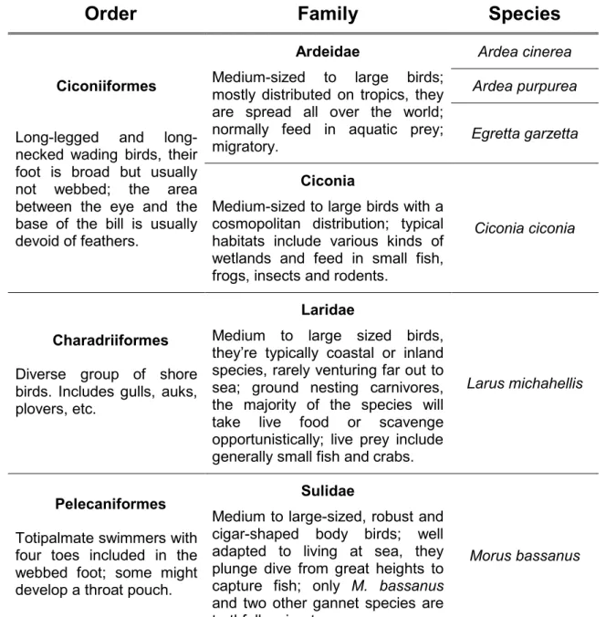
Table 1.1. Overview of the species studied, with summarized information of the Order and Family characteristics. Adapted from [53-57].
Order
Family
Species
Ciconiiformes
Long-legged and long-necked wading birds, their foot is broad but usually not webbed; the area between the eye and the base of the bill is usually devoid of feathers.
Ardeidae
Medium-sized to large birds; mostly distributed on tropics, they are spread all over the world; normally feed in aquatic prey; migratory.
Ardea cinerea
Ardea purpurea
Egretta garzetta
Ciconia
Medium-sized to large birds with a cosmopolitan distribution; typical habitats include various kinds of wetlands and feed in small fish, frogs, insects and rodents.
Ciconia ciconia
Charadriiformes Diverse group of shore birds. Includes gulls, auks, plovers, etc.
Laridae
Medium to large sized birds, they’re typically coastal or inland species, rarely venturing far out to sea; ground nesting carnivores, the majority of the species will take live food or scavenge opportunistically; live prey include generally small fish and crabs.
Larus michahellis
Pelecaniformes Totipalmate swimmers with four toes included in the webbed foot; some might develop a throat pouch.
Sulidae
Medium to large-sized, robust and cigar-shaped body birds; well adapted to living at sea, they plunge dive from great heights to capture fish; only M. bassanus and two other gannet species are truthfully migratory.
Chapter 1 – General Introduction, Objectives and Relevance of the Work
12
1.3. Relevance of the study
Aquatic ecosystems such as wetlands and coastal lagoons are highly productive environments that provide a wide range of ecosystem services. Nonetheless, due their limited ability to recover from disturbance, these habitats are particularly vulnerable to pollution which leads to environmental degradation with consequent inability to perform ecological services and ecosystem impairment [58].
Biomonitorization studies, such as the ones that aim to relate physicochemical analysis with the effects observed in the biological systems, play an important role in the assessment of contaminants’ effects upon ecosystems [59]. A significant part of Portuguese biomonitoring studies has focused on organisms at lower trophic levels such as larvae, crustacean and mollusks, but failed to address contaminants’ effects upon organisms at higher trophic levels such as mammals or birds.
Birds, including birds accepted in wildlife recovery units, reunite a multiple array of characteristics that makes them ideal indicators in environmental risk assessment studies. Functionally relevant components of ecosystems and highly sensitive towards environmental contaminants [27,30], studying birds allows an easy and effective way to evaluate effects of bioaccumulative contaminants and toxins at higher trophic levels.
Using birds accepted for rehabilitation at wildlife recovery centres does not only enables to screen for temporal and spatial trends of contamination in Portuguese populations of wild birds, but it also enables to assess whether background contamination could be related with their illness and at what extent it could impair bird recuperation. Moreover, using ecotoxicological tools to monitor birds during rehabilitation could be a valuable approach to help in birds’ recovering. Thus, in a world with an increasing concern over environmental contamination and its role upon the actual decline of biodiversity, this type of approaches may play a crucial role in the conservation, rehabilitation and management of threatened bird species.
Chapter 1 – General Introduction, Objectives and Relevance of the Work
13
1.4. Organization of the thesis
The present thesis is organized into five chapters. The first one is the present “Introduction”, the second, third and fourth chapters are structured as scientific papers and describe and discuss the results obtained. By order and contents, these are:
Chapter 1 - General Introduction, Objectives and Relevance of the work
Introductory chapter, describing wildlife toxicology and how this field emerged; the role of birds as bioindicator species and historical case studies of contaminant effects upon bird populations; lastly the main aims and relevance of the study are depicted.
Chapter 2 - Characterization of Cholinesterases in Plasma of Three Portuguese Native Bird Species: Application to Biomonitoring
Chapter describing plasma characterization of cholinesterases of three native species: the white stork (Ciconia ciconia), the grey heron (Ardea cinerea) and the northern gannet (Morus bassanus). In this chapter pseudocholinesterase is referred as butyrylcholinesterase.
Chapter 3 - Brain Cholinesterase Reactivation as a Marker of Pesticide Exposure in the Yellow-legged Gull Larus michahellis (Naumann, 1840): A Case Study.
Chapter describing the application of chemical and spontaneous reactivation assays in brain cholinesterase of the yellow-legged gull to investigate a possible case of pesticide poisoning.
Chapter 4 - Ciconiiformes as Sentinels of Chemicals’ Genotoxic Effects: A 5-year Study on Portuguese Birds.
Chapter describing the evaluation of genotoxic damage in Ciconiiformes using the micronucleus test and the erythrocyte nuclear abnormalities (ENA) Assay.
Chapter 5: General Discussion and Conclusions
Chapter 1 – General Introduction, Objectives and Relevance of the Work
14
1.5. References
1. Wijbenga A, Hutzinger O (1984) Chemicals, man and the environment. Naturwissenschaften 71: 239-246.
2. Leblanc GA (2004) Basics of Environmental Toxicology. In: Hodgson E, editor. A Textbook of Modern Toxicology. New Jersey: John Wiley & Sons, Inc. pp. 463-467.
3. Hoffman D, Rattner B, Burton GA, Cairns J (2003) Handbook of Ecotoxicology; Hoffman D, Rattner B, Burton GA, Cairns J, editors. Boca Raton (FL): Lewis Publishers.
4. Fox GA (2000) Perturbations in terrestrial vertebrate populations: Contaminants as a cause. Environmental Contaminants and Terrestrial Vertebrates: Effects on Populations, Communities, and Ecosystems: 19-59.
5. Kendall RJ (2010) Introduction and Overview. In: Kendall RJ, Lacher TE, Cobb GP, Cox SB, editors. Toxicology: Emerging Contaminantsand Biodiversity Issues. Boca Raton: CRC Press.
6. Rattner BA (2009) History of wildlife toxicology. Ecotoxicology 18: 773-783.
7. Pattee OH, Pain DJ (2003) Lead in the Environment. In: Hoffman DJ, Rattner BA, Burton GA, Cairns J, editors. Handbook of Ecotoxicology. Second ed. Boca Raton (FL): Lewis Publishers. pp. 373-408.
8. Alberts PH (2003) Petroleum and Individual Polycyclic Aromatic Hydrocarbons. In: Hoffman DJ, Rattner BA, Burton GA, Cairns J, editors. Handbook of Ecotoxicology. Second ed. Boca Raton (FL): Lewis Publishers. pp. 341-371.
9. Coburn DR, Dewitt JB, Derby JV, Ediger E (1950) Phosphorus Poisoning in Waterfowl. Journal of the American Pharmaceutical Association-Scientific Edition 39: 151-158.
10. Sotherton N, Holland J (2003) Indirect effects of pesticides on farmland wildlife. In: Hoffman DJ, Rattner BA, Burton GA, Cairns J, editors. Handbook of Ecotoxicology. Second ed. Boca Raton (FL): Lewis Publishers. pp. 373-408.
11. Fossi MC, Massi A, Leonzio Ceg (1994) Blood esterase inhibition in birds as an index of organophosphorus contamination: field and laboratory studies. Ecotoxicology: 11-20.
12. Smith PN, Cobb GP, Godard-Codding C, Hoff D, McMurry ST, et al. (2007) Contaminant exposure in terrestrial vertebrates. Environmental Pollution 150: 41-64.
13. Sheffield SR, Sullivan JP, Hill EF (2012) Identifying and Handling Contaminant-Related Wildlife Mortality or Morbidiy. Volume 1: Research. In: Silvy NJ, editor. The Wildlife Thecniques Manual Research 7th ed. Baltimore: Johns Hopkins University Press. pp. 154-180.
Chapter 1 – General Introduction, Objectives and Relevance of the Work
15
14. Parsons KC, Matz AC, Hooper MJ, Pokras MAeg (2000) Monitoring Wading Bird Exposure to Agricultural Chemicals Using Serum Cholinesterase Activity. Environmental Toxicology and Chemistry 19.
15. Koster MD, Ryckman DP, Weseloh DVC, Struger J (1996) Mercury levels in great lakes herring gull (Larus argentatus) eggs, 1972-1992. Environmental Pollution 93: 261-270.
16. Poppenga RH (2007) Avian toxicology. In: C. GR, editor. Veterinary Toxicology: Basic and Clinical Principles. USA: Academic Press. pp. 663-688.
17. Lundholm CE (1997) DDE-induced eggshell thinning in birds: Effects of p,p'-DDE on the calcium and prostaglandin metabolism of the eggshell gland. Comparative Biochemistry and Physiology C-Pharmacology Toxicology & Endocrinology 118: 113-128.
18. Henny CJ, Grove RA, Kaiser JL, Johnson BL (2010) North American Osprey Populations and Contaminants: Historic and Contemporary Perspectives. Journal of Toxicology and Environmental Health-Part B-Critical Reviews 13: 579-603. 19. Grasman KA, Scanlon PF, Fox GA (1998) Reproductive and physiological effects of
environmental contaminants in fish-eating birds of the Great Lakes: A review of historical trends. Environmental Monitoring and Assessment 53: 117-145.
20. Ratcliff.Da (1967) Decrease in Eggshell Weight in Certain Birds of Prey. Nature 215: 208-&.
21. Hickey JJ, Anderson DW (1968) Chlorinated Hydrocarbons and Eggshell Changes in Raptorial and Fish-Eating Birds. Science 162: 271-&.
22. Soler F, Oropesa AL, Perez-Lopez M, Hernandez D, Garcia JP, et al. (2007) Acetylcholinesterase activity in seabirds affected by the Prestige oil spill on the Galician coast (NW Spain). Science of The Total Environment 372: 532-538. 23. Grau CR, Roudybush T, Dobbs J, Wathen J (1977) Altered Yolk Structure and
Reduced Hatchability of Eggs from Birds Fed Single Doses of Petroleum Oils. Science 195: 779-781.
24. Fry DM, Swenson J, Addiego LA, Grau CR, Kang A (1986) Reduced Reproduction of Wedge-Tailed Shearwaters Exposed to Weathered Santa-Barbara Crude-Oil. Archives of Environmental Contamination and Toxicology 15: 453-463.
25. Karr JR (2008) Ecological Health Indicators. In: Jorgensen SE, Fath BD, editors. Encyclopedia of Ecology. Amsterdam: Elsevier. pp. 1037-1041.
26. Holt EA, Miller SW (2011) Bioindicators: Using Organisms to Measure Environmental Impacts. Nature Education Knowledge 2: 8.
27. Mistry J, Berardi A, Simpson M (2008) Birds as indicators of wetland status and change in the North Rupununi, Guyana. Biodiversity and Conservation 17: 2383-2409.
Chapter 1 – General Introduction, Objectives and Relevance of the Work
16
28. Goldstein MI, Lacher TE, Zaccagnini ME, Parker ML, Hooper MJ (1999) Monitoring and Assessment of Swainson’s Hawks in Argentina Following Restrictions on Monocrotophos Use, 1996–97. Ecotoxicology 8: 215-224.
29. Monteiro LR, Furness RW (1995) Seabirds as monitors of mercury in the marine environment. Water, Air, & Soil Pollution: 851-870.
30. Stolen ED, Breininger DR, Frederick PC (2005) Using waterbirds as indicators in estuarine systems: successes and perils. Estuarine Indicators: 409-422.
31. WHO/IPCS (1993) Biomarkers and Risk Assessment: concepts and principles. Environmental Health Criteria 155. Geneva: World Health Organization.
32. Gil F, Pla A (2001) Biomarkers as biological indicators of xenobiotic exposure. Journal of Applied Toxicology 21: 245-255.
33. Tavares PC, Monteiro LR, Lopes RJ, Correia Santos MM, Furness RW (2004) Intraspecific Variation of Mercury Contamination in Chicks of Black-Winged Stilt (Himantopus himantopus) in Coastal Wetlands from Southwestern Europe. Bulletin of Environmental Contamination and Toxicology 72: 437-444.
34. García-Hernández J, Sapozhnikova YV, Schlenk D, Mason AZ, Hinojosa-Huerta O, et al. (2006) Concentration of contaminants in breeding bird eggs from the Colorado River delta, Mexico. Environmental Toxicology and Chemistry 25: 1640-1647. 35. Radic Z, Taylor P (2006) Structure and Function of Cholinesterases. In: Gupta RC,
editor. Toxicology of Organophosphate and Carbamate Pesticides. Waltham: Elsevier Academic Press. pp. 161-186.
36. Fleming WJ (1981) Recovery of Brain and Plasma Cholinesterase Activities in Ducklings Exposed to Organo-Phosphorus Pesticides. Archives of Environmental Contamination and Toxicology 10: 215-229.
37. Hunt KA, Hooper MJ, Littrell EE (1995) Carbofuran Poisoning in Herons - Diagnosis Using Cholinesterase Reactivation Techniques. Journal of Wildlife Diseases 31: 186-192.
38. Goldstein MI, Lacher TE, Woodbridge B, Bechard MJ, Canavelli SB, et al. (1999) Monocrotophos-induced mass mortality of Swainson's hawks in Argentina, 1995-96. Ecotoxicology 8: 201-214.
39. Mayer FL, Versteeg DJ, McKee MJ, Folmar LC, Graney RL, et al. (1992) Physiological and nonspecific biomarkers. In: Huggett RJ, Kimerie RA, Mehrle PMJ, Bergman HL, editors. Biomarkers: Biochemical, Physiological, and Histological Markers of Anthropohenic Stress. Boca Raton: Lewis Publishers. pp. 5-85.
40. Sultatos LG (2006) Interactions of organophosphorus and carbamate compounds with cholinesterases. In: Gupta RC, editor. Toxicology of Organophosphate and Carbamate Pesticides. Waltham: Elsevier Academic Press. pp. 209-218.
41. Shugart L, Bickham J, Jackim G, McMahon G, Ridley W, et al. (1992) DNA alterations. In: Huggett RJ, Kimerie RA, Mehrle PMJ, Bergman HL, editors. Biomarkers:
Chapter 1 – General Introduction, Objectives and Relevance of the Work
17
Biochemical, Physiological, and Histological Markers of Anthropohenic Stress. Boca Raton: Lewis Publishers. pp. 125-153.
42. Skarphedinsdottir H, Hallgrimsson GT, Hansson T, Hagerroth PA, Liewenborg B, et al. (2010) Genotoxicity in herring gulls (Larus argentatus) in Sweden and Iceland. Mutation Research-Genetic Toxicology and Environmental Mutagenesis 702: 24-31.
43. Quiros L, Ruiz X, Sanpera C, Jover L, Pina B (2008) Analysis of micronucleated erythrocytes in heron nestlings from reference and impacted sites in the Ebro basin (NE Spain). Environmental Pollution 155: 81-87.
44. Costa LG, Richter RJ, Li WF, Cole T, Guizzetti M, et al. (2003) Paraoxonase (PON1) as a biomarker of susceptibility for organophosphate toxicity. Biomarkers 8: 1-12. 45. Li WF, Costa LG, Furlong CE (1993) Serum Paraoxonase Status - a Major Factor in
Determining Resistance to Organophosphates. Journal of Toxicology and Environmental Health 40: 337-346.
46. Jemec A, Drobne D, Tisler T, Trebse P, Ros M, et al. (2007) The applicability of acetylcholinesterase and glutathione S-transferase in Daphnia magna toxicity test. Comparative Biochemistry and Physiology C-Toxicology & Pharmacology 144: 303-309.
47. Bouwman H, Polder A, Venter B, Skaare JU (2008) Organochlorine contaminants in cormorant, darter, egret, and ibis eggs from South Africa. Chemosphere 71: 227-241.
48. Malik RN, Rauf S, Mohammad A, Eqani S, Ahad K (2011) Organochlorine residual concentrations in cattle egret from the Punjab Province, Pakistan. Environmental Monitoring and Assessment 173: 325-341.
49. Stegeman JJ, Brouwer M, Di Giulio RT, Förlin L, Fowler BA, et al. (1992) Molecular responses to environmental contamination. In: Huggett RJ, Kimerie RA, Mehrle PMJ, Bergman HL, editors. Biomarkers: Biochemical, Physiological, and Histological Markers of Anthropogenic Stress. Boca Raton: Lewis Publishers. pp. 125-153.
50. Rattner BA, Melancon MJ, Rice CP, Riley W, Eisemann J, et al. (1997) Cytochrome P450 and organochlorine contaminants in black-crowned night-herons from the Chesapeake Bay region, USA. Environmental Toxicology and Chemistry 16: 2315-2322.
51. Kubota A, Iwata H, Tanabe S, Yoneda K, Tobata S (2005) Hepatic CYP1A Induction by Dioxin-like Compounds, and Congener-Specific Metabolism and Sequestration in Wild Common Cormorants from Lake Biwa, Japan. Environmental Science & Technology 39: 3611-3619.
52. Kubota A, Watanabe M, Kunisue T, Kim EY, Tanabe S, et al. (2010) Hepatic CYP1A Induction by Chlorinated Dioxins and Related Compounds in the Endangered Black-Footed Albatross from the North Pacific. Environmental Science & Technology 44: 3559-3565.
Chapter 1 – General Introduction, Objectives and Relevance of the Work
18
53. Hoyo J, Elliot A, Sargatal J, others a (1992) Handbook of the Birds of the World. Volume 1- Ostrich to Ducks. Barcelona: Lynx Edicions.
54. Cramp S, Simmons KEL, Brooks DJ, Collar NJ, Dunn E, et al. (1978) Handbook of the Birds of Europe, the Middle East and North Africa: The Birds of the Western Palaearctic. ; Cramp S, editor. Oxford: Oxford University Press
55. Cramp S, Simmons KEL, Brooks DJ, Collar NJ, Dunn E, et al. (1983) Handbook of the Birds of Europe, the Middle East and North Africa: The Birds of the Western Palaearctic. ; Cramp S, editor. Oxford: Oxford University Press
56. Cramp S, Simmons KEL, Brooks DJ, Collar NJ, Dunn E, et al. (1985) Handbook of the Birds of Europe, the Middle East and North Africa: The Birds of the Western Palaearctic. ; Cramp S, editor. Oxford: Oxford University Press
57. Heinzel H, Fitter R, Parslow J (1998) The birds of Britain and Europe with North Africa and the Middle East London: Harper Collins.
58. Machado DA, Imberger J (2012) Managing wastewater effluent to enhance aquatic receiving ecosystem productivity: A coastal lagoon in Western Australia. Journal of Environmental Management 99: 52-60.
59. Monteiro MSSCA (2003) Avaliação da toxicidade de contaminantes ambientais em populações naturais de Pomatoschistus microps (Krøyer, 1938). Aveiro: University
Chapter 2
Characterization of Cholinesterases in Plasma of Three Portuguese
Native Bird Species: Application to Biomonitoring
Published in PLoS ONE (
doi:10.1371/journal.pone.0033975)
Santos CSA, Monteiro MS, Soares AMVM, Loureiro S (2012). Characterization of Cholinesterases in Plasma of Three Portuguese Native Bird Species: Application to Biomonitoring. PLoS ONE 7(3): e33975.
Chapter 2 – Characterization of Cholinesterases in Plasma of Three Portuguese Native Bird Species: Application to Biomonitoring
20
2. Characterization of Cholinesterases in Plasma of Three
Portuguese Native Bird Species: Application to Biomonitoring
2.1 Abstract
Over the last decades the inhibition of plasma cholinesterase (ChE) activity has been widely used as a biomarker to diagnose organophosphate and carbamate exposure. Plasma ChE activity is a useful and non-invasive method to monitor bird exposure to anticholinesterase compounds; nonetheless several studies had shown that the ChE form(s) present in avian plasma may vary greatly among species. In order to support further biomonitoring studies and provide reference data for wildlife risk-assessment, plasma cholinesterase of the northern gannet (Morus bassanus), the white stork (Ciconia ciconia) and the grey heron (Ardea cinerea) were characterized using three substrates (acetylthiocholine iodide, propionylthiocholine iodide, and S-butyrylthiocholine iodide) and three ChE inhibitors (eserine sulphate, BW284C51, and iso-OMPA). Additionally, the range of ChE activity that may be considered as basal levels for non-exposed individuals was determined. The results suggest that in the plasma of the three species studied the main cholinesterase form present is butyrylcholinesterase (BChE). Plasma BChE activity in non-exposed individuals was 0.48±0.11 SD U/ml, 0.39±0.12 SD U/ml, 0.15±0.04 SD U/ml in the northern gannet, white stork and grey heron, respectively. These results are crucial for the further use of plasma BChE activity in these bird species as a contamination bioindicator of anti-cholinesterase agents in both wetland and marine environments. Our findings also underscore the importance of plasma ChE characterization before its use as a biomarker in biomonitoring studies with birds.
Keywords: Biomarkers, butyrylcholinesterase, Ardea cinerea, Ciconia ciconia, Morus bassanus.
Chapter 2 – Characterization of Cholinesterases in Plasma of Three Portuguese Native Bird Species: Application to Biomonitoring
21
2.2 Introduction
Cholinesterase (ChE) activity has been routinely used as a biomarker to diagnose exposure to anticholinesterase compounds such as organophosphate (OP) and carbamate (CB) pesticides. These pesticides are broadly used to control insect pests and disease vectors; nonetheless, they can be extremely toxic to non-target organisms like mammals and birds [1,2,3,4]. They act by inhibiting the activity of cholinesterases, which causes an over accumulation of acetylcholine at the synapses and consequent disruption of nerve function, leading to subsequent physiologic disorders and ultimately death [5]. In addition to OPs and CBs, other environmental contaminants such as metals, detergents and petroleum-derived products have been found to generate similar inhibitory effects [6,7,8]. Serum or plasma has been broadly used to measure ChE activity as a non-invasive method to monitor exposure of wildlife to pesticides in the field due to its sensitivity to ChE-inhibiting compounds [9,10,11,12]. Nonetheless, its use requires the characterization of the enzyme form(s) present in the tissue assayed and the determination of the normal range of activity in non-exposed individuals [13].
Two enzymes form the family of cholinesterases: acetylcholinesterase (AChE; EC 3.1.1.7) and butyrylcholinesterase (BChE; EC 3.1.1.8). Both catalyze the hydrolysis of the neurotransmitter acetylcholine, but differ in substrate specificity and inhibitor susceptibility [14,15]; tissue distribution can also vary, depending on the organism measured. AChE is predominantly found in the neuromuscular junctions and central nervous system, playing a key-role in the cholinergic neurotransmission, while BChE is mainly found in serum and liver, but its primary physiological role remains unknown [14].
Regarding wildlife exposure to environmental contaminants, waterbirds, such as wading birds and seabirds, are useful indicators of environmental variation upon short and long temporal scales [16]. Wading birds are primarily indicators of wetland quality as they can occupy a wide variety of foraging niches, including agricultural ponds, which makes them often non-target species to OP and CB exposure through ingestion and dermal contact [12]. In the case of seabirds, they are widely used to monitor the occurrence and ecological impacts of contaminants such as oil and mercury in the marine environment [17]. All these possible exposures may lead into an impairment of ChE activities in birds, and therefore biomarkers like this may be also a useful indicator to detect contamination in birds’ habitats. In birds, AChE is found in the brain while BChE is mainly present in plasma; nonetheless, several studies had shown that AChE and other esterases (e.g.
Chapter 2 – Characterization of Cholinesterases in Plasma of Three Portuguese Native Bird Species: Application to Biomonitoring
22
carboxylesterase- CbE) might also occur in avian plasma with wide interspecies differences [3,18].
In order to use plasma ChE as a biomarker of exposure in three Portuguese native bird species, the main aim of this study was to: (i) characterize the ChE form(s) present in birds’ plasma, (ii) determine the basal levels of ChE activity in non-exposed individuals and (iii) establish the appropriate assay conditions for the use of plasma ChE activity, using as bird species: the grey heron (Ardea cinerea) and the white stork (Ciconia ciconia), two wading bird species resident in Portugal, and the northern gannet (Morus bassanus), a migratory seabird common along the Portuguese coast during winter.
2.3 Materials and methods
2.3.1 Sample collection
All the species of birds used to characterize plasma ChE were adult individuals inhabiting the Gaia Biological Park, a nature reserve located in Avintes (Porto, Portugal). C. ciconia individuals were free-living in the park while individuals of A. cinerea and M. Bassanus were in captivity. The disturbance stress caused by the animal handling was minimized by limiting the visit length, avoiding any sampling during extreme weather conditions (e.g. heavy rain, low temperatures) and using a small mantle to cover the head. Blood was drawn from the brachial vein with sterile 1-ml syringes and 25-ga needles, and it was collected into a capillary tube with EDTA (Microvette® CB 300, Sarstedt). Following centrifugation, plasma was extracted and stored at -80ºC until analysis.
2.3.2 Sample preparation and ChE determinations
Plasma samples were diluted in phosphate buffer (0.1M, pH 7.2), and ChE activity was determined in quadruplicate according to the Ellman method [19] adapted to microplate [20] using a microplate reader (Thermo Scientific Multiskan® Spectrum). For all species, plasma dilutions for each individual were prepared using 2µl of plasma (2-µl micropipette, Gilson®) for a final assay volume of 1 ml. The enzymatic activity was expressed in units (U) per ml of plasma (1U is a µmol of substrate hydrolyzed per minute).