Correlation among scapular positioning, functional
postural analysis and upper extremities disability
degree in sonographers (DASH Brazil)*
Correlação entre posicionamento escapular, análise postural funcional e grau de incapacidade dos membros superiores (DASH Brasil) em médicos ultrassonografistas
Geraldo Fabiano de Souza Moraes1, Fernanda Moreira Gonçalves2, Júnia Darth Silva2, Natália Spindola Soares2
OBJECTIVE: To evaluate and measure the scapular positioning, and establishing its correlation with functional postural analysis and degree of upper extremities disability in sonographers. MATERIALS AND METHODS: Eighteen physicians divided into two groups – symptomatic individuals with pain for more than six months, and asymptomatic individuals – were interviewed and submitted to physical assessment with application of the Brazilian version of the DASH questionnaire. Functional measurements of scapular inclination angles and upper limbs positioning were obtained during the ultrasound scanning. The Mann-Whitney U test was utilized
for analyzing the disability questionnaires; the t-Student test, for upper limbs positioning; ANOVA, for scapular
inclination angles, and the Spearman’s test for correlation between the functional upper limbs positioning and disability degree. RESULTS: A significant difference between symptomatic and asymptomatic groups was observed in the first 30 items of the DASH questionnaire as well as in the four study-related questions. Measurements of scapular positioning presented significant differences between the groups in the frontal plane at 90° and 120°. No significant difference was found for the analysis of functional upper limbs positioning. CONCLUSION: Changes in the scapular positioning may result in upper limbs dysfunction, and the application of the DASH questionnaire can indicate the level of pain and disability of an individual.
Keywords: Biomechanics; Shoulder joint complex; Scapular positioning; Scapulohumeral rhythm; Sonographer. OBJETIVO: Avaliar e mensurar o posicionamento escapular e correlacioná-lo com o grau de incapacidade e avaliação funcional de ultrassonografistas. MATERIAIS E MÉTODOS: Dezoito médicos, divididos em grupos sintomático e assintomático em relação à presença de dor por mais de seis meses, responderam a uma entrevista e foram submetidos a avaliação física e aplicação do DASH Brasil. Foi realizada a medida do ân-gulo de inclinação escapular e avaliação do posicionamento dos membros superiores durante a ultrassono-grafia. Foram utilizados testes U Mann-Whitney para avaliação do questionário de incapacidade, t de Student
para posicionamento do membro superior, ANOVA para inclinação escapular e coeficiente de Spearman para correlação do posicionamento funcional de membros superiores e grau de incapacidade. RESULTADOS: Nas 30 primeiras questões do DASH Brasil houve diferença significativa entre os grupos sintomático e assinto-mático. No módulo opcional, relacionado ao trabalho, houve diferença significativa entre os grupos. As medidas do inclinômetro apresentaram diferenças significativas entre os grupos no plano frontal em 90° e 120°. Para o ângulo funcional dos membros superiores não houve diferença significativa entre os grupos. CON-CLUSÃO: Alterações no ângulo de inclinação escapular podem levar à disfunção dos membros superiores e a aplicação do DASH Brasil pode indicar o grau de dor e incapacidade do indivíduo.
Unitermos: Biomecânica; Complexo articular do ombro; Posicionamento escapular; Ritmo escapuloumeral; Médico ultrassonografista.
Abstract
Resumo
* Study developed at Centro Universitário Newton Paiva, Belo Horizonte, MG, Brazil.
1. Master, Physiotherapist, Associate Professor and Coordi-nator for the Course of Physiotherapy, Centro Universitário New-ton Paiva, Belo Horizonte, MG, Brazil.
2. Physiotherapists, Bachelors in Physiotherapy, Centro Universitário Newton Paiva, Belo Horizonte, MG, Brazil.
Mailing address: Dr. Geraldo Fabiano de Souza Moraes. Avenida Silva Lobo, 1718, sala 11011, andar térreo, Nova Granada. Belo Horizonte, MG, Brazil, 30480-230. E-mail: [email protected]
Received June 25, 2007. Accepted after revision August 20, 2008.
20% of the general population(2). Multifac-torial situations cause overload of this joint complex, as follows: marked repetitiveness of movements; upper limb positioning, especially with shoulder abduction; mus-cular fatigue; work organization factors and annulling of mechanisms of work regula-tion, such as overload (reduction or lack of intervals) and psychosocial factors which generate tension and stress(3).
Moraes GFS, Gonçalves FM, Silva JD, Soares NS. Correlation among scapular positioning, functional postural analysis and upper extremities disability degree in sonographers (DASH Brazil). Radiol Bras. 2009;42(1):31–36.
INTRODUCTION
Currently, there is a great concern about preventing diseases and lesions involving the shoulder joint as a result from postural alterations and that lead to musculoskeletal system compensation to the detriment of the functionality(1).
One has observed the relevance of per-ception for the quality of life and health of a great part of the population which are fre-quently impaired by acute and chronic musculoskeletal disorders(4).
The association of scapular and humeral movements allows amplitude of the move-ment of flexion and abduction of the arm between 150° and 180°. The typical mobil-ity in asymptomatic individuals generally occurs with 2° of glenohumeral movement for 1° of scapular rotation, determining a 2:1 ratio for the scapulohumeral rhythm(5,6). Several athletes, particularly swim-mers(7), and practitioners whose activities involve abduction of the upper limbs present a higher predisposition to develop alterations in the mobility-stability relation-ship(8). For example, the professional activ-ity of the sonographer is characterized by the high probability of altered mobility-sta-bility relationship because of long-lasting sustained upper limb abduction(9,10).
Diagnosis by ultrasonography is a rela-tively new medical activity introduced early in the 1940’s(9). With the introduction of new technologies and the recognition of the ultrasound diagnostic capabilities, the number of US procedures and examination times have increased from year to year(9,10). Despite technological developments allow-ing the acquisition of high-resolution im-ages, ergonomic conditions observed dur-ing examinations are not so favorable for the musculoskeletal system of the sono-grapher(11).
Approximately 80% of these practitio-ners report discomfort or incapacity to per-form an appropriate examination because of the presence of symptoms in their upper limbs. One of the factors that apparently in-terferes in the origin of symptoms experi-mented by the sonographer is the high number US examinations which started being performed on a routine basis, besides risks resulting from electric shock, low il-lumination levels, stress and musculoskel-etal overload in upper limbs and vertebral spine(2,12).
Static and dynamic overloads imposed on the muscles of the scapular cingulum and on the upper limbs occur as a result from the necessity of sustaining the shoul-der abduction for a better function of the wrist and hand as the transducer is
posi-tioned perpendicularly to the skin of the patient over the examination(2,9,11,13).
Impairment of the functionality and time spent by the professional away from his/her daily activities leads to an overload of the health system in several countries. It is important that preventive measures be developed and for this purpose it is neces-sary to identify and understand the occupa-tional overload and its consequences in order to minimize the effects on the func-tional capacity of the individual(1). Al-though it is recognized that occupational diseases result from a set of three factors, namely, physical load, psychological load, and cognitive load, the present study was aimed primarily at evaluating and measur-ing the scapular positionmeasur-ing and correlat-ing it with the degree of upper limbs dis-ability and postural functional analysis of sonographers, focusing on their physical workload.
MATERIALS AND METHODS
Study design and sampling
Cross-sectional study approved by the Committee for Ethics in Research of Cen-tro Universitário Newton Paiva under No. 40/2005.
The sample included 18 sedentary vol-unteers in the age range between 27 and 52 years, nine of them included in the asymp-tomatic group and nine in the sympasymp-tomatic group. All the physicians performed ab-dominal ultrasonography. Time of profes-sion ranged from 18 months to 20 years and the amount of examinations/day ranged from 10 to 30. All the eighteen patients had both shoulders evaluated (36 shoulders). Among the individuals included in the symptomatic group, five were women (55.6%) and four were men (44.4%), with mean age of 33.67 ± 8.89 years (27 to 52 years), mean body mass of 69.66 ± 13.10 kg (57.0 to 95.0 kg) and mean height of 1.73 ± 0.07 m (1.63 to 1.83 m). For the sample as whole, the mean body mass in-dex was 22.97 ± 3.5 kg/m2 (19.14 to 31.04
kg/m2). Among the individuals included in
the asymptomatic group, four were women (44.4%) and five were men (55.6%) with mean age of 38.78 ± 7.79 years (30 to 49 years), mean body mass of 76.11 ± 22.7 kg (49.0 to 115.0 kg), and mean height of 1.69
± 0.07 m (1.54 to 1.85 m). For the sample as whole, the mean body mass index was 26.17 ± 6.5 kg/m2 (18.9 to 38.46 kg/m2).
Digital protractor
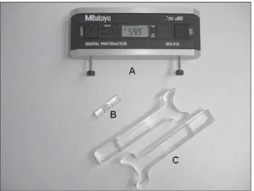
A Mitutoyo Digital Protractor (Mitu-toyo Evaluation Instruments; Chicago, USA) was utilized for measuring the scapu-lar positioning. Two extension arms made of acrylic, each of them measuring 10.0 cm in length, were adapted aiming at a correct accommodation of the device on the scapula and reading of the scapular incli-nation degrees. Additionally, a water level was coupled perpendicularly to the device to assure the correct alignment in relation to the planes of evaluation (Figure 1). The validity and reliability of the utilization of this digital protractor as a means for mea-suring the scapular positioning were de-scribed by Johnson et al.(6), who have uti-lized the same tool and measurements.
The Brazilian version of the Disability Arm Shoulder Hand questionnaire
The authors utilized the Brazilian ver-sion of the Disability Arm Shoulder Hand questionnaire transculturally adapted and translated into Brazilian Portuguese by Orfale et al.(14). This questionnaire was originally developed in English and called DASH Questionnaire by Hudak et al. in 1996, with the objective of measuring the physical disability and symptoms of upper limbs in a heterogeneous population. Ad-ditionally, it was proposed for evaluating disabilities and symptoms of a single or several conditions in upper limbs.
Evaluation of the functional angle of the upper limb
Aiming at measuring the functional angle of the upper limb, skin markers made of acrylic were attached with double-sided tape over the following anatomical refer-ences: 7th cervical vertebra (C7), 7th tho-racic vertebra (T7), acromion and lateral epicondyle. A digital photo camera Sony Mavica MVC-FD 200 (Sony Electronics, Inc.; San Diego, USA) was utilized for the functional positioning image recording.
sag-ittal planes. The most frequently position-ing adopted while performposition-ing US scans was adopted for evaluation.
Data collection
All the volunteers were informed about the objectives and procedures and signed a term of free and informed consent. Sub-sequently, a single observer who had been duly trained for this purpose performed individual interviews, physical evaluation and applied the Brazilian version of the DASH questionnaire with all the volun-teers included in the present study. The data were recorded for later analysis.
With the individual in the seated posi-tion, a second observer who had also been duly trained for this purpose, positioned the digital protractor on the scapular spine. The
extension arms were coupled to the medial border of the scapular spine and inferiorly to the acromion for a correct positioning and accommodation of the device to the anatomical reference (Figure 2). A third observer performed the reading and re-corded the values observed in the rest po-sition of the upper limb (anatomical posi-tion) at 30°, 60°, 90° and 120° of shoulder elevation in the frontal, scapular and sag-ittal planes. A support was utilized for nor-malizing the positioning of the upper limb during the examination in order to assure its correct positioning in these planes.
Then, skin markers were positioned on the anatomical references for photographic recording of the functional angle of the upper limb positioning during the US ex-amination. The volunteer was asked to
simulate his/her functional positioning adopted while performing an US scan. The upper limb positioning was recorded in-structing the volunteer to adopt his/her most frequent positioning while perform-ing US scans. The images were stored and the upper limbs positioning was evaluated with the aid of the software AutoCAD 2004 (Autodesk, Inc.; San Rafael, USA). Straight lines were traced to connect the points from C7 to T7 and from the acromion to the lat-eral epicondyle. The functional angle of the upper limb positioning for the examination was determined by the angle formed by this two straight lines (Figure 3).
Statistical analysis
Descriptive statistics, normality tests (Shapiro-Wilk) and variance equality tests
Figure 2. Positioning of the digital protractor for measurement of the scapular inclination angle.
Figure 1. Apparatus for measurement of the scapular inclination angle. A, digital protractor; B, water level; C, extension arms.
Figure 3. Functional angle of upper limb; asymptomatic (A) and symptomatic (B) positions.
(Levene) were applied for all of the vari-ables utilizing the SPSS (Statistical Pack-age for Social Sciences). The ANOVA analysis of variance was utilized for inves-tigating statistically significant differences between groups and the sides for all the variables, scapular inclination angles and functional evaluation of the upper limbs. The Student’s t-test was utilized to compare the values found by the Brazilian version of the DASH questionnaire. The Spear-man’s coefficient was utilized for deter-mining the correlation between the evalu-ation of upper limbs positioning and dis-ability degree. The significance level estab-lished was α= 0.05.
RESULTS
The mean profession time in the asymp-tomatic group corresponded to ten years, and four years and nine months. The aver-age number of US scans/day in the atomatic group was of 17.8, and in the symp-tomatic group, 22.7. No statistically signifi-cant difference was observed in time of profession (p = 0.092) and number of US scans/day (p = 0.186), between the symp-tomatic and asympsymp-tomatic groups.
Variables analyzed
Brazilian version of the DASH ques-tionnaire – Statistically significant differ-ence was observed for the first 30 questions (p = 0.001) and for the optional work mod-ule (p = 0.012), in the comparison between the symptomatic and the asymptomatic groups, as shown respectively on Figures 4 and 5. The mean score for the first 30 questions of the DASH questionnaire in the symptomatic group was 16.16 ± 13.18 points (2.5 to 45.5 points) and in the four study-related questions was 27.08 ± 20.96 (0 to 56.25 points). The mean score for the first 30 questions in the asymptomatic group was 1.11 ± 1.81 points (0 to 5 points) and 4.16 ± 8.83 points (0 to 25 points) in the study-related questions.
Scapular inclination angle – There was a statistically significant difference of 6.65° (p = 0.016) and 5.87° (p = 0.033) in the scapular inclination angle in the fron-tal plane at 90° and 120°, respectively, in the comparison between the right shoulder in the symptomatic group and the right
shoulder in the asymptomatic group. Also, a statistically significant difference of 4.43° (p = 0.028) was observed in the asymptom-atic group, in the comparison of the left shoulder in relation to the right shoulder in rest position (0), frontal plane.
Functional angle of the upper limb – According to Figure 6, the functional po-sitioning of the upper limb did not present any statistically significant difference be-tween the symptomatic and asymptomatic groups (p = 0.765), with a movement am-plitude > 50° in both groups. The mean upper limb positioning angle in the symp-tomatic group during the US scan simula-tion was of 51.78 ± 15.11° (38 to 78°). The
mean upper limb positioning angle in the asymptomatic group during the US scan simulation was of 53.89 ± 14.3° (34 to 76°). As regards the measurement of the scapular inclination angle (functional po-sitioning) of upper limbs, and between the disability degree for the symptomatic group, the correlation as mildly negative (r = –0.473; p = 0.199) and for the asymptom-atic group the correlation was weakly nega-tive (r = –0.092; p = 0.814).
DISCUSSION
In the present study no statistically sig-nificant difference was observed between
Figure 6. Comparison symptomatic and as-ymptomatic groups (mean and standard deviation) for the angle of upper limb position-ing.
GRUPO
Figure 5. Comparison between symptomatic and asymptomatic groups (mean and standard deviation) for the disability degree evaluated by the op-tional module for work-ers of the Brazilian ver-sion of the DASH ques-tionnaire.
GRUPO
time of profession and number of exami-nations performed per day, comparing the asymptomatic group with the symptomatic one. So, no direct relationship was ob-served between time of profession and symptoms. One can infer that this may be occurred due to a physiological adaptation of the musculature as a result of necessities of the occupational activity.
In the evaluation of the Brazilian ver-sion of the DASH questionnaire, the first 30 questions presented statistically signifi-cant differences between the symptomatic and asymptomatic groups. In the present study, the results obtained with the ques-tionnaire indicated pain and/or discomfort in the shoulder of the individuals of the symptomatic group. One can infer that these symptoms result from the sustained shoulder abduction by the sonographer during the US scan corroborated by the measurement of the upper limb positioning angle during the US scan simulation.
The necessity of holding the transducer with arm abduction positioning with no support, particularly for visualizing struc-tures in the left side half of the patient’s body, and the extended time of the US scan-ning indicate a direct relationship with the symptoms reported by the volunteers en-rolled in the present study.
According to the results of the Brazil-ian version of the DASH questionnaire, the authors could establish a parallel with the approach of the International Classification of Functionality, Disability and Health (ICF). The authors observed that the activ-ity and participation in the symptomatic group was impaired because of the struc-tural and functional alteration of upper limb itself, which may have led to a certain degree of disability for both occupational and daily life activities. However, the dis-ability should ever be seen in a bio-psycho-social context, considering that these three factors are directly related with the ICF topics(15,16).
In the present study, the upper limb po-sitioning angle during the US scan simu-lation did not present any statistically sig-nificant difference between the symptom-atic and asymptomsymptom-atic groups. The authors observed that in the majority of ultrasonog-raphy clinics evaluated, the US equipment was in compliance with a certain
standard-ization in relation to the height of the sono-grapher’s chair and the patient’s bed and distance of the monitor screen. This is not an ideal standardization, considering that each sonographer has a different biotype, and this factor may have interfered in the abduction angulation among individuals.
Jakes(17) and Muir et al.(18) have demon-strated that the posture adopted for the upper limb during the occupational activ-ity represents an indicator for overload and risk for development of shoulder disorders. Jakes(17) reports that the main causes of musculoskeletal lesions in sonographers are: the necessity of sustained shoulder abduction for a better positioning of the transducer on the patient’s skin; the US scanning performance associated with the manipulation of the monitor screen, and ergonomic conditions such as inappropri-ate height and tilt of the monitor screen, height of the sonographer’s chair and height of the patient’s bed during examina-tion. All these factors associated with re-duced intervals between examinations, may contribute to functional alterations and permanent disability(12,18,19).
A mean abduction of 20° is considered as acceptable for continuous activities with a little overload(15). Considering that the av-erages observed in the present study were higher than the recommended 20°, the risk for development of muscular imbalance as well as postural alterations and dynamic overload, is high for the individuals in-cluded in the present study.
As far as the measurements performed with the protractor are concerned, statisti-cally significant differences were found be-tween the symptomatic and asymptomatic groups in the frontal plane at 90° and 120°. The difference observed at 90° was of 6.65° in the right shoulder of the symptom-atic volunteers as compared with the right shoulder of the asymptomatic ones; and at 120°, a difference of 5.87° was observed in the right shoulder of the symptomatic in-dividuals as compared with the right shoul-der of the asymptomatic ones. Also, there was a significant difference in the asymp-tomatic group in the frontal plane at rest position corresponding to 4.43° of the left shoulder in relation to the right shoulder.
With a sustained abduction at approxi-mately 90°, the musculature may present
fatigue with consequential incapacity for sustaining this position for long periods, related to the occupational activity (ultra-sonography). This factor can lead to a mus-cular adaptation to assure the functionality; moreover, this may trigger muscular imbal-ances in muscular arcs where there is a dif-ficulty for muscular groups to sustain an appropriate stabilization or positioning of bodily segments during the development of a specific activity such as sports and occu-pational activities.
Barbosa et al.(11) have affirmed that dur-ing the US scanndur-ing, the sonographer must sustain shoulder abduction without any support, which causes an isometric contrac-tion of the upper limb muscles, mainly the scapular cingulum, in an attempt to provide a stabilization capable of allowing a precise movement of the wrist and hand, increas-ing the effectiveness in the accomplish-ment of the required motor task.
In the present study, the difference ob-served in the comparison between groups of shoulders utilized during US scanning may be due to the muscular fatigue result-ing from the posture adopted to sustain the upper limb abduction for performing the examination. In association with the exami-nation time and the compensation required to sustain the system functionality, this imbalance may affect the movements’ con-tinuity in upper muscular arcs.
The authors observed a scapular dyski-nesia at 120° abduction, that seems to be directly associated with the difficulties re-ported by the volunteers participating in the present study through their answers to some items of the Brazilian version of the DASH questionnaire related to daily life activities (DLA), for example, “wash the back”, “change an overhead bulb”. As re-gards the difference observed in the asymp-tomatic group, at rest (0) in frontal plane, one can infer that the symptomatic indi-viduals may have adopted an antalgic po-sitioning during the scapular measurement, which may have interfered in the values of angles observed between the right and left shoulders. Such adaptations change both the passive and active forces which act on the shoulder during the movement.
of the individual to his/her professional activity. Also, it is important to note that the objective evaluation is not always coinci-dent with individual’s perception of his/her own disability.
Therefore, postural alterations may im-pair the musculoskeletal system ability to perform precise movements so, with the time and frequency of repetition of the task, the pain arises as a result of these impre-cise movements, which may specifically impair the system functionality depending on the capacity of each individual and of the system apparatus to cope with and adapt to this new context.
CONCLUSION
Based on the results of the present study, the authors could observe that there is a relationship between changes in the scapu-lar inclination angle and the degree of up-per limbs disability in sonographers during US scans. Alterations in the scapular incli-nation complicate the upper limbs stability, impairing their functionality during occu-pational and daily life activities. It is ideal that priority be given to the performance of the US scanning in the scapular plane, where the scapular muscles act in mechani-cal advantage, which could avoid the mus-cular imbalance between these muscles and the glenohumeral muscles. Also, ergo-nomic changes must be implemented be-sides emphasizing the strengthening of the
stabilizing musculature of the scapula, aim-ing at preventaim-ing these alterations to allow the physicians a better functional perfor-mance in their daily occupational activities.
REFERENCES
1. Moraes GFS, Faria CDCM, Teixeira-Salmela LF. Scapular muscle recruitment patterns and iso-kinetic strength ratios of the shoulder rotator muscles in individuals with and without impinge-ment syndrome. J Shoulder Elbow Surg. 2008; 17(1 Suppl):48S–53.
2. Pope DP, Croft PR, Pritchard CM, et al. Preva-lence of shoulder pain in the community: the in-cidence of case definition. Ann Rheum Dis. 1997; 56:308–12.
3. Côté JN, Raymond D, Mathieu PA, et al. Differ-ences in multi-joint kinematic patterns of repeti-tive hammering in healthy, fatigued and shoulder-injured individuals. Clin Biomech (Bristol, Avon). 2005;20:581–90.
4. Svendsen SW, Bonde JP, Mathiassen SE, et al. Work related shoulder disorders: quantitative ex-posure-response relations with reference to arm posture. Occup Environ Med. 2004;61:844–53. 5. Faria CDCM, Teixeira-Salmela LF, Goulart FRP, et al. Scapular muscular activity with shoulder im-pingement syndrome during lowering of the arms. Clin J Sports Med. 2008;18:130–6.
6. Johnson MP, McClure PW, Karduna AR. New method to assess scapular upward rotation in subjects with shoulder pathology. J Orthop Sports Phys Ther. 2001;31:81–9.
7. Cunha GM, Marchiori E, Ribeiro EJ. Avaliação ultra-sonográfica da articulação do ombro em nadadores de nível competitivo. Radiol Bras. 2007;40:403–8.
8. Downar JM, Sauers EL. Clinical measures of shoulder mobility in the professional baseball player. J Athl Train. 2005;40:23–9.
9. Wihlidal LM, Kumar S. An injury profile of prac-ticing diagnostic medical sonographers in Alberta. Int J Ind Ergonomics. 1997;19:205–16.
10. Schoenfeld A, Goverman J, Weiss DM, et al. Transducer user syndrome: an occupational haz-ard of the ultrasonographer. Eur J Ultrasound. 1999;10:41–5.
11. Barbosa LH, Coury HJCG. A atividade do médico ultra-sonografista apresenta riscos para o sistema músculo-esquelético? Radiol Bras. 2004;37:187– 91.
12. David S. Importance of sonographers reporting work-related musculoskeletal injury: a qualitative view. J Diag Med Sonography. 2005;21:234–7. 13. Matias R, Pascoal AG. The unstable shoulder in arm elevation: a three-dimensional and elec-tromyographic study in subjects with gleno-humeral instability. Clin Biomech (Bristol, Avon). 2006;21(Suppl 1):S52–8.
14. Orfale AG, Araújo PMP, Ferraz MB, et al. Trans-lation into Brazilian Portuguese, cultural adapta-tion and evaluaadapta-tion of the reliability of the dis-abilities of the arm, shoulder and hand question-naire. Braz J Med Biol Res. 2005;38:293–302. 15. Dahl TH. International Classification of
Function-ing, Disability and Health: an introduction and discussion of its potential impact on rehabilita-tion services and research. J Rehab Med. 2002; 34:201–4.
16. Sampaio RF, Mancini MC, Gonçalves GGP, et al. Aplicação da Classificação Internacional de Fun-cionalidade, Incapacidade e de Saúde (CIF) na prática clínica do fisioterapeuta. Rev Bras Fisio-ter. 2005;9:129–36.
17. Jakes C. Sonographers and occupational overuse syndrome: cause, effect, and solutions. J Diag Med Sonography. 2001;17:312–20.
18. Muir M, Hrynkow P, Chase R, et al. The nature, cause, and extent of occupational musculoskel-etal injuries among sonographers – recommenda-tions for treatment and prevention. J Diag Med Sonography. 2004;20:317–25.