Validation of the Sysmex sp-1000i automated slide preparer-stainer in a clinical
laboratory
Eberson Damião dos Santos de Bitencourt1,2 Carlos Franco Voegeli2
Gabriela dos Santos Onzi3 Sara Cardoso Boscato3 Carine Ghem4 Terezinha Munhoz1
1Pontifícia Universidade Católica do Rio Grande do Sul – PUCRS, Porto Alegre, RS, Brazil
2Laboratório Central de Analises Clínicas da Santa Casa de Porto Alegre – ISCMPA, Porto Alegre, RS, Brazil
3Universidade deCaxiasdo Sul – UCS, Caxiasdo Sul, Brazil
4Instituto de Cardiologia do Rio Grande do Sul – IC/FUC, Porto Alegre, RS, Brazil
Conlict-of-interest disclosure:
The authors declare no competing inancial interest
Submitted: 3/13/2013 Accepted: 8/23/2013
Corresponding Author: Carine Ghem
Instituto de Cardiologia – Unidade de Pesquisa
Av. Princesa Isabel, 370, Santana 90.620-000 Porto Alegre, RS, Brazil Phone: 55 51 3230 3600 [email protected]
www.rbhh.org or www.scielo.br/rbhh
DOI: 10.5581/1516-8484.20130121
Introduction
Nowadays, the speed and quality of information have become essential items in the release of laboratory reports and without the advancement of technology, clinical laboratories could not perform a large volume of tests and provide reliable results in a useful time(1-3). In recent years, automated blood cell counters have undergone major upgrades with the introduction of new methodological principles with progressive evolution of cell analysis and progressive evolution of software(4,5).
Among the numerous advantages of using automated equipment are the reduce time to release results, high sensitivity, greater accuracy with reduced coeficient of variation, better reproducibility and higher productivity in laboratory testing(2,6). However, for a device to be cleared for use in the laboratory routine it must pass through a validation process. Equipment validation in the clinical laboratory is of great importance in establishing reliable and documented evidence that the process was carried out in accordance with pre-determined speciications and necessary quality requirements(6,7).
The complete blood count (CBC) is the most used tool in the diagnosis of hematologic diseases, and also serves as a complement exam in the diagnosis of other diseases; it is thus one of the most requested tests in clinical investigations(3,8). Conventional microscopic evaluations of blood cells is of great importance, since the observation of speciic cellular changes may be clinically relevant, contributing to the patient’s diagnosis(2,9,10). However morphological and differential analysis of blood cells is subjective and depends on factors such as the experience of the observer and the quality of the blood smear and staining(3,8-10).
The Sysmex® SP-1000i and CellaVision® DM-96 devices developed by Sysmex Corporation® and CellaVision AB, respectively are intended to minimize this subjectivity, increasing the quality and accuracy in hematological analysis.
The Sysmex® SP-1000i device is an automated slide preparer-stainer that can be used to prepare standard slides for reading in the CellaVision® DM-96 with uniform and high quality blood smears, regardless of the hematocrit value of the patient(11). These systems aim to improve the technical eficiency and consistency of results, increasing the speed, quality, speciicity and sensitivity of hematological analysis and contributing to early and accurate diagnoses of diseases. Background:The speed and quality of information have become essential items in the release of laboratory reports. The Sysmex®SP1000-I device has been developed to prepare and stain smear slides. However, for a
device to be cleared for use in the laboratory routine it must pass through a validation process.
Objective: To evaluate the performance and reliability of the Sysmex® SP-1000i slide preparer-stainer
incorporated into the routine of a hospital laboratory in Porto Alegre.
Methods: Peripheral blood samples of patients attending the laboratory for ambulatory exams with leukocyte
counts between 7000/μL and 12,000/μL were evaluated, independent of gender and age. Two slides were
prepared for each sample using the Sysmex® SP-1000i equipment; one of the slides was used to perform quality
control tests using the CellaVision® DM96 device, and the other slide was used to compare pre-classiication by
the same device and the classiication performed by a pharmacist-biochemist.
Results:Theresults of all the slides used as controls were acceptable according to the quality control test as
established by the manufacturer of the device. In the comparison between the automated pre-classiication and the classiication made by the professional, there was an acceptable variation in the differential counts of leukocytes for 90% of the analyzed slides. Pearson correlation coeficient showed a strong correlation for band neutrophils (r = 0.802; p-value < 0.001), segmented neutrophils (r = 0.963; p-value < 0.001), eosinophils (r = 0.958; p-value < 0.001), lymphocytes (r = 0.985; p-value < 0.001) and atypical lymphocytes (r = 0.866; p-value < 0.001) using both methods. The red blood cell analysis was adequate for all slides analyzed by the
equipment and by the professional.
Conclusion: The new Sysmex®SP1000-i methodology was found to be reliable, fast and safe for the routines of
medium and large laboratories, improving the quality of microscopic analysis in complete blood counts.
Some studies have shown that the automated CellaVison® DM96 digital system is an eficient tool to perform leukocyte count and differential(8,12,13), a morphological analysis of erythrocytes(14-16) and platelet counts(17) providing high-deinition images of blood cells. To perform the microscopic analysis of slides with this equipment it is necessary that the blood smears present with a gradual transition in thickness, with square or straight edges, showing no ripples, bubbles or spaces; these requirements can only be achieved by automated blood slide preparation(8,18,19).
The objective of this study is to evaluate the performance and reliability of the Sysmex® SP-1000i device incorporated into the routine of a hospital laboratory in the city of Porto Alegre.
Methods
Samples
Eighty peripheral blood samples, with leukocyte counts between 7000/μL and 12,000/μL, from patients of both genders and independent of age were submitted for hematologic evaluation at the Central Laboratory of the Análises clínicas da Irmandade da Santa Casa de Misericórdia de Porto Alegre (ISCMPA), Rio Grande do Sul, from October to November 2011. The samples were collected and stored in 4-mL tubes with ethylenediaminetetraacetic acid (EDTA)-K3 (Vacuette ®, Greiner bio-one).
This study was approved by the Research Ethics Committee of ISCMPA (no. 008939/2011).
Preparation of Blood Smears
The Sysmex® SP-1000i equipment prepares blood smears according to the hematocrit value of each sample, adjusting the level of the blood smear by the angle of inclination and the speed of the spreader slide (Table 1)(11). Ten samples for each level of blood smear performed by the equipment were selected.
For each patient two slides were prepared by the Sysmex® SP-1000i device, using May-Grunwald-Giemsa stain. The irst slide was designed to control the quality, using the CellaVision® DM96 equipment to evaluate the quality of staining and the percentage of artifacts in the blood smear through a cell localization test. This test checks the ability of the equipment to focus and distinguish leukocytes, erythrocytes and artifacts
in blood smears; the quality control test is only considered adequate when cell localization is greater than 97% and artifacts are less than 30%.
The second slide was used to perform the differential analysis and qualitative assessments of the white and red blood cells (RBC) and platelets in the CellaVision® DM96 device. The pre-classiication test by the equipment was evaluated by a pharmacists-biochemist and in cases of disagreement between the results, the cells were re-classiied.
Statistical Analysis
The sample size calculation was performed using the program OpenEpi, version 2, considering an error of 5% and a power of 80%. Statistical analysis was performed with the aid of the Statistical Package for Social Sciences (SPSS), version 15.0, and GraphPad Prism 5. Numerical variables are described as means and standard deviations and categorical variables are described as proportions. Correlations were made by the Pearson correlation test and for all comparisons a level of 5% (p-value ≤ 0.05) was considered statistically signiicant.
The comparative analysis between the assessments made by the CellaVision® DM96 and the classiication by the professional was made using the Variability Statistics Table for manual leukocyte counts(20,21). The cell count performed by the professional was used as the reference in the comparisons.
Results
The quality of the slides produced by the Sysmex® SP-1000i device was assessed by the CellaVision® DM 96 device taking into account the quality of the staining, the cell localization test and the percentage of artifacts in the blood smear for each hematocrit level (Table 2). All slides evaluated had a quality considered appropriate, following the technical speciications for quality of the CellaVision® DM96 equipment.
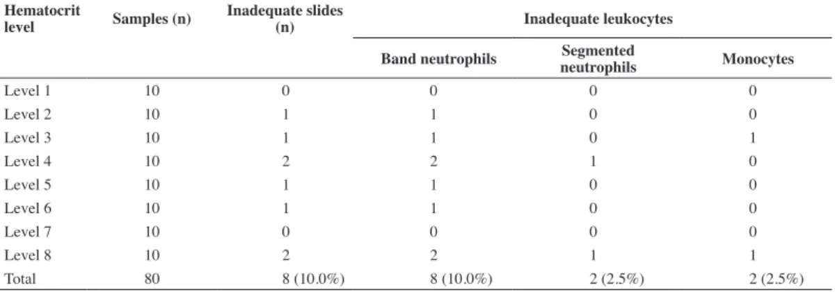
The pre-classiication of the leukocyte count obtained by the CellaVision® DM96 device was compared with the professional classiication after agreement or the re-classiication of the leukocytes. This demonstrated that the variation was acceptable in 90% of the slides. One or more leukocytes were incorrectly classiied for only eight (10%) slides, with the band neutrophil being the most common wrongly classiied cell (Table 3).
Qualitative analysis of the RBC proved to be adequate for the integrity of cells, uniform distribution on the slide, morphology and staining for all of the slides produced by the Sysmex® SP-1000i device. The RBC analysis provided by CellaVision® DM96 was not used for comparison, leaving the qualitative evaluation of the RBC as well as qualitative changes in leukocytes to the professional. It was also possible to perform an estimation of the platelet count and to assess the presence of platelet aggregates and ibrin.
A good correlation was obtained between the automated pre-classiication and the professional classiication for segmented neutrophils, eosinophils and lymphocytes (Table 4). The correlation coeficient (r2) between the two evaluations was determined for the leukocyte count (Figure 1). The Table 1 - Settings for blood smear performed by the Sysmex® SP-1000i
equipment calculated from the hematocrit of the patient
Hematocrit (%) Angle (°) Speed (mm/s)
Level 1 < 20 31 180
Level 2 20-25 30 175
Level 3 25-30 29 165
Level 4 30-35 29 160
Level 5 35-40 29 155
Level 6 40-45 29 150
Level 7 45-50 29 120
The present study demonstrates the validation process for the implementation of the Sysmex® SP-1000i equipment in the laboratory routine, where the quality of blood smears and slide staining were evaluated using the CellaVision® DM96 equipment which had previously been validated in the laboratory routine.
The preparation and staining of the blood smear on a microscope slide are fundamental steps to perform a qualiied microscopic examination, however the manual preparation of CBC slides, despite being traditional and satisfying, has certain limitations that can directly inluence the differential count and morphological analysis of cells.
The ideal blood smear for a qualiied CBC analysis should be uniform, free of bubbles and laws and with well-stained cells that enable their correct identiication and classiication. These requirements in the preparation of a blood smear are achieved by automated equipment.
The Sysmex® SP-1000i equipment automates the preparation and staining of slides to standardize the preparation of blood smears using the hematocrit value of the sample, regulating the amount of blood, the velocity and the angle to improve the quality of the smear. In this study the smear and staining of slides prepared in the Sysmex® SP-1000i proved to be of good quality as all of the slides provided acceptable results in the cell localization and artifacts tests when processed for quality control in the CellaVision® DM96 equipment.
The pre-classiication of leukocyte differential counts by the CellaVision® DM96 device showed that the cells were correctly Table 2 - Evaluation of the quality of slides produced by the Sysmex® SP-1000i, using quality control by the
CellaVision® DM96 for each hematocrit level
Table 3 - Number of slides and leukocytes improperly classiied by the CellaVision® DM96 using slides prepared by the
Sysmex® SP-1000i
n Hematocrit (%) Leukocyte count
(x103/µL)
Cell Localization
Test (%) Artifacts (%)
Level 1 10 16.68 ± 1.87 9.9 ± 1.3 99.85 ± 0.24 15.24 ± 5.530
Level 2 10 22.96 ± 1.13 10.1 ± 2.10 100 15.72 ± 11.42
Level 3 10 26.96 ± 1.39 9.4 ± 1.5 99.85 ± 0.24 0006 ± 3.68
Level 4 10 32.49 ± 1.38 9.6 ± 1.8 99.81 ± 0.32 13.25 ± 11.21
Level 5 10 037.2 ± 1.47 8.7 ± 1.5 99.9 ± 0.21 8.35 ± 4.81
Level 6 10 41.16 ± 0.78 9.6 ± 1.1 100 10.67 ± 6.150
Level 7 10 46.44 ± 1.12 9.1 ± 1.4 100 15.64 ± 13.62
Level 8 10 52.27 ± 1.95 9.5 ± 1.5 100 14.2 ± 7.75
Hematocrit
level Samples (n)
Inadequate slides
(n) Inadequate leukocytes
Band neutrophils Segmented
neutrophils Monocytes
Level 1 10 0 0 0 0
Level 2 10 1 1 0 0
Level 3 10 1 1 0 1
Level 4 10 2 2 1 0
Level 5 10 1 1 0 0
Level 6 10 1 1 0 0
Level 7 10 0 0 0 0
Level 8 10 2 2 1 1
Total 80 8 (10.0%) 8 (10.0%) 2 (2.5%) 2 (2.5%)
metamyelocytes, myelocytes and promyelocytes were grouped for analysis because of the low frequency in the sample.
Discussion
The task to change or introduce new technologies into the clinical laboratory is the responsibility of the professionals working in the technical department along with the managers in order to ensure high standards in the new service being provided. Even if a method or device has already been tested and validated by the manufacturer, it is important that its performance is veriied in the laboratory to check whether the new equipment works as expected thereby providing adequate results(21).
Table 4 - Correlation for the pre-classiication of cells performed by the
CellaVison® DM96 and the classiication by the professional.
Leukocytes Correlation (r) p-value
Band neutrophils 0.802 < 0.001
Segmented neutrophils 0.963 < 0.001
Eosinophils 0.958 < 0.001
Basophils 0.568 < 0.001
Lymphocytes 0.985 < 0.001
Monocytes 0.791 < 0.001
Myelocytes 0.465 < 0.001
Metamyelocytes 0.456 < 0.001
Plasmocytes 0.373 < 0.001
Atypical Lymphocytes 0.866 < 0.001
identiied in 90% of slides. The low percentage of incorrectly classiied slides demonstrates the quality of the slides.
Band neutrophils were the cell class most often pre-classiied erroneously by the device. This was also observed by Kratz et al. who reported that the percentage of correct pre-classiications made by this equipment was low for immature and abnormal cells, demonstrating the dificulty of automation to differentiate band neutrophils and segmented neutrophils(8).
All slides were adequate in the evaluation of qualitative changes of white blood cells, RBC and platelets however for samples presenting platelet aggregates, conventional microscopy is essential as the CellaVision® DM96 device selects a ixed area on the slide which cannot be modiied by the user.
The correlations between automated pre-classiication and the professional’s classiication were statistically signiicant, demonstrating the quality of the blood smears.
Our indings demonstrated a good correlation coeficient (r2 > 0.90) between CellaVision® DM96 and the professional’s
classiications for segmented neutrophils, eosinophils and lymphocytes as was also observed by Lee et al. for segmented neutrophils and lymphocytes(22). On the other hand, a weak correlation was observed between the equipment and professional classiications for immature granulocytes, band neutrophils, basophils, monocytes, atypical lymphocytes and erythroblasts. Some studies have also demonstrated a weak correlation between the pre-classiication by CellaVision® DM96 and the analysis made by a professional for monocytes(8,14,22), basophils(14,22) and band neutrophils(22), which demonstrates a tendency of this equipment to incorrectly classify these cells in other cell classes.
The differential count of peripheral blood cells is an important diagnostic tool which requires trained personnel and access to an automated microscopy in the hematology laboratory. The use of three technologies: hematology analyzer, automated slide preparer and automated slide analysis can optimize the routine low, providing rapid and accurate results with excellent cost-beneit.
Figure 1 - Linear regression and scatter plots for band neutrophils, segmented neutrophils, eosinophils, monocytes, lymphocytes, atypical lymphocytes,
A limitation of this study was its restricted sample size to validate the equipment, as well as not performing tests on leukopenic and leukocytosis samples.
Conclusion
Choosing new equipment for the laboratory involves the analysis of numerous factors such as the needs of the laboratory, quality control, cost-beneit, ease of handling by professionals, accessible software, as well as the success of the validation process.
The results obtained in this study show the quality of the Sysmex® SP-1000i device in a laboratory; it provides beneits due to its reliability and speed to prepare blood smears thus it is an important ally in the release of quality CBC reports in clinically useful times.
References
1. Ryan DH. Automated analysis of blood cells. In: Hoffman R, Benz EJ,
Shattil SJ, Furie B, Cohen HJ, Silberstein LE, editors. Hematology: basic principles and practice. 2nd ed. New York: Churchill Livingstone; 1995.
p. 2223-35.
2. Failace R, Pranke P. Avaliação dos critérios de liberação direta dos resultados de hemograma através de contadores eletrônicos. Rev Bras
Hematol Hemoter. 2004;26(3):159.
3. Yu H, Ok CY, Hesse A, Nordell P, Connor D, Sjostedt E, et al. Evaluation of na automated digital imaging system, nextslide digital review network, for examination of peripheral blood smears. Arch Pathol Lab Med.
2012;136(6):660-7.
4. Buttarello M. Plebani M. Automated blood cell counts: state of the art. Am J Clin Pathol. 2008;130(1):104-16.
5. Barnes PW, McFadden SL, Machin SJ, Simson E; International
Consensus Group for Hematology. The international consensus group for hematology review: suggested criteria for action following automated
CBC and WBC differential analysis. Lab Hematol. 2005;11(2):83-90. 6. Bacall NS. Analisador automático hematológico e a importância de
validar novos equipamentos em laboratórios clínicos. Rev Bras Hematol
Hemoter. 2009;31(4):218-20.
7. Borges LF, Siqueira LO. Validação de tecnologia 5diff do analisador hematológico Sysmex XS-1000i para laboratório de pequeno/médio
porte. Rev Bras Hematol Hemoter. 2009;31(4):247-51.
8. Kratz A, Bengtsson HI, Casey JE, Keefe JM, Beatrice GH, Grzybek
DY, et al. Performance evaluation of the CellaVision DM96 system: WBC differentials by automated digital image analysis supported by an
artiicial neural network. Am J Clin Pathol. 2005;124(5):770-81.
9. Andrade SR, Kramer DG. Estudo comparativo entre os procedimentos manual e automatizado na contagem diferencial dos leucócitos. Rev Bras Anal Clin. 2001;33(1):35-7.
10. Lewis SM, Bain BJ, Bates I. Hematologia prática de Dacie e Lewis. 9th
ed. Porto Alegre: Artmed; 2006.
11. Automated Hematology Slide Preparation Unit SP-1000i. Instructions for use. Kobe, Japan: Sysmex Corporation; 2009.
12. Yamamoto T, Tabe Y, Ishii K, Itoh S, Maeno I, Matsumoto K, et al. [Performance evaluation of the CellaVision DM96 system in WBC differentials]. Rinsho Byori. 2010;58(9):884-90. Japanese.
13. Billard M, Lainey E, Armoogum P, Alberti C, Fenneteau O, Da Costa L.
Evaluation of the CellaVision DM automated microscope in pediatrics.
Int J Lab Hematol. 2010;32(5):530-8.
14. Briggs C, Longair I, Slavik M, Thwaite K, Mills R, Thavaraja V, et al. Can automated blood ilm analysis replace the manual differential? An
evaluation of the CellaVision DM96 automated image analysis system.
Int J Lab Hematol. 2009;31(1):48-60.
15. Cornet E, Perol JP, Troussard X. Performance evaluation and relevance of the CellaVision DM96 system in routine analysis and in patients with
malignant hematological diseases. Int J Lab Hematol. 2008;30(6):536-42.
16. Surcouf C, Delaune D, Samson T, Foissaud V. [Automated cell recognition in hematology: CellaVision DM96 TM system]. Ann Biol Clin (Paris). 2009;67(4):419-24. French.
17. Gao Y, Mansoor A, Wood B, Nelson H, Higa D, Naugler C. Platelet count estimation using the CellaVision DM96 system. J Pathol Inform. 2013;4:16.
18. Briggs C, Longair I, Slavik M, Thwaite K, Mills R, Thavaraja V, et al. Can automated blood ilm analysis replace the manual differential? An
evaluation of the CellaVision DM96 automated image analysis system.
Int J Lab Hematol. 2009;31(1):48-60.
19. CellavisionTM DM 96. Manual for user [Internet]. Suécia: CellaVision
AB; 2009. [cited 2011 Jun 21]. Available from: http://www.cellavision.
com/?id=4103
20. Henry JB. Diagnósticos clínicos e tratamento por métodos laboratoriais.
20th ed. Barueri, SP: Manole; 2008.
21. Oliveira CA, Mendes ME. Gestão da fase analítica do laboratório: como assegurar a qualidade na prática. Rio de Janeiro: ControlLab; 2010. 22. Lee LH, Mansoor A, Wood B, Nelson H, Higa D, Naugler C.
Performance of CellaVision DM96 in leukocyte classiication. J Pathol
Inform. 2013;4:14.