jcoloproctol(rioj).2015;35(3):175–177
w w w . j c o l . o r g . b r
Journal
of
Coloproctology
Case
Report
Endoanal
pilonidal
sinus:
case
report
and
literature
review
Carolina
Talini
a,
Geanine
Baggio
Fracaro
a,b,
Allan
Cezar
Faria
Araújo
a,c,
Doryane
Maria
dos
Reis
Lima
b,d,
André
Pereira
Westphalen
a,c,∗aHospitalUniversitáriodoOestedoParaná(HUOP),Cascavel,PR,Brazil bSociedadeBrasileiradeColoproctologia,RiodeJaneiro,RJ,Brazil
cUniversidadeEstadualdoOestedoParaná(UNIOESTE),Cascavel,PR,Brazil dFaculdadeAssisGurgacz,Cascavel,PR,Brazil
a
r
t
i
c
l
e
i
n
f
o
Articlehistory:
Received2February2015 Accepted20February2015 Availableonline29April2015
Keywords:
Pilonidalsinus Rectalfistula
Transrectalultrasound
a
b
s
t
r
a
c
t
PilonidalsinusisatermfirstusedbyHodgesin1880todescribegranulomatouslesions containinghairsinitsinterior.Thepresenceofendoanalpilonidalsinusisrareandonly ninecaseshavebeenreportedinmedicalliterature.Thisarticledescribesamale,42years, initiallysubmittedtofistulotomywithsetonplacementthatfouryearslaterevolvedwith complaintsofanalpurulentdischargeassociatedwithpainfulanalgroin.Duringthefirst evaluationasmallendoanaltumorwasfound.Itwaslocatedinposterolateralanusposition andcontainedhairinitsinterior,withoutpurulentdischargeatthatmoment.Underwent surgerythatconfirmedthepresenceofendoanalpilonidalsinus.Thesinuswasopened andlefttohealbysecondaryintention.Thepatienthadgoodoutcomewithnosignsof recurrence.
©2015SociedadeBrasileiradeColoproctologia.PublishedbyElsevierEditoraLtda.All rightsreserved.
Cisto
pilonidal
endoanal:
relato
de
caso
e
revisão
de
literatura
Palavras-chave:
Cistopilonidal Fístulaanal Ultrassomendoanal
r
e
s
u
m
o
Otermo cistopilonidalfoi descritopela primeiravezpor Hodgesem1880 para descr-everlesõesgranulomatosascontendopêlosemseuinterior.Apresenc¸adecistopilonidal endoanaléraraeexistemapenasnovecasosrelatadosnaliteraturamédica.Descreve-se umcasomasculino,42anos,emacompanhamentohá5anosnoservic¸o.Submetido inicial-menteàfistulotomiaemdoistemposcomsedenho,evoluindo4anosdepoiscomqueixasde secrec¸ãopurulentaintermitenteporviaanalassociadaapresenc¸adetumorac¸ãodolorosa. Noexamefísicoconstatou-sepresenc¸adeorifíciocontendopêlosemseuinterior,sem
∗ Correspondingauthor.
E-mail:[email protected](A.P.Westphalen).
http://dx.doi.org/10.1016/j.jcol.2015.04.002
176
jcoloproctol(rioj).2015;35(3):175–177secrec¸ãopurulentanomomentodoexame.Foiencaminhadoparatratamentocirúrgicoque confirmoudiagnósticodecistopilonidalendoanal.Noprocedimentofoirealizadaabertura docistoquefoideixadoparacicatrizarporsegundaintenc¸ão.Opacienteapresentouboa evoluc¸ão,semsinaisderecidivalocal.
©2015SociedadeBrasileiradeColoproctologia.PublicadoporElsevierEditoraLtda. Todososdireitosreservados.
Introduction
Pilonidal sinus is a term first used by Hodges in 1880 to describe granulomatous lesions containing hairs in its interior.1SuchlesionswerefirstdescribedbyWarrenin1854
andcalledhairycyst.2Isacommonentityinyounghairyman
andmostofthemcontainlonghairwithnofollicles.1
Similar lesions are being described inother body parts suchasabdominalwall,ears,hands,armpit,gluteal, interdig-italregion,nipple,occipital,perineumandumbilicus.1–3This
diversityoftopographyisafactorthatsupportsatheory,and insupportofthattheory,someauthorssuggestthatpilonidal cystsaretheresultoflosthairsthatarepushedintotheskin. Thepresenceofendoanalpilonidalsinusisrare4andonly
ninecaseshavebeenreportedinmedicalliterature.5Thereare
severalwaystoexplainthegenesisofthosesinusintheanal canal.Oneofthemregardsanextensionofasacro-coccygeal cysttotheperianalregionandotherlesswidespreadtheories arethatthepilonidalcystcouldbebornthroughtheentryof hairsintoanopenanal fissureorthathaircangetintothe analcanalregionastheydoelsewhereinthebody.1,3,4
Nowa-daysthemostacceptedtheoryconcernstheacquireddisease afterperforminganalfistulotomyorothersurgicalprocedure inwhichhairmayenterthesubcutaneoustissuethroughthe healingwoundor throughthenewlyformedscar andmay serveasthecradleofapilonidalsinus.1,4,5
The pilonidal sinus can often be confused with anal fistula. Usually the patient has multiple recurrences and underwentseveralsurgicalproceduresbutwithoutsymptoms improvement.6
Thisarticleaimstoreportanendoanalpilonidalsinuscase thatappearedaftersurgicaltreatmenttocorrectananal fis-tula.
Clinical
presentation
GF,male,42years,smoker,diagnosedwithHIV10yearsago (usingEfavirenz,LamivudineandZidovudine),whofirstcame tothisColoproctologyservice5yearsago,complainingabout afastgrowinganaltumor,painful,whichevolvedto sponta-neousdrainageofpus.Afterfirstserviceadmissionananal posterolateralfistulawasdiagnosedinassociationwithgrade Ihemorrhoidsand multipleperianalskintags. Thepatient underwentsurgicalprocedurewithexcisionofskintags(that pathologicalanatomyconfirmedthatitwassquamous papil-loma with hyperkeratosis associated with viral cytopathic default),fistulotomywithsetonplacementandcauterization ofhemorrhoidalnipples.
Fig.1–3Dendoanalultrasoundshowingthinningofthe analcanal,withoutanylesions.
Aboutayearagostartedpresentinganorectalcomplaints again.Relateddrainageofpurulentandfetidsecretion dur-ingthisperiodandpainduringevacuationwithoutbleeding, denyingincreasingonthenodulesize.Duringthefirst eval-uationasmallendoanaltumorwasfound.Itwaslocatedin posterolateralanuspositionandcontainedhairinitsinterior, withoutpurulentdischargeatthatmoment.
Patient was submitted to 3D endoanal ultrasound that found no lesions despitethe thinning ofthe left dorsolat-eralinternalandexternalanalsphincterintheposterolateral quadrant ofthemiddle anal canal(Fig. 1). Itwasclinically diagnosedaspilonidalsinusoftheanalcanalandthepatient underwentsurgery.
jcoloproctol(rioj).2015;35(3):175–177
177
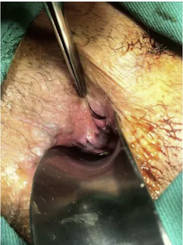
Fig.2–Transoperatorydemonstratingposterolateralsinus containinghairinitsinterior.
Discussion
Differentpilonidalsinuspresentationsaredescribedin litera-ture.Regardingthoselocatedinendoanalregion,eightcases werereportedinmanandoneoftheminawoman.1–5When
itcomestotheage,thosewereallyoungpatients–23,27,29, 30,42,46,55and58yearsforthemenand42yearsforthe woman.
Fourofthempresentedrecurrentpurulentdischargeand weresubmittedtothemostdiversesurgicalprocedures with-outclinicalimprovement.1,3–5Twoofthemhadnosymptoms
and were diagnosedduringa surgicalprocedure to correct hemorrhoids4 andthreeofthemweresymptomaticforthe
firsttime.2Noneoftheevaluatedpatientshadfoundhairin
endoanalregionbythemselves.Asinthepresentedcase,the sinuswereuniqueineightofthereportedcasesandoneother patientpresentedtwoendoanalsinus.1
When it comesto the surgicalprocedure, infour cases thecysttrajectorywasopenedandlefttohealbysecondary intention1,4justlikethecasepresentedhereandontheother
fourcasesthelesionwascompletelyremoved.2–5
Oneofthereportedcases5hadasimilaroutcomeastheone
wepresenthere.Bothpatientswereinitiallysurgicallytreated tocorrectananalfistulawithseton,evolvedwithsymptoms recurrenceanddevelopedendoanalpilonidalsinus.
Oneofthepreviouscasereportshadatwoyearsfollow-up4
and none of the five patients presented disease recur-rence. Other author presentedoneyear follow up with no recurrence.5Allothercasereportspresentednoinformation
aboutthefollow-upperiod.1–3
Conclusion
Endoanalpilonidalsinusisarareentitywithveryfewreported casesinliterature.Intherelatedcasesmostpatientsareyoung menwhoaresymptomaticforalongtimeuntilthetrue dis-easeisdiagnosed.Surgicaltreatmentseemstobeeffectivefor healingwithlowrecurrencerates.
Conflicts
of
interest
Theauthorsdeclarenoconflictsofinterest.
r
e
f
e
r
e
n
c
e
s
1.WilsonE,FailesDG,KillingbackM.Pilonidalsinusesoftheanal canal:reportofcase.DisColonRectum.1971;14(6):468–70.
2.WestonSD,SchlachterIS.Pilonidalcystoftheanalcanal:case report.In:ReadatthemeetingoftheAmericanProctologic Society.1962.
3.AccarpioG,DaviniMD,FazioA,SenussiOH,YakubovichA. Pilonidalsinuswithananalcanalfistula:reportofcase.Dis ColonRectum.1988;31(12):965–7.
4.OrtizH,MartiJ,DeMiguelM,CarmonaA,Caba ˜nasIP. Hair-containinglesionswithintheanalcanal.IntJColorectal Dis.1987;2:153–4.
5.AlrawashdehW,AjazS,HammondTM,PorrettTRC,LunnissPJ. Primaryanalpilonidaldisease.ColorectalDis.2008;10:303.