Letters to the Editor
Radiol Bras. 2016 Jul/Ago;49(4):267–276
268
http://dx.doi.org/10.1590/0100-3984.2015.0037
Felipe Welter Langer1, Gustavo Suertegaray1, Daiane dos Santos1, Giordano Rafael Tronco Alves1, Carlos Jesus Pereira Haygert1
1. Hospital Universitário de Santa Maria (HUSM) – Universidade Federal de Santa Maria (UFSM), Santa Maria, RS, Brazil. Mailing address: Dr. Felipe Welter Langer. Departamento de Radiologia e Diagnóstico por Imagem, Hospital Universitário de Santa Maria – Universidade Federal de Santa Maria. Santa Maria, RS, Brazil, 97105-900. E-mail: [email protected].
restricted diffusion. There is typically no gadolinium enhancement. After glycemic correction, similarly to the clinical findings, such regions tend to return to normal signal intensity.
It is important to highlight the role of susceptibility-weighted imaging (SWI) in differentiating between changes seen in HCHB and areas of calcification or hemorrhage, which represent the most common differential diagnoses. Calcium and blood deposits both generally manifest as hyperintensities on T1-weighted images with corresponding hypointensities on T2*-weighted images and SWI; conversely, HCHB changes tend to present as unilateral hyperintensities on T1-weighted images with no matching changes on T2*-weighted images or SWI(7,8).
REFERENCES
1. Shan DE, Ho DMT, Chang C, et al. Hemichorea-hemiballism: an ex-planation for MR signal changes. AJNR Am J Neuroradiol. 1998;19:863– 70.
2. Postuma RB, Lang AE. Hemiballism: revisiting a classic disorder. Lan-cet Neurol. 2003;2;661–8.
3. Narayanan S. Hyperglycemia-induced hemiballismus hemichorea: a case report and brief review of the literature. J Emerg Med. 2012;43:442–4. 4. Wintermark M, Fischbein NJ, Mukherjee P, et al. Unilateral putaminal
CT, MR, and diffusion abnormalities secondary to nonketotic hypergly-cemia in the setting of acute neurologic symptoms mimicking stroke. AJNR Am J Neuroradiol. 2004;25:975–6.
5. Hawley JS, Weiner WJ. Hemiballismus: current concepts and review. Parkinsonism Relat Disord. 2012;18:125–9.
6. Zaitout Z. CT and MRI findings in the basal ganglia in non-ketotic hyperglycaemia associated hemichorea and hemi-ballismus (HC-HB). Neuroradiology. 2012;54:1119–20.
7. Chavhan GB, Babyn PS, Thomas B, et al. Principles, techniques, and applications of T2*-based MR imaging and its special applications. Radiographics. 2009;29:1433–49.
8. Hansford BG, Albert D, Yang E. Classic neuroimaging findings of nonketotic hyperglycemia on computed tomography and magnetic reso-nance imaging with absence of typical movement disorder symptoms (hemichorea-hemiballism). J Radiol Case Rep. 2013;7:1–9.
Anterior cerebral artery aneurysm rupture presenting as hemorrhage in the splenium of the corpus callosum
Dear Editor,
A 43-year-old, right-handed male presented with a three-day history of severe, holocranial headache. Three weeks prior, he had experienced another series of severe, pulsatile headaches accom-panied by fever, malaise, and paresthesia of the second and third digits of the left hand. The neurologic examination revealed apraxia of the left hand and constructional apraxia of the right hand, with-out sensorimotor or cerebellar deficits, consistent with callosal disconnection syndrome.
Non-contrast computed tomography and magnetic reso-nance imaging demonstrated a hematoma in the splenium of the corpus callosum (Figure 1). No subarachnoid blood was visual-ized. Cerebral angiography revealed evidence of recent aneurysm rupture at the junction of the A1 and A2 segments of the right anterior cerebral artery (ACA) and vasospasm of the distal right ACA (Figure 2A). The decision was made to embolize the aneu-rysm with detachable coils (Figure 2B). At the conclusion of the procedure, there was complete embolization of the aneurysm sac,
without disruption of the integrity of the intracranial arteries or defect in the brain parenchyma. The remainder of the hospital stay was uneventful, and the patient was discharged on post-ad-mission day 11 with a prescription for a 6-day tapered course of nimodipine. Angiography performed at 6 months of follow-up demonstrated that the coils remained in place within the aneu-rysm sac (i.e., the aneuaneu-rysm sac continued to be occluded).
Reports of remote intraparenchymal hemorrhage as a pre-senting finding of aneurysm rupture are rare(1). For example, in a group of 460 patients with subarachnoid hemorrhage, Abbed et al.(2) reported 116 cases of intraparenchymal hematoma forma-tion, none of which appeared to be proximal to the site of aneu-rysm rupture. In fact, our search of the literature revealed only isolated cases of remote focal hemorrhage. In 2002, Friedman et al.(3) described a ruptured anterior communicating artery aneu-rysm associated with a perisylvian frontotemporal hematoma. Also in 2002, Lee et al.(4) described the case of a patient with ruptured saccular ACA aneurysm that evolved to hemorrhage of the left putamen. In 2005, Paus et al.(5) reported an even more perplex-ing case of anterior communicatperplex-ing artery aneurysm rupture, with adjacent subarachnoid hemorrhage and focal hematoma in the
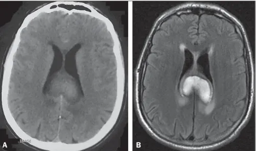
Figure 1. Non-contrast computed tomography (A) and T2-weighted fluid attenuated inversion recovery mag-netic resonance imaging (B) demonstrating a large, heterogeneously enhancing mass in the splenium of the corpus callosum, consistent with a focal collec-tion of intraparenchymal blood. No evidence of sub-arachnoid hemorrhage is apparent.
Letters to the Editor
Radiol Bras. 2016 Jul/Ago;49(4):267–276
269
http://dx.doi.org/10.1590/0100-3984.2015.0130 left posterior temporal lobe that was distant from the aneurysm
and from any subarachnoid cisterns.
The case presented here is important because it establishes a mechanism for remote bleeding. In previous reports, a variety of explanations for distant hemorrhage have been proposed, includ-ing hypertensive crisis, the formation of jets through subarach-noid cisterns, venous infarction, intraluminal thrombosis, hem-orrhagic infarction secondary to vasospasm, and occult vascular anomaly. However, none of those reports provided direct evidence to support any of the proposed mechanisms. In contrast, in our case, we observed definite angiographic evidence of vasospasm in the vessels between the aneurysm and the site of hemorrhage. That constitutes a strong indication that vasospasm-associated hemorrhagic infarction is a mechanism of remote hematoma formation following cerebral aneurysm rupture.
In conclusion, we have described a case of ACA aneurysm rupture presenting as remote intraparenchymal hemorrhage in the splenium of the corpus callosum and have demonstrated that vasospasm-induced hemorrhagic infarction is a plausible mecha-nism for distant bleeding. Neuroradiologists and neurosurgeons should be aware of this rare phenomenon in order to reduce the likelihood of inappropriate treatment.
REFERENCES
1. Abla AA, Wilson DA, Williamson RW, et al. The relationship between
Thiago Giansante Abud1, Andrew D. Nguyen2, Lucas Giansante Abud3, Emmanuel Houdart4
1. Department of Interventional Neuroradiology, Hospital Israelita Albert Einstein, São Paulo, SP, Brazil. 2. Division of Neuro-Interventional Radi-ology, University of California-San Diego, San Diego, CA, USA. 3. De-partment of Neuroradiology, Documenta – Hospital São Francisco, Ri-beirão Preto, SP, Brazil. 4. Department of Interventional Neuroradiology, Hôpital Lariboisière, Paris, France. Mailing address: Dr. Thiago Giansante Abud. Rua da Consolação, 2840, ap. 12, Cerqueira César. São Paulo, SP, Brazil, 01416-000. E-mail: [email protected].
ruptured aneurysm location, subarachnoid hemorrhage clot thickness, and incidence of radiographic or symptomatic vasospasm in patients en-rolled in a prospective randomized conten-rolled trial. J Neurosurg. 2014;120:391–7.
2. Abbed KM, Ogilvy CS. Intracerebral hematoma from aneurysm rupture. Neurosurg Focus. 2003;15:E4.
3. Friedman JA, Rabinstein AA, Meyer FB. Perisylvian frontotemporal he-matoma due to rupture of an anterior communicating artery aneurysm. Case illustration. J Neurosurg. 2002;97:493.
4. Lee JK, Lee JH, Kim IY, et al. Simultaneous occurrence of subarach-noid hemorrhage due to ruptured aneurysm and remote hypertensive intracerebral hemorrhage: case report. J Korean Med Sci. 2002;17: 144–6.
5. Paus C, Daniel RT, Regli L. Posterior temporal haematoma associated with anterior communicating artery aneurysm rupture. J Clin Neuro-sci. 2005;12:182–4.
Fat necrosis associated with the use of oral anticoagulant therapy: atypical mammographic findings
Dear Editor
We report the case of a 54-year-old female with systemic lu-pus erythematosus, lulu-pus nephritis, antiphospholipid syndrome, and deep vein thrombosis, who was being treated with an oral an-ticoagulant and a corticosteroid, as well as receiving immunosup-pressive therapy. Her international normalized ratio was between 2 and 3, and she presented with recurrent spontaneous hemato-mas. She had been diagnosed 20 months prior with miliary pulmo-nary tuberculosis, which had been treated for 12 months. After the patient had undergone mammography (Figure 1), we reviewed the clinical data: she reported a recent spontaneous left-sided he-matoma, with palpable nodules and ecchymosis, in the superolateral quadrant. As can be seen in Figure 2, ultrasound with Doppler flow imaging showed correspondence between this findings and
an irregular hypoechoic nodule with indistinct margins without vascularization, measuring 6.0 × 3.0 cm, associated with archi-tectural distortion, in the superolateral quadrant—together with images suggestive of lipid cysts. Initially undetermined, the le-sion was considered likely benign, suggestive of fat necrosis, prob-ably associated with anticoagulant use and hematoma formation. To avoid biopsy, we opted for a strategy of observation only.
Fat necrosis is often silent, appearing only as an abnormal mammographic finding. In rare cases, it can manifest as a pal-pable mass without associated mammographic findings. It is typi-cally secondary to incidental or iatrogenic trauma and can occur in patients who are using anticoagulants or even in those without a relevant history.
Mammography is the most important test in the assessment of fat necrosis. Depending on the stage and the amount of fibro-sis, it can manifest as a lipid cyst or as features that simulate ma-lignancy: spiculated hyperdense areas; nodules accompanied by Figure 2.A: Digital subtraction angiography of the
cerebral vessels, demonstrating an aneurysm (black arrow) at the junction of the A1 and A2 segments of the right ACA. The aneurysm is irregular in ap-pearance, with a rupture sac and Murphy’s test sug-gestive of recent rupture. The right A2 segment is characterized by an irregular caliber and a beaded appearance (open arrows), consistent with arterial vasospasm. B: Complete embolization of the an-eurysm sac after coiling.