T
ABSTRACT
EVALUATION OF THE EFFECTS OF PROCESSING DELAYS
AND PROTECTIVE PLASTIC CASES ON IMAGE QUALITY
OF A PHOTOSTIMULABLE PHOSPHOR PLATE SYSTEM
Clóvis Monteiro BRAMANTE1, Alexandre Silva BRAMANTE2, Rogério Emílio de SOUZA3, Ivaldo Gomes MORAES4, Norberti BERNARDINELI1, Roberto Brandão GARCIA5
1- DDS, MSc, PhD, Professor, Department of Operative Dentistry, Endodontics and Dental Materials, Bauru School of Dentistry, University of São Paulo, Bauru, SP, Brazil.
2- DDS, MSc, Brasiliense School of Dentistry, Brasília, DF, Brazil. 3- DDS, MSc, PhD, Dental School, FACIMP, Imperatriz, MA, Brazil.
4- DDS, MSc, PhD, Associate Professor, Department of Operative Dentistry, Endodontics and Dental Materials, Bauru School of Dentistry, University of São Paulo, Bauru, SP, Brazil.
5- DDS, MSc, PhD, Assistant Professor, Department of Operative Dentistry, Endodontics and Dental Materials, Bauru School of Dentistry, University of São Paulo, Bauru, SP, Brazil.
Corresponding address: Prof. Dr. Clovis Monteiro Bramante - Faculdade de Odontologia de Bauru - USP - Alameda Octávio Pinheiro Brisolla, Nº 9-75 - 17012-901 Bauru, SP, Brasil - Phone +55-14-3235-8344 - email: [email protected]
Received : October 03, 2007 - Modification : January 28, 2008 - Accepted : May 07, 2008
his ex vivo study evaluated the quality of digital radiographic images obtained with the photostimulable phosphor plate system (Digora) according to the processing delay and maintenance of optical plates in either opaque (supplied with the system) or transparent protective plastic cases during this period. Five radiographs were obtained from the mandibular molar region of a dry human mandible using optical plates. These plates were placed in the protective plastic cases before obtaining the radiographs and were processed immediately or after processing delays of 5, 60 and 120 min, when the case was removed. The results revealed a reduction in image quality when processing was delay 120 min compared to the other times. The opaque case provided better protection to the sensor than the transparent case. In conclusion, a 120-min processing delay for the Digora system caused a reduction in image quality, yet without interfering with the quality of diagnosis. The opaque case supplied by the system’s manufacturer provided better protection to the optical plate than the transparent case.
Key words: Radiography. Digital images. Phosphor storage plates. Digora.
INTRODUCTION
Digital radiograph or radiovisiography was first described in 1987 and was commercially introduced by
Trophy Radiologie in collaboration with Mouyen12 in 1993.
Three systems are employed for radiovisiography: the CCD sensor (Charge-Coupled Device), CMOS sensor (Complementary Metallic Oxide Sensor), and PSPs (PSPs – Phosphor Storage Plate – optical plate); the latter includes
the Digora, Digident, Denoptix and Den Ortix systems11.
Some studies have found better outcomes of digitized images for endodontic diagnosis and treatment5-9,13,15-18.
However, some doubts remain in the use of radiovisiography based on the optical plate system as to the durability of the plate3, image quality according to the
processing delay1,2,14, and storage conditions10. No study
has yet investigated the influence of utilization of opaque cases on the quality of digital image.
This ex vivo study evaluated the quality of digital
radiographic images obtained with the Digora system according to the processing delay and maintenance of optical plates in either opaque (supplied with the system) or transparent protective plastic cases during this period.
MATERIAL AND METHODS
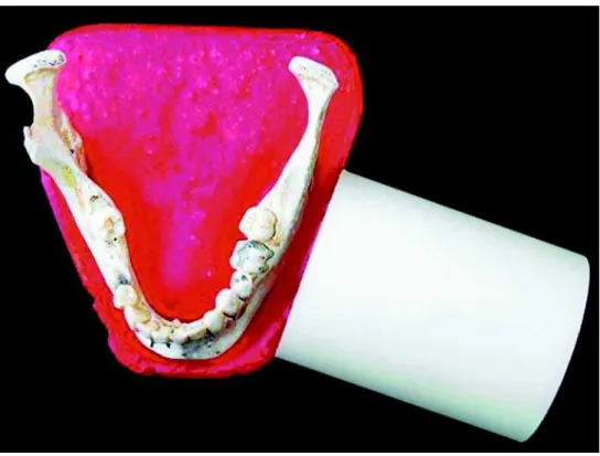
The study was conducted on a dry human mandible fixed on an acrylic resin base. A plastic tube was fixed frontally to the mandible to standardize the position of the X-ray source, thus standardizing the vertical and horizontal angles (Figure 1). A groove was prepared on this resin base to standardize the position of the optical plate.
set at 60 kVp and 10 mA, with open tube, total filtration of 1.5 mm Al, exposure time of 0.3 s, and optical plate of the Digora system (Soredex, Orion Corporation, Finland). Two groups were constituted for x-ray exposure: in group A, the optical plate was inserted and sealed in the opaque plastic
case supplied by the manufacturer and kept as such until processing; in group B, the optical plate was stored in a transparent plastic case (Figure 2). Five optical plates were exposed for each period of processing delay.
The optical plates were processed in the Digora scanner
FIGURE 1- Mandible on acrylic base for standardization of x-ray incidence angle and position of the optical plate
immediately and 5, 60 and 120 min after X-ray exposure, and were transferred to the computer for analysis of digitized images. Analysis was performed directly on the computer screen by three examiners using the Digora for Windows software. The images were analyzed as to brightness, contrast and resolution of tooth structures (enamel, dentin, root and pulp cavity) and periapical structures (periodontal space, cortical bone, alveolar bone), which received scores 0 to 2, as follows: 0- Poor image quality; 1- Good image quality; 2- Excellent image quality.
Data were submitted to the Kruskal-Wallis non-parametric test, and comparison of results between opaque and transparent cases were submitted by the Mann-Whitney test. Significance level was set at 5%.
RESULTS
Table 1 presents the scores assigned by examiners to the images, according to processing delays and utilization of opaque or transparent plastic cases.
There was statistically significant difference (p<0.05) regarding the processing delays, namely immediate, 5 and 60 min with utilization of opaque case (Table 2). This difference was observed between the 120-min delay and the other periods (Table 3). No statistically significant difference (p>0.05) was observed among processing delays with utilization of transparent case (Table 4). Comparison of results between opaque and transparent cases did not reveal significant differences (p>0.05) (Table 5).
Processing delay (min)
Immediate 5 60 120
Images Images Images Images
1 2 3 4 5 1 2 3 4 5 1 2 3 4 5 1 2 3 4 5
A
Opaque Examiner I 2 2 2 2 2 2 2 2 2 2 2 2 1 2 2 1 1 1 2 2
case Examiner II 2 2 2 2 2 2 2 2 2 1 2 1 2 2 1 1 1 1 2 2
Examiner III 2 2 2 2 2 2 2 2 2 2 2 2 2 2 2 2 0 2 2 2
B
Transparent Examiner I 2 2 2 2 2 2 1 2 2 2 2 2 2 1 2 0 1 0 1 2
case Examiner II 2 2 2 2 2 2 2 1 2 2 2 2 2 1 2 2 0 1 0 2
Examiner III 2 2 2 2 2 2 1 2 2 2 2 1 1 2 2 2 1 0 1 1
TABLE 1- Scores(0 to 2) assigned to radiographic images (1 to 5) by examiners (I, II and III), according to the type of plastic case (A-opaque and B-transparent) and processing delay (min.)
Processing delay Median Sum of ranks Mean ranks Number of values
Immediate 2.0 60.0 12.0 5
5 min 2.0 60.0 12.0 5
60 min 2.0 60.0 12.0 5
120 min 1.0 30.0 6.0 5
TABLE 2- Kruskal Wallis test for analysis of the processing delay of optical plates protected with opaque cases
Hc= 10.05882; Chi-square with 3 degrees of freedom; Probability = 0.018072 (significant)
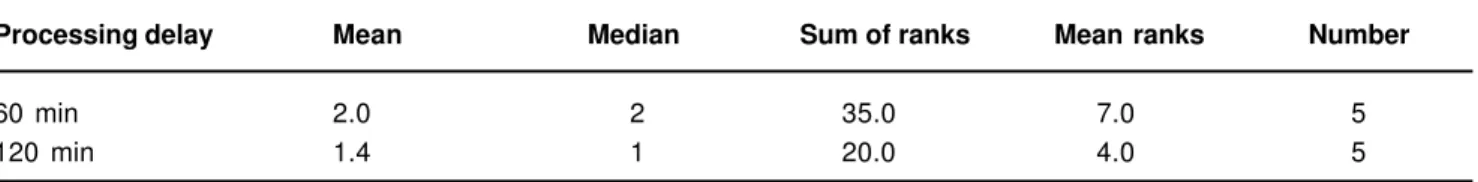
Processing delay Mean Median Sum of ranks Mean ranks Number
60 min 2.0 2 35.0 7.0 5
120 min 1.4 1 20.0 4.0 5
TABLE 3- Mann Whitneytest for analysis of the processing delay of optical plates protected with opaque cases
DISCUSSION
Digital radiography was introduced to dental practice with a view to replace the conventional radiography due to the shorter exposure to radiation, achievement of high-quality images, possibility of adjustments with aid of software, and comparable reliability to conventional radiography, for both endodontic diagnosis and treatment 3,5-7,19.
The main disadvantage of the optical plate system is the need of additional time for processing and possible loss of quality when the sensitized optical plate is exposed to light. Some authors have emphasized that, after exposure to x-ray, the optical plates should be processed within 1 h in order to avoid loss of image quality1,2. Other authors believe that the
plates can be processed within 6 h if stored in appropriate cases10.
In the present study, a processing delay of 120 min caused a mild reduction in image quality, yet without compromising the quality of diagnosis (Table 1). The three examiners were able to interpret radiographic details in all study periods. It should be highlighted that the images were not altered as to the brightness and contrast provided by the system, being analyzed as produced on the computer screen.
Together with the Digora system, the manufacturer supplies opaque plastic cases that offer protection against contamination and the deleterious effect of light. Images of optical plates maintained in the original opaque cases after exposure to x-ray maintained their image quality up to 120 min (Table 2), with significant difference between the 120-min processing delay and the other periods (Table 3). When the optical plates were maintained in transparent cases, there was also a progressive reduction of image quality (Table 1), which was more accentuated at 120 min (Table 4), though without statistical difference among periods. This is due to
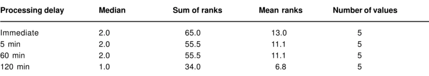
Processing delay Median Sum of ranks Mean ranks Number of values
Immediate 2.0 65.0 13.0 5
5 min 2.0 55.5 11.1 5
60 min 2.0 55.5 11.1 5
120 min 1.0 34.0 6.8 5
TABLE 4- Kruskal Wallis test for analysis of the processing delay of optical plates protected with transparent cases
Hc= 5.164999; Chi-square with 3 degrees of freedom; Probability = 0. 160106 (non significant)
Case Mean Median Sum of ranks Mean ranks Number
Opaque 1.85 2.0 431.5 21.6 20
Transparent 1.70 2.0 388.5 19.4 20
TABLE 5- Mann Whitneytest for comparison between opaque and transparent cases used for protection of optical plates
U=178.5. Normal approximation: Z=0.83688636; Probability = 0.40265645 (non significant)
the fact that the optical plate protected with transparent cases is subjected to the continuous action of light, with progressive loss of image quality. However, images obtained with optical plates protected with both opaque and transparent cases and with processing delays of up to 120 min provided good quality for diagnosis.
CONCLUSIONS
The findings of the present study revealed that the processing delay of 120 min for the Digora system caused a reduction in image quality, yet without interfering with the quality of diagnosis. The opaque case supplied by the system’s manufacturer provided better protection to the optical plate than the transparent case.
REFERENCES
1- Akdeniz BG, Grondahl HG, Kose T. Effect of delayed scannig of storage phosphor plates. Oral Surg Oral Med Oral Pathol Oral Radiol Endod. 2005;99:603-7.
2- Akdeniz BC, Grondahl HG. Degradation of storage phosphor images due to scanning delay. Dentomaxillofacial Radiol. 2006;35:74-7
3- Bedard A, Davis TD, HG. Angelopoulos C. How durable are they as a digital dental radiographic system? J Contemp Dent Pract. 2004;5:57-69.
4- Bramante AS, Bramante CM, Bernardineli N, Moraes I, Garcia RB. Diagnóstico de defeitos ósseos por meio da radiografia convencional, digital e tomografia helicoidal. Rev Port Estom Medicina Dentária Cir Maxilofac. 2007;48:15-21.
6- Hedrick RT, Dove SB, Peters DD, McDavid WD. Radiographic determinations of canal length: direct digital radiography versus conventional radiography. J Endod. 1994;20:320-6.
7- Holtzmann DJ, Johnson WT, Southard TE, Khademi JA, Chang PJ, et al. Storage-phosphor computed radiography versus film radiography in the detection of pathologic periradicular bone loss in cadavers. Oral Surg Oral Med Oral Pathol. 1998;86:90-7.
8- Huda W, Rill LN, Benn DK, Pettigrew JC. Comparison of a photostimulable phosphor system with film for dental radiology. Oral Surg Oral Med Oral Pathol Oral Radiol Endod. 1997;83:725-31.
9- Kaeppler G, Vogel A, Axmann-Krcmar D. Intra-oral storage phosphor and conventional radiography in the assessment of alveolar bone structures. Dentomaxillofacial radiol. 2000;29:362-7.
10- Martins MGMQ, Haiter F Neto, Whaites EJ. Analysis of digital images acquired using different phosphor storage plates (PSPs) subjected to varying reading times and storage conditions. Dentomaxilofacial Rad. 2003;32:186-90.
11- Miles DA, The deal on digital: the status of radiographic imaging. Compedium 2001;22:1057-64.
12- Mouyen F, Benz C, Sonnabend E, Lodter JP. Presentation and physical evaluation of radiovisiography. Oral Surg Oral Med Oral Pathol. 1989;68:238-42.
13- Nair MK, Nair UP. Digital and advanced imaging in endodontics: a review. J Endod. 2007;33:1-6.
14- Oliveira AE, Almeida SM, Paganini GA, Haiter F Neto, Bóscolo FN. Comparative study of two digital radiographic storage phosphor systems. Braz Dent J. 2000;11:11-6.
15- Parissis N, Kondylidou-Sidira A, Tsirlis A, Patias P. Conventional radiographs vs digitized radiographs: image quality assessment. Dentomaxillofacial Radiol. 2005;34:353-6.
16- Shearer AC, Mullane E, Macfarlane TV, Grondahl HG, Horner K. Three phosphor plate systems and film compared for imaging root canals. Int Endod J. 2001;34:275-9.
17- Yoshiura K, Kawazu T, Chikui T, Tatsumi M, Tokumori K, Tanaka T, et al. Assessment of image quality in dental radiography, part 2: optimum exposure conditions for detection of small mass changes in 6 intraoral radiography systems. Oral Surg. Oral Med. Oral Pathol. Oral Radiol Endod. 1999;87:123-9.