* Universidade Bandeirante de São Paulo (UNIBAN, campus Maria Cân-dida, São Paulo, Brasil) – Departamento de Pós-Graduação / Mestrado em Ciências da Reabilitação Neuromotora.
Received in 22/11/05. Final version received in 28/3/06. Approved in 16/5/06.
Correspondence to: Ademir da Costa Lana, Rua Dr. Francisco Mesquita, 215 – apto. 44 – 04304-050 – São Paulo, SP. E-mail: [email protected]
Influence of low and high intensity physical exercise on
hypernociception threshold and other parameters of rats
*
Ademir da Costa Lana, Célia Aparecida Paulino and Ivair Donizeti Gonçalves
O
RIGINALA
RTICLEKeywords: Physical exercises. Treadmill. Hipernociception. Rats. ENGLISH VERSION
ABSTRACT
Physical exercise practice is an important habit to maintain the physical and emotional well-being of those who regularly adopt it, and can bring great benefits to health. However, depending on the type, intensity, frequency and duration of the exercises, they can also do a certain degree of harm to the organism. As a benefit, the role of physical exercise is described in the specific and non-speci-fic immune function and, in the latter, the inflammatory process stands out. Therefore, the purpose of this study was to show the effects of low and high intensity physical exercises on hypernoci-ception, and to evaluate the body mass and the relative weight of some organs in rats. For the study, male adult Wistar rats were used and submitted (trained group) or not submitted (non-trained group) to physical exercises on an ergometric treadmill. The acute inflammation was induced by an injection of carrageenin-0.5% into the plantar tissue of the left hind paw of each rat and the nocicep-tion was measured by the plantar test before and after 1, 2, 3, 4, 6, 8 and 24 hours. At the end of the evaluations, the rats were sub-mitted to deep anesthesia until their euthanasia, to collect and weigh the adrenal glands, heart, spleen and kidneys, and to subse-quent histopathological study of those tissues. The statistical analy-sis of the results showed a significant increase (P < 0.05) of the hypernoniception threshold in H2, H3, H4 and H6moments in ani-mals trained at high intensity. There was also a significant reducti-on (P < 0.05) in body mass besides hypertrophy of the adrenal glands and heart and an increase in the kidneys relative weight of the rats trained at high intensity, in addition to adrenal hypertrophy of the animals trained at low intensity. There were no statistically significant changes in the other parameters evaluated in this stu-dy. We concluded that physical exercises on a treadmill were ca-pable of changing the nociception, body mass and the relative weight of some organs, but in a way that was dependent on the exercises protocol applied to the animals.
INTRODUCTION
Physical activity is considered an important factor for physical and emotional well-being promotion, besides improving life quality of whom adopt it regularly(1), improving the mood and being the
best weapon against obesity, a growing concern in our world now-adays(2). On the other hand, physical inactivity is associated to a
considerable increase of the risk development of a series of de-generative and chronic diseases(1).
According to Caspersen et al.(3), physical activity is defined as
any body movement performed with the skeletal muscles partici-pation involving a bigger energetic use, when compared to resting indices. It is performed through physical exercises, that is, repeti-tive, structured and planned body movements, resulting in
improve-ment of one or more physical ability components. Thus, during and after exercises a great number of changes in the neuro endo-crinal systems occurs (indices increase of the adrenalin, noradren-alin, cortisol, corticotrophin liberator hormone, adrenocorticotropi-cal hormone, among other endogenous substances) and immune-system (changes in the concentration and functions of leukocytes, the natural killer cells and the T and B lipocytes, be-sides alterations in the immunoglobins, cytokines and other solu-ble factors indices), although the quality and quantity of these al-terations and the necessary time for them depend on the intensity and duration of these exercises(4-5).
Regular physical exercises are also able to promote an accelera-tion of the repairing processes in inflammaaccelera-tion(6). Some studies
therefore, showed that exercises interfere in several steps of the inflammatory process, promoting leukocytes migration towards the inflammation site (chemiotaxis) and increase of the fagocitosis ability of these cells in humans and animals(7), besides increasing
the macrophages anti-tumor activity(8).
Inflammation is a response from the vascularized conjunctiva tissue to a chemical, physical or biological aggression(9-10). The
in-flammatory response is characterized by redness, heat, edema, pain and function loss. Moreover, it is the expression of functional and structural phenomena in the microcirculation and in the inter-stitial tissue close to the lesion, with the participation of the local sensitive enervation(9-11). Inflammation is a process commonly
lim-iting which may have different origins and, among them, the trau-matisms (contused or penetrating)(10).
Pain is mainly a body protection mechanism against tissular ag-gression and may be triggered by several types of stimuli (or agents) which stimulate the pain receptors (nociceptors). In some occa-sions, the pain fibers excitation becomes progressively higher, especially in the slow pain, as the painful stimulus continues. Such sensibility increase in the nociceptors is named hyperalgesia or hypernociception(12). According to Tribioli(13), pain is characterized
by a sensory and emotional experience, of unpleasant characteris-tic, associated to a tissue injury. The pain perception and the body response to the painful stimuli depend on the nervous system com-plexity and this process is named nociception.
The results of the majority of the specific studies show that an improvement in pain may occur after physical exercises practice; therefore, some researchers have been referring to this phenome-non as exercise-induced analgesia(14), with several reports in
hu-mans(15-17) and animals(18-20). However, further research is still
need-ed in order to fully understand the antinociceptor responses after exercise, once the involved mechanisms in this process are rather complex and are not totally clear, despite the studies performed so far(14).
recep-tors(21-22). In some stress situations, such as during physical
exer-cise, the plasmatic index of the β-endorphins increases in 3 to 10 times(23). Moreover, its plasmatic concentration increases in
re-sponse to extended physical exercises, if the intensity is above 50% of the maximal VO2and with duration higher than three min-utes, while exercises in lower intensities do not cause increase in the plasmatic indices of these peptides, even in long duration ex-ercises(24).
It is also reported that acute physical exercises may lead to re-sponses that involve the activation of the hypothalamus-hypophy-sis-adrenal axis, reaction similar to the stress one, which induces liberation of ACHT (adrenocorticotrophic hormone) and stimulation of the adrenal glands. Consequently, synthesis and secretion of glucocorticoid hormones are observed, which stimulate the body metabolic adaptations(25); among them the inflammatory response
inhibition(26). During light exercises, the cortisol secretion (in
hu-mans) is slightly changed, while in exhaustive exercises signifi-cant increase of this hormone is seen(25). Actually, several
stress-ing clinical factors, such as surgeries, traumas, burns and infectious processes, induce a model of hormonal and immunological re-sponse similar to the one during physical exercise(27).
Once again, according to Parízková and Stanková(28) the adrenal
glands weight and the final body weight in rats, are reliable indica-tors in order to verify the stressor effect of physical exercises, especially the ones performed on treadmill. Such weight is directly related to the intensity of the exercises performed by the animals, that is, higher intensities demand bigger energy use and result in lower weight gain(29).
Furthermore, physical exercises produce a state of relative hy-pox due to the increase of metabolic demand, and induce cardio-vascular adaptations(30) As a result, daily physical exercises may
lead to a cardiac hypertrophy, observed in many laboratory animals species, in which different physical training protocols were ap-plied(31). However, the heart is not the only organ that may present
alterations after physical exercises; the kidneys and the spleen may also suffer alterations, probably due to adaptation processes trig-gered by exercises(31).
Thus, the literature on analgesia associated to physical exercis-es is conflicting, once some rexercis-esearchers report alterations in the pain threshold after exercises sessions, in humans and animals, while others do not. In addition, the research on the antinoceptive effect in rats is scarce, using physical exercises protocols on tread-mill, once the majority of them uses swimming as protocol. Ac-cordingly, the aim of this work was to study and compare the ef-fects of programmed and controlled physical exercises of low and high intensity on the nociception, besides to evaluate the effects of these exercises on the body mass and the relative weight of rats’ adrenal glands, heart, spleen and kidneys.
METHODS
Animals
Wistar male rats with approximately 60 days of age and body weight ranging from 180 and 250 grams (at the beginning of the experiments), were used in this work. The animals came from the Central Animal Morgue of the Medicine School of São Paulo (EPM/ UNIFESP, São Paulo). These animals were used according to the regulations and procedures relative to the animal use in laboratory, described by the Committee on Care and Use of Laboratory Re-sources National Research Council (EUA). This study was previ-ously analyzed and approved by the Ethics Committee of the Ban-deirante University of São Paulo – UNIBAN, São Paulo (protocol 024/2004).
The animals were placed in polypropylene plastic boxes mea-suring 41 x 34 x 16 cm, containing five rats per box and kept in room with temperature and humidity controlled. There was
artifi-cial light with photoperiod of 12 hours of light and 12 hours of dark, with the light phase starting at 8:00 hr. The rats were fed with standard food and water ad libitum; identified, weighted and ran-domly divided in groups of trained animals and groups of non-trained animals. These animals were kept in these conditions for an adap-tation period of at least seven days prior to the experiments begin-ning.
Rats low and high intensity physical training
In order to apply the physical exercises of low and high intensi-ty, the animals of the 2 trained groups (TR, n = 12 in each group) were submitted to experimental protocols performed on program-mable and adapted treadmill for rats (10400-Inbramed®), with 8
stainless steel individual lanes, with holes for ventilation and indi-vidually covered by transparent acrylic movable lids, with ventila-tion holes as well. The rats of the 2 non-trained groups (NTR, n = 8 in each group) were only manipulated and placed on the treadmill (without movement), with the aim to expose them to the exercis-es site (treadmill), which may reprexercis-esent a strexercis-ess source to thexercis-ese animals.
The physical exercises protocol of low intensity consisted of a 12-week training, 5 times per week. In the two first weeks, the sessions’ duration was gradually increased until daily 60 minutes and kept until the end of the training. The velocity was 5 m/min in the two first weeks and was also gradually increased until reach-ing the 15 m/min velocity. The treadmill inclination was kept at 1% during the entire training.
The physical exercises protocol of high intensity also consisted of a progressive training with 11 weeks duration, 5 times per week. In the first week the animals were placed on the treadmill and trained during 15 minutes, 5 m/min, with 0% of inclination. From the first week until the end of the sixth week, the duration, veloc-ity and treadmill inclination were gradually increased until 75 min-utes of duration, 25 m/min velocity and 15% of inclination, being the inclination kept for a period of 5 weeks.
Although they were not directly measured, the low and high physical exercises intensities were based on previous studies, which estimated for the low intensity exercises an oxygen maxi-mal consumption (maximaxi-mal VO2) of approximately 50%(32-33), and
for the high intensity ones, a consumption of approximately 70% for the rats(32-34).
Nociception induction
An acute inflammatory response was provoked in order to in-duce the nociception. A 0,5% carrageenan injection type Lambda (Sigma®) diluted in Ringer solution (Aster®) was injected in the
plan-tar tissue of the left hind-paw of each rat, in the 0,1 ml volume per animal. The remaining solution was not used in another experi-ment.
Measuring of the nociception threshold
The nociception threshold induced by the carrageenan (hyper-nociception) was evaluated through the Hargreaves method, us-ing the Plantar Test, a device that measures the time for the rat’s paw removal (threshold) facing a thermal stimulus (Plantar Test – Ugo Basile®)(35). In order to justify such test, the measurements
occurred before and 1, 2, 3, 4, 6, 8 and 24 hours after the carrag-eenan injection or (not)(35-36). The final results obtained, that is, the
latency for the paw’s removal from the heat site, was directly ex-pressed in seconds (sec).
Body weight evaluation
The animals from both groups, TR and NTR, were weighted three times per week, on mechanical scale for rats (Marte®), during the
Collection and weighting of the adrenal glands, heart, spleen and kidneys
At the end of the experiment, the animals were submitted to euthanasia through general deep anesthesia. Later, the adrenalec-tomia and the adrenal glands weighting were performed, as well as the removal and weighting of other organs such as heart, spleen and kidneys of the rats from the TR and NTR groups, on analytical scale (Marte®).
The final calculation of the organs relative weight of each rat was performed dividing the weight of each organ (in grams) by the body weight of each animal on the collection day, and multiplying the result by 100. The result was then expressed in grams/100 grams of live weight (g/100 g l.w.).
Anatomopathological study of the tissues
The collected and weighted adrenal glands, heart, spleen and kidneys were later set and conserved in 10% formaldehyde; frag-ments of these organs were blocked, cut and stained with Eosin-Hematoxylin (E-H), routine histological technique, for observation of possible microscopic lesions in these tissues.
Statistical analysis
The obtained results were submitted to the Bartlett test for def-inition of the test type (parametric or non-parametric). The
Vari-ance Analysis test (ANOVA) was used for analysis of possible dif-ferences among the several studied groups, followed by the Tukey-Kramer test of multiple comparisons (parametric data). The Stu-dent t-test (parametric data) and the Mann-Whitney test (non-parametric data) were also used for detection of possible dif-ferences between the 2 distinct groups (TR and NTR). The results were expressed as average ± standard deviation and the statisti-cal significance index adopted was the 5% one (P < 0,05).
RESULTS
Table 1 presents the effects of low intensity physical exercises on the hypernociception threshold. Similar response is observed between the animals from the TR and NTR groups during the en-tire evaluation period, that is, statistically significant differences (P > 0,05) of this threshold in the two groups of animals were not detected at any of the analyzed moments. The hypernociception peak (low intensity training) was observed at the H4moment in both groups. Table 2 presents the effects of high intensity physical exercises on the hypernociception threshold and, differently from the low intensity physical training, the statistical analysis revealed an increase of the hypernociception threshold at the H2, H3, H4(P < 0,05) and H6 (P < 0,01) moments in the animals of the TR group in relation to the ones from the NTR group. The hypernociception
TABLE 1
Hypernociception threshold in Wistar rats submitted or not to low intensity physical exercises, on treadmill, and measured at different moments (from 0 to 24 hours)
Animal Average and standard-deviation of the latency for paw removal (in seconds) groups
(n = 8-12)a H
0 H1 H2 H3 H4 H6 H8 H24
TR 10,5 ± 3,0 6,3 ± 2,8A 3,2 ± 1,4AB 2,3 ± 0,5AB 1,9 ± 0,6AB 2,7 ± 1,0AB 4,0 ± 2,0AB 8,8 ± 3,4D NTR 11,0 ± 2,4 7,2 ± 3,9A 3,3 ± 1,9AB 2,0 ± 0,6AB 1,8 ± 0,4AB 2,9 ± 1,2AB 2,7 ± 1,0AB 8,8 ± 2,0C
a number of animals per group; A P < 0,05 in relation to the H
0moment (ANOVA followed by Tukey-Kramer); B P < 0,05 in relation to the H1 moment (ANOVA followed
by Tukey-Kramer); C P < 0,05 in relation to the H
1 moment until H8 (ANOVA followed by Tukey-Kramer);
D P < 0,05 in relation to the H
2 moment until H8 (ANOVA
followed by Tukey-Kramer).
Ps.: There were not statistically significant differences between the TR and NTR groups (not paired ‘’t’’ Student test).
TABLE 2
Hypernociception threshold in Wistar rats submitted or not to high intensity physical exercises, on treadmill, and measured at different moments (from 0 to 24 hours)
Animal Average and standard-deviation of the latency for paw removal (in seconds) groups
(n = 8-12)a H
0 H1 H2 H3 H4 H6 H8 H24
TR 9,6 ± 0,6 5,9 ± 1,1 2,8 ± 1,1AB* 3,1 ± 1,0AB* 2,7 ± 0,6AB* 3,9 ± 1,2AB** 5,3 ± 1,9AC 10,1 ± 2,7E NTR 9,2 ± 0,8 5,3 ± 1,0A 1,9 ± 0,4AB 2,2 ± 0,9AB 2,2 ± 06AB 2,4 ± 0,7AB 4,8 ± 1,4AD 09,1 ± 2,8E
a number of animals per group; * P < 0,05 in relation to the NTR group (not paired ‘’t’’ Student test); ** P < 0,01 in relation to the NTR group (not paired ‘’t’’
Student test); AP < 0,05 in relation to the H
0 moment (ANOVA followed by Tukey-Kramer); B P < 0,05 in relation to the H1 moment (ANOVA followed by
Tukey-Kramer);C P < 0,05 in relation to the H
2 moment until H4 (ANOVA followed by Tukey-Kramer);
D P < 0,05 in relation to the H
2 moment until H8 (ANOVA followed by
Tukey-Kramer); E P < 0,05 in relation to the H
2moment until H8 (ANOVA followed by Tukey-Kramer).
peak (high intensity training) occurred between the H2 and H6 mo-ments in both groups.
Concerning the final body weight of the trained animals under low intensity protocol, there was not statistically significant differ-ence between the animals from TR group in relation to the NTR group ones (P > 0,05). However, a lower final body weight of rats from TR group under high intensity protocol in relation to those animals from NTR group (P < 0,01) was detected (figure 1).
Moreover, the statistical analysis of the results did not demon-strate significant differences (P > 0,05) in the relative weight of heart, spleen and kidneys of animals from TR group in low intensi-ty (TR-LI) in relation to those animals from NTR group (NTR-LI). Nonetheless, after high intensity exercises, heart hypertrophy (P < 0,05) and relative kidneys weight increase (P < 0,01) were
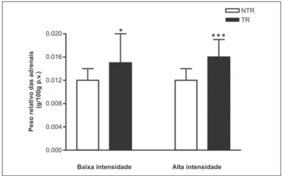
detect-ed in the organs weighting and anamopathological study in the rats from the TR group TR (TR-HI) in relation to those from the NTR group (NTR-HI). No statistically significant differences (P > 0,05) in the relative weight of spleen of animals from the TR group in high intensity (TR-HI) occurred in relation to those in the NTR group (NTR-HI) (figure 2). Likewise, adrenal glands hypertrophy in the rats from the TR groups in low intensity (TR-LI) (P < 0,05) and in high intensity (TR-HI) (P < 0,001) was observed in relation to those animals from the NTR group (NTR-HI) (figure 3).
DISCUSSION
maintenance and these adaptations are also important to the con-trol of several diseases, especially the ones with cardiovascular and endocrine-metabolic nature(37).
Thus, several studies have shown that physical exercises are able to promote changes in many functions of the human and rats‘ bodies. However, the research published so far mentions several results discrepancies for many reasons, such as the use of differ-ent forced or voluntary physical training protocols, the variation in the duration and intensity of the physical exercises and the multi-plicity of used tests for evaluation of the influence of exercises in the investigated parameters(38). Therefore, the comparison of the
effects of different intensities of physical exercises, such as the ones used here, in the modulation of the nociception and other biological parameters, is important in order to try to contribute to the clarification of issues still obscure concerning this subject.
Hence, this paper evaluated the influence of low and high inten-sity exercises on treadmill on the hypernociception modulation, the body mass and the relative weight of the adrenal glands, heart, kidneys and spleen of rats, considering the physical exercises as a means of stressing stimulus capable of inducing changes in the homeostasis state and reorganize responses of several systems, among them the neuroendocrinal system(39). Nonetheless, although
the experimental models used here are standardized and the eval-uations quantified, the research was conducted in animals, which may limit its extrapolation to humans.
The study showed that low intensity physical exercises did not alter the hypernociception threshold at any of the evaluation
mo-ments, once there were not statistically significant differences in this parameter, between the rats in the trained groups (TR) and non-trained ones (NTR). It is known that acute exercises may trig-ger several body’s responses, among them the stress reactions involving the hypothalamus-hypophysis-adrenal axis, which induc-es the CRH liberation (corticotrophin release hormone) and conse-quently stimulates the ACTH secretion and increases synthesis and secretion of glucocorticoid hormones by the adrenal glands. Moreover, these facts generate metabolic adaptations to physical exercise(25). In addition, the animals used in this work were trained
from a protocol whose training intensity corresponds to 50% of the maximal VO2(32-33) and thereby, the obtained results in this
exer-cise intensity corroborate those by Pertovaara and Kemppainen(40)
and by Droste et al.(16), from studies with humans which questioned
whether the low intensity of physical exercise would be sufficient in order to cause analgesic effect.
Characteristically, the induced analgesia by stress may occur through one of the two systems, that is, the opiod and the non-opioid systems(41). It is mentioned in the literature then, that during
inflammatory pain, the CRH may stimulate the opioid peptides re-lease (especially β-endorphins) of several types of immune-cells, producing antinociception in inflamed sites, due to the activation of opioid receptors present in peripheral sensory nervous termina-tions(42).
In this work, the results of hypernociception after low intensity physical training suggest that the neuroendocrine axis (hypothala-mus-hypophysis-adrenal) stimulation was not sufficient so that the circulating CRH indices could promote such effects. Moreover, the non-alteration of the nociception threshold in this exercise intensi-ty may be indicative that such threshold may have balanced if this endogenous opioid system has been activated, once these ani-mals were tested 16 hours after the last physical exercises ses-sion. In research conducted by Shyu et al.(18) with spontaneous
training of rats in metal wheel, increase in the nociception thresh-old after the end of the exercises was observed, which gradually decreased in the six first hours of inactivity followed by the physi-cal exercise.
Besides that, according to Terman et al.(19) and Gamaro et al.(43),
repeated exposition to the same aversive stimulus may lead to an adaptation process to this stimulus. In this work, the rats trained under a low intensity protocol during 12 weeks, may have devel-oped an adaptation to the not very severe exercises and conse-quently, a lower activation of the neuroendocrine axis as well, what, at a certain extent, would explain the fact that an increase in the nociception threshold in this training intensity did not occur.
Figure 1 – – – – – Final body mass (in grams) of rats submitted or not to low and high intensities physical exercises
** P < 0,01 (in relation to the NTR group).
Results expressed as average and standard-deviation, n = 8 to 12 per group.
Figure 2 – – – – – Relative weight of organs (g/100 g l.w.) of rats submitted or not to low (LI) and high (HI) intensities
** P < 0,05 (in relation to the NTR group). ** P < 0,01 (in relation to the NTR group).
Results expressed as average and standard-deviation, n = 8 to 12 per group.
Figure 3 – – – – – Relative weight of the adrenal glands (g/100 g l.w.) of rats sub-mitted or not to low and high intensities physical exercises.
*** P < 0,05 (in relation to the NTR group). *** P < 0,001 (in relation to the NTR group).
On the other hand, Kemppainen et al.(44) reported a significant
increase in the dental pain threshold only after physical exercises using 74% of the individuals‘ maximal aerobic ability Yet, Koltyn(17)
reported significant increase in the pain threshold when a single exercise session is used in bicycle ergometric test respecting a protocol with around 75% of the individuals‘ maximal aerobic abil-ity. The results of this study show that high intensity physical exer-cises increase the hypernociception threshold in the H2, H3, H4 and H6, moments, reinforcing the data already published.
Concerning this topic, maybe high intensity physical exercises here used, differently from the low intensity ones, are a stressor stimulus more intense to animals, and, hence, are able to trigger more evident neuroendocrine responses in the body, with increase in the seric indices of CRH, ACTH and glucocorticoid hormones. Such hypothesis can be corroborated by the bigger hypertrophy of the adrenal glands of the rats under high intensity training than in the ones under low intensity training. Therefore, it is possible that a rats‘ body adaptation to the high intensity exercises would be more difficult in comparison to low intensity ones. According to this hypothesis, the results of other research in rats, are conclu-sive in the demonstration that exercises promote hypertrophy of the adrenal glands(31-45) and that the physical training intensity
influ-ences the intensity-dependent response of the hypothalamus-hy-pophysis-adrenal axis activation(46). Therefore, the results obtained
here suggest the participation of these hormones in the antinoci-ceptor effects, even if the rats have been evaluated after 16 hours of the end of physical training.
Another justification to be considered for the results obtained in this work about hypernociception after high intensity exercises, is the activation of the peripheral opioid system (activation of opioid receptors present in peripheral sensory nervous terminations) through the release of opioid peptides of inflamed tissues, espe-ciallyβ-endorphins, stimulated by the plasmatic high indices of CRH and IL-1 (Interleukin 1), triggered by such exercises. These find-ings support the ideas by Schäfer et al.(47) and Mousaet al.(42), who
reported that, in the inflammatory pain, the endogenous and exog-enous CRH and IL-1 are able to release opioid peptides of inflamed tissues and that these immune derivate opioids would be released by conditions that involve environmental stress, resulting in noci-ception inhibition.
In this research, no alteration in the final body mass of the ani-mals submitted to low intensity physical exercises was observed, although the TR group animals were visually smaller in relation to the control group (NTR). The fact that no statistically significant differences were observed in the end of the training may be indi-cation of a substitution of adipose tissue (fat) for muscular mass in the TR group animals, according to indication by Mayer et al.(48).
On the other hand, it was observed that the final body mass of rats trained with high intensity exercises was smaller after 11 weeks period of physical training, suggesting increase in the energy use in these animals. These results corroborate the literature with trained rats on treadmill under high intensity protocols, which re-duce the food intake with consequent weight loss(31-48). As
men-tioned before and reinforcing such idea, physical exercises repre-sent a type of stress and promote CRH release by the hypothalamus due to the dependence of the stressor agent intensity. Actually, Rivest and Richard(49) demonstrated anorexigenic effects for the
CRH, simulating hence, the effects triggered by exercises. In this context, the high intensity physical exercises applied in this study seem to actually represent a stressor stimulus sufficiently capable to activate the hypothalamus-hypophysis-adrenal axis, consequently increasing the seric indices of CRH.
Likewise, Stevenson et al.(50) mentioned that the degree of
ap-petite decrease may be related to the stress degree provoked by the physical exercise intensity in animals and that such anorexia
may be mediated by the high indices of catecholamines derived from this stress. Accordingly, the histopathological finding of this work showing hypertrophy of the medullar and cortical areas of the adrenal glands is indicative of a stress situation in these ani-mals.
Furthermore, countless variables may induce cardiac hypertro-phy in rats submitted to hypertro-physical exercises, such as the frequency, intensity and duration of the physical training sessions, besides the training program duration. Other factors include the age at the beginning of the training, sex and type of animal used in the phys-ical training(51). Thus, the results found in this research related to
the heart’s relative weight after low and high intensities training, agree with the authors mentioned above. In addition, low intensity exercises did not cause increase in the heart weight of trained rats, in relation to the non-trained ones. However, there was in-crease in the relative weight of these organs and myocytes hyper-trophy, detected through the histopathological analysis of the rats trained at high intensity.
Despite everything, the heart is not the only organ that presents adaptation alterations after physical exercises; the spleen’s rela-tive weight may also present alteration, but the results are rather conflicting(31). Bloor et al.(45) did not detect significant alterations in
the relative weight of spleen in adult rats trained with swimming exercises, 1 daily hour for 10 weeks. Likewise, decrease in the spleen weight in exercised rats has been reported, possibly due to a higher blood expelling stored in the organ(52) or still, due to the
fact that physical exercises promote an increase in the synthesis and secretion of corticosterone (glucocorticoid hormone), which would lead to a decrease of the splenic tissue(31). The
anatomopatho-logical findings of this study did not reveal alterations in the spleen’s relative weight and, therefore, do not agree with the results in the literature.
Furthermore, the histopathological analyses showed increase in the relative weight and moderate congestion of the kidneys of trained animals at high intensity and moderate renal congestion, without alteration in the kidney weight of rats trained at low inten-sity. According to Shizuru et al.(53), the renal responses to physical
exercises are related to their intensity, therefore, exercises per-formed at low intensities increase the urinary flow and the sodium excretion, while at high intensities, these two parameters consid-erably decrease. Maybe such decrease is due to the high plasmat-ic indplasmat-ices of aldosterone, hormone whplasmat-ich progressively increases, reaching up to six times more than the indices observed in resting bodies, as means of keeping the body liquids and the homeosta-sia. Actually, Oliveira et al.(54) reported that the primary function of
the kidneys is to regulate the volume and composition of the extra cellular liquid, and hence, these alterations that occur during the performance of physical exercises may generate hemodynamic changes and changes in the sodium and water excretion. The find-ings of this study concerning the kidneys weight of the trained animals do not confirm the findings of the literature(45-55), showing
decrease in this organ’s weight, maybe by the decrease of the number of glomerulus caused by the hypoxia conditions imposed by the exercises. One of the possible causes of this results diver-gence may be the fact that the research was performed with rats too young for the physical training, differently from the age of the rats from this study.
As a result, one may suggest that the type, intensity, frequency and duration of the physical exercises, besides other factors, may affect the nociception and other biological variables in rats, depend-ing on the adopted exercises protocol.
REFERENCES
1. Minor MA, Hewert JE, Webel RR. Anderson SK, Kay DR. Efficacy of physical conditioning exercise in patients with rheumatoid arthritis and osteoarthritis. Arthritis Rheum 1989;32:1396-405.
2. Gurwitz D. Physical activity: good for your health, very good for your gene ex-pression. Clin Genet 2000;57:249-56.
3. Caspersen CJ, Powell KE, Christenson GM. Physical activity, exercise and phys-ical fitness: definitions and distinctions for health-related research. Public Health Rep 1985;100:126-31.
4. Nieman DC. Exercise immunology: practical applications. Int J Sports Med 1997; 18:S91-100.
5. Marcus BH, King TK, Clark MM, Pinto BM, Bock BC. Theories and techniques for promoting activity behaviours. Sports Med 1996;22:321-31.
6. Nieman DC. Influence of carbohydrate on the immune response to intensive prolonged exercise. Exerc Immunol 1998;4:64-76.
7. Fehr HG, Lotzerich H, Michna H. Human macrophage function and physical ex-ercise: phagocytic and histochemical studies. Eur J Appl Physiol 1989;58:613-7.
8. Woods JA, Davis JM, Mayer EP, Ghaffar A, Pate RR. Exercise increases inflam-matory macrophage anti-tumor cytotoxicity. J Appl Physiol 1983;75:879-86.
9. Phillips J, Murry P, Crocker J. The biology of disease. England: Blackwell Sci-ence, 1995;22-9.
10. Kumar V, Abbas AK, Fausto N. Robbins e Cotran: bases patológicas das doen-ças. Rio de Janeiro: Elsevier, 2005;49-89.
11. Gallin JI, Goldstein MI, Snyderman R. Inflammation: basic principles clinical cor-relates. New York: Raven Press, 1992;1-4.
12. Guyton AC, Hall JE. Tratado de fisiologia médica. Rio de Janeiro: Guanabara Koogan, 2002;516-26.
13. Tribioli RA. Análise crítica atual sobre a TENS envolvendo parâmetros de estim-ulação para o controle da dor (dissertação de mestrado). Ribeirão Preto: Univer-sidade de São Paulo, 2003.
14. Koltyn KF. Analgesia following exercise: a review. Sports Med 2000;29:85-98.
15. Kemppainen P, Paalasmaa P, Pertovaara A. Dexamethasone attenuates exer-cise-induced dental analgesia in man. Brain Res 1990;519:329-32.
16. Droste C, Greenlee MW, Schrek M. Experimental pain thresholds and plasma beta-endorphin levels during exercise. Med Sci Sports Exerc 1991;23:334-42.
17. Koltyn KF, Wertz AL, Gardiner RL. Perception of pain following aerobic exercise. Med Sci Sports Exerc 1996;28:1418-21.
18. Shyu BC, Andersson SA, Thorén P. Endorphin mediated increase in pain thresh-old induced by long-lasting exercise in rats. Life Sci 1982;30:833-40.
19. Terman GW, Morgan MJ, Liebskind JC. Opioid and non-opioid stress analgesia from cold water swim: importance of stress severity. Brain Res 1986;372:167-71.
20. Hoffman PL, Terenius L, Thorén P. Cerebrospinal fluid immunoreactive beta-endorphin concentration is increased by long-lasting voluntary exercise in the spontaneously hypertensive rat. Regul Pept 1990;28:233-9.
21. Janal MN, Colt EWD, Clark WC, Glusman M. Pain sensitivity, mood and plasma endocrine levels in man following long-distance running: effects of naloxone. Pain 1994;19:13-25.
22. Thorém P, Floras JS, Hoffmann P, Seals DR. Endorphins and exercise: physio-logical mechanism and clinical implications. Med Sci Sports Exerc 1990;22:417-28.
23. Moerch H, Pedersen BK. Beta-endorphin and the immune system: possible role in autoimmune diseases – A review. Autoimmunity 1995;21:161-71.
24. Pestell RG, Hurley DM, Vandongen R. Biochemical and hormonal changes dur-ing 1000 km ultramarathon. Clin Exp Pharmacol Physiol 1989;16:353-61.
25. Sothmann MS, Hart BA, Horn TS. Exercise training and cross-stressor adapta-tion hypothesis. Exerc Sports Sci Rev 1996;24:267-87.
26. Prada FJ, Carneiro EM, Azevedo JRM, Luciano E. Respostas endócrino-metabó-licas em ratos diabéticos. Rev Bras Ativ Fís Saúde 1997;2:22-9.
27. Pedersen BK, Hoffman-Goetz L. Exercise and the immune system: regulation, integration and adaptation. Physiol Rev 2000;80:1055-81.
28. Parízkova J, Stanková L. Influence of physical activity on a treadmill metabolism of adipose tissue in rats. Br J Nutr 1964;18:25-32.
29. Thomas BM, Miller AT. Adaptation to forced exercise in the rat. Am J Physiol 1958;193:350-4.
30. Bloor CM, Leon AS, Pasyk S. The effect of exercise on organ and cellular devel-opment in rats. Lab Invest 1968;19:675-80.
31. Ostman-Smith I. Exercise and organ size. Acta Physiol Scand 1979;477:1-40. 32. Brooks GA, White TP. Determination of metabolic and heart rate responses of
rats to treadmill exercise. J Appl Physiol. 1978;45(6):1009-15.
33. Silva GJJ. Efeito do treinamento físico de baixa intensidade sobre a sensibilida-de dos aferentes pressorreceptores, barorreflexo e reflexo cardiopulmonar em ratos espontaneamente hipertensos (dissertação de mestrado). São Paulo: Uni-versidade de São Paulo, 1999.
34. Ramires PR. Efeitos do treinamento físico e da dieta suplementada com glutati-ona na resposta à isquemia-reperfusão cardíaca in vivo no rato (tese de doutora-do). São Paulo: Universidade de São Paulo, 1999.
35. Hargreaves KM, Dubner R, Brow F, Flores C, Joris J. A new and sensitive meth-od for measuring thermal nociception in cutaneous hyperalgesia. Pain 1988;32: 77-88.
36. Costello AH, Hargreaves KM. Suppression of carrageenan-induced hyperalge-sia, hyperthermia and edema by bradykinin antagonist. Eur J Pharmacol 1989; 71:259-63.
37. Cartee GD, Farrar RP. Exercise training induces glycogen sparing during exer-cise by old rat. J Appl Physiol 1988;64:259-65.
38. Burghardt PR, Fulk LJ, Hand GA, Wilson MA. The effects of chronic treadmill and wheel running on behavior in rats. Brain Res 2004;1019:84-96.
39. Rosa LPBC, Vaisberg MW. Influências do exercício na resposta imune. Rev Bras Med Esporte 2002;8:167-72.
40. Pertovaara A, Kemppainen P. Comments on Padawer and Levine. Pain 1992;50: 239-40.
41. Tsuda A, Ida Y, Satoh H, Tsujimmaru S, Tanaka M. Stressor predictability and rat brain noradrenaline metabolism. Pharmacol Biochem Behav 1989;32:69-72.
42. Mousa A, Bopaiah CP, Stein C, Schafer M. Involvement of corticotropin-releas-ing hormone receptor subtypes 1 and 2 in peripheral opioid-mediated inhibition of inflammatory pain. Pain 2003;106:297-307.
43. Gamaro GD, Xavier MH, Denardin JD, Pilger J, Ely DR, Ferreira MBC, et al. The effects of acute and repeated restraint stress on the nociceptive response in rats. Physiol Behav 1998;63:693-97.
44. Kemppainen P, Pertovaara A, Huopaniemi T, Ohansson G, Karonen S. Modifica-tion of dental pain and cutaneous termal sensitivity by physical exercise in man. Brain Res 1985;360:33-40.
45. Bloor CM, Pasyk S, Leon AS. Interaction of age and exercise on organ and cellu-lar development. Am J Pathol 1970;5:185-98.
46. Chennaoui M, Merino D, Lesage J, Drogou C, Guezennec CY. Effects of moder-ate and intensive training on the hypothalamo-pituitary-adrenal axis in rats. Acta Physiol Scand 2002;175:113-21.
47. Schäfer M, Carter L, Stein, C. Interleucin 1β and corticotropin-releasing factor inhibit pain by releasing opioids from immune cells in inflamed tissue. Proc Natl Acad Sci 1994;91:4219-23.
48. Mayer J, Marshall NB, Vitale JJ, Christensen JH, Mashayekhi MB, Stare EJ. Exercise, food intake and body weight in normal rats and genetically obese adult mice. Am J Physiol 1954;177:544-48.
49. Rivest S, Richard D. Hypothalamic paraventricular nucleus lesions do not pre-vent anorectic effect of exercise in male rats. Am J Physiol 1990;259:579-84. 50. Stevenson JAF, Box BM, Feleki V, Beaton JR. Bouts of exercise and food intake
in the rat. J Appl Physiol 1966;2:118-22.
51. Scheuer J, Tripton CM. Cardiovascular adaptations to physical training. Ann Rev Physiol 1977;39:221-51.
52. Gollnick PD, Struck PJ, Bogyo TP. Lactic dehydrogenase activities of rat heart and skeletal muscle after exercise and training. J Appl Physiol 1967;22:623-7. 53. Shizuru EM, Freud BJ, Hashiru GM, Clay-Baugh JR. Hormonal electrolyte and
renal responses to exercise are intensity dependent. J Appl Physiol 1991;70: 900-6.
54. Oliveira AO, Fileto C, Melis MS. Effect of strenuous maternal exercise before and during pregnancy on rat progeny renal function. Braz J Med Biol Res 2004; 37:907-11.