Rat hindlimb jo int immo bilizatio n
with acrylic re sin o rtho se s
Programa de Pós-Graduação, Departamento de Fisioterapia, Universidade Metodista de Piracicaba, Piracicaba, SP, Brasil C.A. da Silva, R.R.J. Guirro,
M.L.O . Polacow, K.M. Cancelliero and J.L.Q . Durigan
Abstract
The objective of the present study was to propose an orthosis of light material that would be functional for the animal and that would maintain only the ankle joint immobilized. Male Wistar rats (3 to 4 months old, 250-300 g) were divided into 2 groups (N = 6): control and immobilized for 7 days. Rats were anesthetized with sodium pentobarbital (40 mg/kg weight) and the left hindlimb was immobi-lized with the orthoses composed of acrylic resin model, abdominal belt and lateral supports. The following analyses were performed: glycogen content of the soleus, extensor digitorum longus, white gastrocnemius, red gastrocnemius, and tibialis anterior muscles by the phenol sulfuric method, and the weight, fiber area and intramuscular connective tissue of the soleus by the planimetric system. Data were analyzed statistically by the Kolmogorov-Smirnov, Student t and Wilcoxon tests. Immobilization decreased glycogen in all muscles (P < 0.05; soleus: 31.6%, white gastrocnemius: 56.6%, red gastrocne-mius: 39%, extensor digitorum longus: 41.7%, tibialis anterior: 45.2%) in addition to reducing soleus weight by 34% (P < 0.05). Furthermore, immobilization promoted reduction of the fiber area (43%, P < 0.05) and increased the connective tissue (200%, P < 0.05). The orthosis model was efficient comparing with another alternative immobiliza-tion model, like plaster casts, in promoting skeletal muscle alteraimmobiliza-tions, indicating that it could be used as a new model in other studies related to muscle disuse.
Co rre spo nde nce
R.R.J. Guirro
Universidade Metodista de Piracicaba Rodovia do Açúcar, km 156 13400-911 Piracicaba, SP Brasil
E-mail: rjguirro@ unimep.br
Research supported by FAP/UNIMEP.
Received O ctober 21, 2005 Accepted April 10, 2006
Ke y words
•Immobilization
•Hypotrophy
•Joint
•O rthoses
•Physical therapy
Intro ductio n
Muscular hypotrophy induced by disuse is a condition frequently found in the physio-therapeutic clinic which can occur in asso-ciation with orthopedic disorders such as ligament ruptures, bone fractures, muscular and medulla lesions, inflammatory processes, degenerative joint and muscular pathologies, as well as in situations in which patients are
confined to bed for long periods of time for medical or surgical reasons (1).
Although immobilization has been ex-tensively studied, more investigation is still required because of the wide variation in responses resulting from joint position, num-ber of joints immobilized, application time, material used, and whether or not there is load on the limb.
A frequently used model is limb suspen-sion, in which there is no weight load, and several studies have associated this model with other techniques. Nemirovskaya and Shenkman (4) associated hindlimb suspen-sion with ankle immobilization in a neutral position and a platform to lean the limb on. In the Tanaka et al. (5) study, suspension was associated with fixing the ankle with steel wire.
The models described in the literature induce a joint position that permits the muscle to remain in a stretched or shortened condi-tion. Some studies have used the plantar flexion position to maintain the soleus muscle in the shortened position, with the extensor digitorum longus and tibialis anterioris muscles thus remaining in a stretched posi-tion (6-8). Wagatsuma et al. (9) induced ankle plantar flexion by cast application, maintaining the joint in the maximum posi-tion. Sakakima et al. (10) used 70º position-ing and Ahtikoski et al. (6), in addition to studying a group that maintained plantar flexion (150-160º), also used a group main-taining ankle dorsiflexion (30-40º). The neu-tral position of the ankle joint is also used to keep the muscles in a resting condition with-out muscular tension (3,11,12).
These studies also differed in terms of the type of material type used for joint im-mobilization. In the Wagatsuma and Yamada (13) study, epoxy resin was used to maintain ankle plantar flexion. In other studies, the neutral position of the ankle differed accord-ing to the choice of material, such as hexcelite or fixed needles (3,11) or plaster casts ap-plied for different periods of time (3,12).
In 2002, Coutinho et al. (14) proposed the use of cotton tissue, steel mesh and
adhe-sive tape to analyze the soleus, which re-mained shortened, and the tibialis anterioris, which was stretched. Jarvinen et al. (8) im-mobilized rat left hindlimbs with casts, main-taining the knee in flexion and the ankle in extension, or vice versa, using the contralat-eral limb as control.
An aspect observed in the immobiliza-tion studies is that more than one joint was immobilized, including the ankle, the knee, the hip, and even the pelvis. Thus, the aim of the present study was to propose an orthosis of light material that would be functional for the animal and that would maintain only one joint immobilized, in this case the ankle kept in a neutral position, leaving the knee and hip joints free, allowing weight load.
Mate rial and Me thods
O rthosis pre paration
Orthosis preparation followed several stages used in dentistry, related to the manu-facturing of dental prostheses. Traditionally, acrylic molding models and temporary pros-theses are made according to the Phillips methodology (15) that involves the follow-ing steps:
1E) was separated from the alginate and its parts were scraped and sanded. 4) Expulsivity: with a lamp and spatula No. 7, wax was placed inside the retention points to create expulsivity areas, but without affecting the anatomical shape of the limb. 5) Acrylic resin application: the plaster model was dipped in an insulator to create an insulator film. After drying, the methyl methacrylate polymer (powder) and the mono-mer (liquid) were mixed in a glass container to produce the following phases: sandy, sticky, plastic, and rubbery. In the sticky phase, the resin was transferred from the container to the surface of the plaster model and fitted, sculpted and adjusted to the model by hand and with a small Lecron spatula. 6) Acrylization: the acrylic resin was applied to the model, chemi-cally activated, and tested for exothermal re-action. 7) Expulsion: after acrylization, the acrylic resin model (Figure 1F) was polished with stone and polishing eraser and cut later-ally with a carbaryl disk. The retention angles were reduced with a number 5 drill and polish-ing stone.
Immobilization proce dure
Male Wistar rats (3 to 4 months old, 250-300 g) were maintained in controlled animal house conditions with free access to food and water and treated in accordance with the recommendations of the Guide on the care and use of laboratory animals (16).The ani-mals were placedin groups of 3 in 40 x 30 cm boxes lined with newspaper in order to prevent sawdust from entering the internal compartment of the orthoses and cause skin lesions.
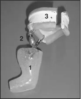
The animals were divided into two groups (N = 6): control and immobilized for 7 days. To prepare the orthoses, the rats were anes-thetized with sodium pentobarbital (40 mg/ kg weight) and their left hindlimbs were immobilized with the acrylic resin models (Figure 2) adapted in combination with poly-vinyl chloride or poly-vinyl belts 40 mm in diam-eter (Figure 1G) covered with latex and
at-A B C
D E F
G H I
Figure 1. Sequence of the process or orthosis preparation. A and B, positioning of the ankle joint in a neutral position; C, limb molding with potassium alginate; D, placement of the plaster in the alginate mold; E, plaster mold; F, acrylic resin model; G, abdominal belt; H, supports; I, bilateral supports coupled to the abdominal belt.
1
2
3
tached to two 15-mm long lateral supports (Figure 1H) which allowed movement (Fig-ure 3).
After the experimental period, the fol-lowing analyses were performed: glycogen content of the soleus, extensor digitorum longus, white gastrocnemius, red gastrocne-mius, and tibialis anterior muscles, as well as the weight, fiber area and intramuscular con-nective tissue of the soleus.
The muscle samples were submitted to digestion with hot 30% KOH and the glyco-gen was precipitated by ethanol to determine muscle glycogen (17). Between the precipi-tation steps the sample was centrifuged at 1612.8 g for 15 min. The precipitated glyco-gen was submitted to acid hydrolysis in the presence of phenol and its values are
re-ported as mg/100 mg wet weight.
For morphometric analysis, the ventral segment of the soleus was fixed in buffered 10% formol solution. The tissue was embed-ded in paraffin, cut into several non-serial 7-µm thick cross-sections and stained with hematoxylin-eosin.
The image analysis system used con-sisted of the Image Pro-plus 4.0 software (Media Cybernetics, Silver Spring, MD, USA) and a digital camera (JVC®
manufac-turer, Lawrenceville, GA, USA) coupled to a microscope (Zeiss, Narberth, PA, USA) and connected to a microcomputer. All the images were captured at 10X magnification. The areas of 375 soleus muscle fibers from each animal were analyzed as follows: 15 fibers per area in 5 areas per section, and a total of 5 sections per animal. A square reticulum was used for randomly choosing 15 fibers per cuts that coincided with the straight intersections.
A planimetry system was used for the analysis of intramuscular connective tissue density by scoring points by means of a reticulum with 2500-µm2 squares containing
56 straight line intersections. The coincident points in the endomysium and perimysium in 5 areas per section in 5 sections per animal corresponded to a total of 1400 points per animal.
The relative area of connective tissue (area density) was calculated by dividing the sum of the number of coincident points in the straight line intersections in connective tissue (endomysium and perimysium) by the total number of points.
Although five hindlimb muscles were chosen for metabolic analysis, only the so-leus was used for morphometric analysis because, by being monoarticular and by pre-dominantly containing type I fibers, it is the muscle that presents the highest degree of atrophy.
Statistical analysis was initially performed by the Kolmogorov-Smirnov normality test. The muscle weight and glycogen data, which
A B
Figure 3. Orthosis adaptation to the animal’s left hindlimb, main-taining the ankle joint in a neu-tral position (A) and placing the weight load on the hindlimb, without interfering with walking (B).
Table 1. Effect of immobilization on muscle glycogen concentration, soleus fiber and connective tissue area of rat submitted to joint immobilization for 7 days.
Control Immobilized
Glycogen (mg/100 mg muscle)
Soleus 0.38 ± 0.06 0.26 ± 0.03* White gastrocnemius 0.46 ± 0.06 0.20 ± 0.05* Red gastrocnemius 0.41 ± 0.04 0.25 ± 0.09* Extensor digitorum longus 0.36 ± 0.08 0.21 ± 0.06* Tibialis anterior 0.31 ± 0.09 0.19 ± 0.07*
Soleus fiber area (µm2)
1st quartile 2180 1197
Median 2496 1423*
3rd quartile 2879 1659
Soleus connective tissue area (%)
1st quartile 7.1 25.0
Median 8.9 26.8
3rd quartile 10.7 30.4
Data are reported as means ± SD (glycogen) and medians (fiber and connective tissue area) for 6 animals per group.
presented normal distribution, were analyzed by the Student t-test, whereas the data con-cerning muscle fiber area and connective tissue density, which did not present normal distribution, were analyzed by the nonpara-metric Wilcoxon test. The level of signifi-cance was set at P < 0.05 for all analyses.
Re sults
The skeletal muscle of rats submitted to hindlimb immobilization for 7 days presented a significant decrease (P < 0.05) in glycogen content indicated by a reduction of 31.6% in the soleus, 56.6% in the white gastrocne-mius, 39% in the red gastrocnegastrocne-mius, 41.7% in the extensor digitorum longus, and 45.2% in the tibialis anterior muscles, suggesting a functional integration between homeostasis in the fiber contractile process and carbohy-drate control (Table 1).
The soleus muscle, which was chosen for weight evaluation, showed a 34% reduction in weight (control: 123.5 ± 5.28 mg, immo-bilized: 81.3 ± 4.63 mg, P < 0.05), suggest-ing proteolysis resultsuggest-ing from disuse and osmotic mobilization of active energy re-serves.
Immobilization also promoted alterations in the morphology of the soleus muscle, characterized by a 43% reduction in fiber area (P < 0.05) and by a 200% increase in connective tissue (P < 0.05; Table 1, Figure 4A,B).
The orthosis, which weighed 22.72 ± 2.25 g (mean ± SD), did not interfere with the animal’s ability to walk since its weight was thrown onto the immobilized limb, with a blocked limb movement and anterior and lateral hip movements. It is important to emphasize that the animals showed no change in fluid or solid intake, or any skin lesions in the limb.
D iscussio n
The effectiveness of the orthoses in
pro-moting metabolic and morphologic alter-ations in the hindlimb was observed after a period of 7 days. Previous studies had dem-onstrated that most changes in the skeletal muscle system occur during the first seven days of muscle disuse (18). It has been ob-served that the greater vulnerability of the slow (type I) compared to the fast muscle fibers (type II) is due to metabolic ences. Immobilization seems to have ent effects on protein synthesis by the differ-ent types of muscle fibers (7). In has been reported that oxidative enzymes respond with decreased activity during immobilization, suggesting that the muscle fibers with pre-dominantly oxidative metabolism (type I) are more vulnerable to muscle atrophy (19). In addition to the susceptibility to inher-ent atrophy of type I fiber metabolism, other factors that determine this condition are the characteristics of postural fibers. Ploug et al. (11) reported that the greater susceptibility of the soleus to atrophy is related to inactiv-ity because it is a postural muscle that has greater basal activity than non-postural muscles. In a recent study, it was observed that immobilization for two weeks, in addi-tion to promoting significant reducaddi-tion of slow fibers (type I), also promoted a signifi-cant increase in fast muscle fibers (type IIc) compared to control (5).
These studies corroborate Lieber’s affir-mation (20) and support the findings of other studies showing that the muscles considered
A
B
50 µm
50 µm Figure 4. Soleus muscle fibers
to be antigravitational, monoarticular and possessing a larger proportion of slow fi-bers, are the most vulnerable to atrophy induced by muscle disuse.
On this basis, we chose the soleus muscle, predominantly composed of type I fibers, for morphological analysis because of its greater susceptibility to muscle atrophy from disuse. The analysis of glycogen content was also done in this muscle, in addition to the others that compose the triceps sural and anterior compartment of the hindlimb.
The immobilized soleus presented a sig-nificant reduction in weight, in glycogen reserves and in fiber area, as well as an increase in connective tissue, showing the interrelation of contractile activity with en-ergy homeostasis and muscle fiber morphol-ogy that would appear to indicate a condi-tion of muscle atrophy. Similar results were reported by Fournier et al. (21) who immobi-lized the soleus and gastrocnemius muscles in shortened, neutral and stretching posi-tions. Their results showed that the muscles immobilized in the shortened and neutral positions lost a significant amount of weight after 28 days. There was a weight reduction of 55% in the soleus muscle in the neutral position and 77% in the shortened position; in the gastrocnemius muscle the reduction was of 54% in the neutral position and of 53% in the shortened position compared to control.
Generally speaking, immobilization for different periods of times promotes atrophy ranging from 15 to 70%, depending on the animal species and on the fibers evaluated (12). Kannus et al. (22) showed a reduction of 69% in the fiber area of the soleus immo-bilized with a plaster cast for 3 weeks.
Regarding the connective tissue of the soleus muscle, the present results are similar to those of other investigations that showed an increase in density in the immobilized muscle condition. Williams et al. (2) ob-served an increase in the amount of connec-tive tissue in the perimysium of the soleus
muscle immobilized in the shortened posi-tion for 2 days. They also observed that the collagen fibers of the perimysium presented a more acute angle than observed in normal muscles, with consequent decreased muscu-lar elasticity and increased passive tension. Other studies have shown morphological alterations caused by immobilization, dem-onstrating that the increase in connective tissue occurs in the endomysium and per-imysium (23). Others also observed that there is increased collagen turnover in the connec-tive tissue during immobilization (22). In-disputably, the amount of intramuscular con-nective tissue increased dramatically in vari-ous disuse models (50-700%) during immo-bilization, tenotomy or denervation (23).
Muscle disuse caused by conditions of long periods in bed, orthoses or fixations on limbs and unweighted conditions induce in-sulin resistance and a catabolic state in the affected skeletal muscles of humans. How-ever, it is still unclear how chronic muscle disuse or immobilization alters insulin sig-naling (3). Insulin resistance explains the compromised glycogen reserves due to im-mobilization in all muscles analyzed in the present study.
Hirose et al. (3) studied the signaled insu-lin pathway in rats that had their left hind limb immobilized by fixation of the knee and ankle to 90º for 7 days, and verified a reduction in the transduction of the intracel-lular signal stimulated by insulin, suggest-ing deficit in insulin receptor activation, and in other molecules including insulin recep-tor substrate 1 phosphorylation and activa-tion of phosphatidylinositol 3-kinase, indi-cating that insulin resistance can also be caused by immobilization.
resistant to the movements of the animal. In addition to promoting skeletal muscle alter-ations, the type of rat hindlimb
immobiliza-tion proposed here, due to it structure, also allowed the animal to be functional, thus deserving further study.
Re fe re nce s
1. Reardon KA, Davis J, Kapsa RM, Choong P, Byrne E. Myostatin, insulin-like growth factor-1, and leukemia inhibitory factor mRNAs are upregulated in chronic human disuse muscle atrophy. Muscle Nerve 2001; 24: 893-899.
2. Williams PE, Catanese T, Lucey EG, Goldspink G. The importance of stretch and contractile activity in the prevention of connective tissue accumulation in muscle. J Anat 1988; 158: 109-114. 3. Hirose M, Kaneki M, Sugita H, Yasuhara S, Martyn JA.
Immobiliza-tion depresses insulin signaling in skeletal muscle. Am J Physiol Endocrinol Metab 2000; 279: E1235-E1241.
4. Nemirovskaya TL, Shenkman BS. Effect of support stimulation on unloaded soleus in rat. Eur J Appl Physiol 2002; 87: 120-126. 5. Tanaka T, Kariya Y, Hoshino Y. Histochemical study on the changes
in muscle fibers in relation to the effects of aging on recovery from muscular atrophy caused by disuse in rats. J Orthop Sci 2004; 9: 76-85.
6. Ahtikoski AM, Koskinen SO, Virtanen P, Kovanen V, Takala TE. Regulation of synthesis of fibrillar collagens in rat skeletal muscle during immobilization in shortened and lengthened positions. Acta Physiol Scand 2001; 172: 131-140.
7. Heslinga JW, te Kronnie G, Huijing PA. Growth and immobilization effects on sarcomeres: a comparison between gastrocnemius and soleus muscles of the adult rat. Eur J Appl Physiol Occup Physiol
1995; 70: 49-57.
8. Jarvinen MJ, Einola SA, Virtanen EO. Effect of the position of immobilization upon the tensile properties of the rat gastrocnemius muscle. Arch Phys Med Rehabil 1992; 73: 253-257.
9. Wagatsuma A, Fujimoto K, Yamada S. Effect of treatment with nifedipine, an L-type calcium channel blocker, on muscular atrophy induced by hindlimb immobilization. Scand J Med Sci Sports 2002; 12: 26-30.
10. Sakakima H, Yoshida Y, Sakae K, Morimoto N. Different frequency treadmill running in immobilization-induced muscle atrophy and ankle joint contracture of rats. Scand J Med Sci Sports 2004; 14: 186-192.
11. Ploug T, Ohkuwa T, Handberg A, Vissing J, Galbo H. Effect of
immobilization on glucose transport and glucose transporter expres-sion in rat skeletal muscle. Am J Physiol 1995; 268: E980-E986. 12. Qin L, Appell HJ, Chan KM, Maffulli N. Electrical stimulation
pre-vents immobilization atrophy in skeletal muscle of rabbits. Arch Phys Med Rehabil 1997; 78: 512-517.
13. Wagatsuma A, Yamada S. Specific protein alteration in the soleus following immobilization-atrophy. Scand J Med Sci Sports 2000; 10: 205-210.
14. Coutinho EL, Gomes AR, Franca CN, Salvini TF. A new model for the immobilization of the rat hind limb. Braz J Med Biol Res 2002; 35: 1329-1332.
15. Phillips RW. Materiais dentários de skinner. Rio de Janeiro: Guanabara Koogan; 2001.
16. National Research Council. Guide for the care and use of laboratory animals. Washington: National Academy Press; 1996.
17. Lo S, Russell JC, Taylor AW. Determination of glycogen in small tissue samples. J Appl Physiol 1970; 28: 234-236.
18. Okita M, Yoshimura T, Nakano J, Motomura M, Eguchi K. Effects of reduced joint mobility on sarcomere length, collagen fibril arrange-ment in the endomysium, and hyaluronan in rat soleus muscle. J Muscle Res Cell Motil 2004; 25: 159-166.
19. Appell HJ. Muscular atrophy following immobilisation. A review.
Sports Med 1990; 10: 42-58.
20. Lieber RL. Skeletal muscle structure, function, and plasticity, the physiological basis of rehabilitation. Philadelphia: Lippincott; 2002. 21. Fournier M, Roy RR, Perham H, Simard CP, Edgerton VR. Is limb
immobilization a model of muscle disuse? Exp Neurol 1983; 80: 147-156.
22. Kannus P, Jozsa L, Jarvinen TL, Kvist M, Vieno T, Jarvinen TA, et al. Free mobilization and low- to high-intensity exercise in immobili-zation-induced muscle atrophy. J Appl Physiol 1998; 84: 1418-1424.