Licenciado sob uma Licença Creative Commons DO): http://dx.doi.org. . / - . . .AO
[T]
Histomorphometric alterations of muscle
soleus provoked by drawn out immobilization:
experimental study with wistar lineage rats
[)]
Alterações histomormofétricas do músculo sóleo
provocadas pela imobilização prolongada: estudo
experimental com ratos da linhagem wistar
[A]
Priscila Daniele de Oliveira[a, b], Célia Regina de Godoy Gomes[c], Rodrigo Franco de Oliveira[b],
Deise A. de Almeida Pires-Oliveira[b], Sonia Maria Marques Gomes Bertolini[c, d]*
[a] Universidade Estadual de Londrina UEL , Londrina, PR, Brazil [b] Universidade Norte do Paraná Unopar , Londrina, PR, Brazil [c] Universidade Estadual de Maringá UEM , Maringá, PR, Brazil [d] Centro Universitário de Maringá Cesumar , Maringá, PR, Brazil
[R]
Abstract
Objective: This study has as objective to analyze the effect of joint immobilization of the soleus muscle
of posterior members of rats on morphometric profile view, at periods of and days. Materials and methods: Ten male Rattus novergicus albinus, Wistar variety, were used, separated into two groups group
290
) and )) , of animals each, with the first group submitted to immobilization for days and the second for days. The experiment control was acquired from the right contralateral member of each animal. The immobilization of the left posterior member was done by adapted orthesis. The morphometric analysis of soleus was by non-serial cross sections, μm thick. Results: From the obtained images, the muscle fibers
areas were analyzed, including the form of the fibers, comparing with the control group. )n both the periods of immobilization cross sections of staple fibres had reduction of the section area, front to the comparison to its respective member has controlled, with significant important p < . , especially it enters the experimental groups of and days. Conclusion: )t can be concluded that immobilization to articulate
of rats per and days modifies the section area more transversa of staple fibres of the sóleo muscle and deleterious effect of more significant the cellular components for drawn out periods.
[P]
Keywords: )mmobilization. Rats. Skeletal muscle.
[B] Resumo
Objetivo: Este estudo teve como objetivo verificar os efeitos da imobilização articular no músculo sóleo de ratos, por meio de uma análise histomorfométrica, em períodos de 21 e 45 dias. Materiais e métodos: Foram utili-zados 10 Rattus novergicus albinus machos, variedade Wistar (3 a 4 meses, massa corpórea entre 250–300 g). A amostra foi dividida em dois grupos (G1 e G2), com 5 animais em cada, sendo o primeiro grupo submetido à imobilização por 21 dias e o segundo por 45 dias. O controle do experimento foi obtido a partir do membro con-tralateral direito do respectivo animal. A imobilização do membro posterior esquerdo foi feita por meio de uma órtese adaptada. A análise histomorfométrica do sóleo foi realizada por meio de cortes transversais não seriados de 8 μm de espessura. Resultados: Foram analisadas, por meio das imagens obtidas, a forma e a área das fibras musculares comparando-as com o grupo-controle. Foi observada a presença de fibras musculares com morf olo-gias distintas, evidenciadas pelos diferentes contornos celulares, predominando fibras com formato poliédrico. Em ambos os períodos de imobilização houve redução da área de secção transversa das fibras, frente à compa-ração ao seu respectivo membro contralateral, com diferenças significativas (p < 0,0001), especialmente entre os grupos experimentais. Conclusão: Pode-se concluir que a imobilização articular de ratos por 21 e 45 dias altera a área de secção transversa das fibras do músculo sóleo, sendo as alterações mais significativas evidenciadas em períodos mais prolongados de restrição ao movimento. [K]
Palavras-chave: Imobilização. Ratos. Músculo esquelético.
Introduction
Generally, in the clinical practice it is observed a tendency to precocious immobilization of a body segment , due to orthopedic or neurological af-fections, such as ligament ruptures, fractures and muscle lesions , .
The immobilization of a body segment compre-hends local rest, continuous and rigid with morpho-logical, physiological and biochemical manifestations
in the skeletal musculature , , , , observed in
animals as well in human beings .
The restriction to movement by immobiliza-tion, because of its non-contribution to homeostatic maintenance in anabolic and catabolic reactions,
converges to alterations of trophic character , and, yet can be distinguished by the strength decreasing,
by the muscle size, the fiber area , the
extensi-bility , the resistance of ligaments and tendons,
contracture formation, electrical activity alteration and reduction of the number of sarcomeres in series resulting in muscle rigidity during the first
week of immobilization .
)n this sense, Gomes et al. add that the muscle
disuse also promotes the decrease in the density of capillaries and the spreading of the intramuscular connective tissue both in the perimysium and in the endomysium, besides promoting an increasing of
col-lagen fibers of the tissue , reduction of glycogen
size and number of mitochondria, increase in lactate
concentration with the work and lastly, skeletal
muscle hypertrophy , .
According to Appel and Glass ,
hyper-trophy may be defined as a consequence of the sub-traction in the muscle protein synthesis and/or de-velopment of it, and the decrease of availability of energetic substrates. Taking into account that the causes are distinguished among age, gender, muscle group extensor/flexor , fiber type, length, immo-bilization time and position in which the muscle is
immobilized .
Caiozzo et al. and Lieber reinforce that
uniarticulate muscles, whose action is anti-gravity, have a higher degree of hypertrophy in situations
of disuse. Simultaneously, Kasper et al. and
Talmadge observe that fibers of type ) have a
better adaptability in relation to fibers of type )), be-ing, therefore, more affected, which makes it possible to confirm that the soleus muscles that are mainly constituted of tonic fibers suffer considerable
com-promising regards mobility restriction .
)n experimental range, some studies were directed to the different periods of muscle inactivity, showing that, only one week is enough to promote important sarcomeres adaptations and morphometric alterations in the mechanism of soleus muscles and
gastrocne-mius of rats , , , , , . )n this sense, there
are researches that show a reduction of almost % of reduction of the gastrocnemius muscle diameter in
immobilizations for days , or .
The literature points out atrophy by immobility
varying from % to % and along two months,
a normal muscle may lose up to half of its volume .
According to these reports and considering that immobilization is still an option of treatment fre-quently used, although it brings undesirable
del-eterious effects to the muscle fibers directly
influencing musculoskeletal lesions rehabilitation, this study had as an objective verifying the effects of articular immobilization in soleus muscles from rats, through histomorphometric analysis in periods of and days.
Materials and methods
The present study received approval from the Committee for Ethical Conduct in Animal
Experimentation of the Universidade Estadual de
Maringá CEAE , report / . A total of male
Rattus navergicus albinus,Wistarvariety three to
four months, body mass between – g . They
were divided into two groups groups ) and )) , five animals each, the first group underwent immobiliza-tion for days and the second one for days. The control of the experiment was made through the right contralateral limb of the animal.
The animals stayed at Centro Universitário de Maringá Cesumar animal house being treated
ac-cording to the recommendations from Guide for the
care and use of laboratory animals , where they
stayed in polypropylene cages cm length x cm
width x cm height , covered with wood shaves. The environment was acclimatized and controlled to periodic photo cycles hours light/dark, with
water and rat food Purina™ for rodents ad libitum.
The general monitoring of the experimented member happened daily.
The animals were anaesthetized with an
anes-thetic association of Ketamine at mg/kg-
com-bined with mg/kg- Xylazine, with the dose of
. mL for each g of weight, so that the animals
were deeply sedated for the realization of the left posterior member immobilization. This, through a molded aluminum orthosis to the segment to be
im-mobilized in ankle dorsal extension and flexion
which permitted the ambulation with weight unloading. This way, when necessary, it was replaced, respecting our procedure. The immobilization didn’t stop the load locomotion with weight unload and the animals feeding inside the cage.
After the experimental period, the animals were sacrificed, according to the immobilization phases, with a lethal dose of thiopental sodium mg/kg g
diluted in water mL , applied via intraperitoneal.
After that, the dissection of soleus muscle occurred for histological verification.
For histomorphometric analysis, the soleus muscle was withdrawn, being careful so it was pos-sible to maintain the muscle fibers longitudinally disposed in relation to the biggest axis of the frag-ment, and frozen in liquid nitrogen, following the
frozen method of non-fixed tissues , for
pos-terior freezer store - C . Each muscle fragment was withdrawn and subjected to microtomy at - C
Leica CM cryostat™ , μm of thickness, they
were flushed with hematoxylin/eosin (E and fixed
292
After the blade preparation, the observation was auxiliated by Olympus BX microscope and ob-tained in photomicroscope BX , with photographic equipment PM -AK. Areas of transverse section of soleus muscle fibers by animal were analyzed, thus determined: fibers by field, being fields by cut, and by animal, in a way which were evaluated
the shape and the muscle fiber area , , .
Both groups were analyzed through the method of average measurement of larger and smaller di-ameters by )mage Pro-Plus . with objective of X
. To statistical analysis the t test of Student, with
significance level of %.
Results
)t was observed in the controlled group the pres-ence of muscle fibers with polygon outlines and reg-ular fascicreg-ular pattern Figures A and C . )t was observed in the experimental group the presence of distinct outlines polymorphism of muscle fibers from soleus muscle. )t was verified in this group the rounding and diameter decrease of muscle fibers, as well as the increase of the space occupied by en-domysium and perymisium demonstrating a clear increase in the occupied area by the connective tissue
Figures B and D .
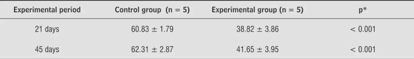
Concerning the histomorphometric analysis, in both periods of immobilization there was a reduc-tion of the transverse secreduc-tion of the fibers under the avarege calculation of larger and smaller diameters in relation to the comparison to its respective con-tralateral member control . This decrease was of
. % . ± . and of . % . ± . in
the animals of and days of immobilization, re-spectively. The values were verified by statistic tests
with significant importance p < . . )t is
im-portant to emphasize the observation p < . of significant importance among the experimental groups of and days, suggesting the progres-sive exacerbation of hypertrophic degree muscle volume when related to the immobilization time
Table .
Discussion
The findings of experimental models done so far do not converge to a general agreement referring to the physiological-histochemical alterations provoked by the muscle disuse, having contradictory conclu-sions. These denote differential percentage of sus-ceptibility to hypertrophy antagonistic functional equivalence in relation to each type of muscle fiber
and its specific location , , , , .
The soleus muscle composed of % of fibers of type ) was used for being a biarticular muscle and for acting in static posture as dynamic creat-ing tensile and compressive strength in knees and ankle articulations.
The results of the present study point out the ex-istent association between the transverse area of the muscle fiber and the articular immobilization, during
and days. Järvinen et al. report that the
longer the exposure period, the bigger the number of collagen deposit between the muscle fibers and the reduction of myofibril volume, interfering in the muscle regeneration process and consequently in the rehabilitation. At articular level, this tissue increase conjunct reduces the articular amplitude of move-ment, which is harmful to synovial fluid production, therefore the articulation lubrication and the nutri-tion of articular cartilage. The results reached with the histomorphometric analysis show and increase in the occupied space by the endomysium and perymis-ium from the animals from the experimental group from and days.
A B
D C
Figure 1 - Photomicroscopes of transverse sections of the middle third from the soleus muscle
As well as Chakravarthy et al. that demon-strated a significant reduction of soleus muscle mass after three weeks of immobilization and Mercier et
al. emphasized that through the suspension of
weight unloading, there is a reduction of % in the soleus muscle weight in the same period. )n a study
from the authors point out a muscle weight
reduction of . % in the soleus and of . % in the long finger extensors during the period of days of articular restriction.
Chingui et al. in their studies conclude that
the biggest homeostatic compromising occurs in the initial phase of disuse. )n this context, it is important to enhance that the determination of the results of studies on immobilization present a multifactorial factor differing regarding the model and used mate-rial to promote disuse, the period of immobilization, the articular position and the type of muscle fibers
analyzed , , , .
Similar to the observation of other authors in the studied samples, we found morphological al-terations presence of diverse outlines of the muscle fibers and the sharp decline of the muscle fibers diam-eters after days of immobilization, which
corrobo-rates with the findings, Okita et al. , Järvinen et al.
and )tai et al. report about hypertrophy in
the period of three, five and eight weeks, respectively. As a limitation of the study, we point out the con-trol of the experiment made through right contralat-eral member from the respective animal, which may have interfered in the movement of the control group initial phase after immobilization.
Conclusion
The articular immobilization of rats for and days alters the transverse section area of the soleus muscle fibers, in other words, it provokes cellular
hypertrophy with significant deleterious effects of cel-lular components. )t is also concluded that the increase of the area of the intermuscular septum indicate a pro-liferation of the connective tissue of muscle covering and that the biggest alterations are found in the muscle fibers of rats’ immobilization for a longer period.
)t is suggest experiment studies with group con-trol composed of samples regardless the experimen-tal group.
References
. Caierão QM, Teodori R, Minamoto VB. A influência da imobilização sobre o tecido conjuntivo muscular: uma revisão. Fisioter Mov. ; : - .
. Lima SC, Caierão QM, Durigan JLQ, Schwarzenbeck A, Silva CA, Minamoto VB, et al. Short-term immobiliza-tion causes morphometric and mechanical alteraimmobiliza-tions on rat muscles. Rev Bras Fisioter. ; : - . . Mercier J, Perez-Martin A, Bigard X, Ventura R. Mus-cle plasticity and metabolism: effects of exercise and chronic diseases. Mol Aspects Med. ; : - . . Delfino GB, Durigan JLQ, Cancelliero KM, Silva CA.
Efeito do sulfato de vanadil sobre o comprometimento metabólico muscular induzido pela imobilização de membro posterior de ratos. Rev Bras Med Esporte.
; : - .
. Abdalla DR, Bertoncello D, Carvalho LC. Avaliação das propriedades mecânicas do músculo gastrocnêmio de ratas imobilizado e submetido à corrente russa. Fisioter Pesqui. ; : - .
. Zarzhevsky N, Coleman R, Volpin G, Fuchs D, Stein (, Reznick AZ. Muscle recovery after immobilisation by external fixation. J Bone Joint Surg Br. ;
: - .
Table 1 - Average and standard deviation of the transverse section area (in micrometers) of the analyzed fibers from the soleus muscle from the analyzed animals (groups: control and experimental) and value of p
Experimental period Control group (n = 5) Experimental group (n = 5) p*
21 days 60.83 ± 1.79 38.82 ± 3.86 < 0.001
45 days 62.31 ± 2.87 41.65 ± 3.95 < 0.001
294
. Okita M, Yoshimura T, Nakano J, Motomura M, Egu-chi K. Effects of reduced joint mobility on sarcomere length, collagen fibril arrangement in the endomy-sium, and hyaluronan in rat muscle. J Muscle Res Cell Motil. ; : - .
. Reardon KA, Davis J, Kapsa RM, Choong P, Byrne E. Myostatin, insulin-like growth factor- , and leu-kemia inhibitory factor mRNAs are upregulated in chronic human disuse muscle atrophy. Muscle Nerve.
; : - .
. Krivickas LS. Treinamento de flexibilidade. )n: Fron-tera WR, Dawson DM, Slovir DM. Exercício físico e reabilitação. Porto Alegre: Artmed; . p. - . . Bodine SC, Latres E, Baumhueter S, Lai VK, Nunez L,
Clarke BA, et al. )dentification of ubiquitin ligas-es required for skeletal muscle atrophy. Science.
; : - .
. Appell (J. Muscular atrophy following immobiliza-tion: a review. Sports Med. ; : - . . Glass DJ. Signaling pathways that mediate skeletal
muscle hypertrophy and atrophy. Nat Cell Biol. ; : - .
. Caiozzo VJ, (addad F, Baker MJ, (enrrick RE, Pritto N, Baldwin KM. Microgravity-induced transformations of myosin isoforms and contractile properties of skeletal muscle. J Appl Physiol. ; : - .
. Lieber RL. Skeletal muscle structure, function, and plasticity, the physiological basis of rehabilitation. nd ed. Philadelphia: Lippincott; .
. Kasper CE, Talbot LA, Gaines JM. Skeletal muscle damage and recovery. AACN Clin )ssues. ; :
- .
. Talmadge RJ. Mechanical properties of rat soleus af-ter long-af-term spinal cord transection. J Appl Physiol.
; : - .
. Tanaka T, Kariya Y, (oshino Y. (istochemical study on the changes in muscle fibers in relation to the effects of aging on recovery from muscular atrophy caused by disuse in rats. J Orthop Sci. ; : - . . Mercier C, Jobin J, Lépine C, Simard C. Effects of
hindlimb suspension on contractile properties of young and old rat muscles and the impact of electri-cal stimulation on the recovery process. Mech Ageing
Dev. ; : - .
. Coutinho EL, Gomes AR, França CN, Salvini TF. A new model for the immobilization of the rat hind limb. Braz J Med Biol Res. ; : - .
. Arruda EJ, Grassi DO, Guirro RRJ, Silva CA. Perfil qui-miometabólico de músculos esqueléticos de ratos submetidos a diferentes modelos de imobilização articular. )n: Anais da . Mostra Acadêmica e do . Congresso de )niciação Científica; Set ; Piraci-caba. Pracicaba: Unimep; . p. - .
. Konno EAB, Alves EPB, Bertolini GRF, Barbieri C(, Mazzer N. Remobilização por alongamento estático cíclico em músculo sóleo de ratos imobilizados em en-curtamento. Rev Bras Med Esporte. ; : - . . Portinho D, Boin VG, Bertolini GRF. Efeitos sobre o
tecido ósseo e cartilagem articular provocados pela imobilização e remobilização em ratos Wistar. Rev Bras Med Esporte. ; : - .
. Millis DL. Responses of musculoskeletal tissues to dis-use and remobilization. )n: Millis DL, Levine D, Taylor RA, editors. Canine rehabilitation & physical therapy. Missouri: Elsevier; . p. - .
. Freitas CLR, Rocha CS, Vaz MA. Comportamento mecânico do músculo esquelético após duas semanas de imobilização. )n: Anais do . Congresso Brasileiro de Biomecânica; ; São Pedro. São Pedro: Socie-dade Brasileira de Biomecânica; . p. - . . Appell (J. Skeletal muscle atrophies during
immobi-lization. )nt J Sports Med. ; : - .
. Jackman RW, Kandarian SC. The molecular basis of skeletal muscle atrophy. Am J Physiol Cell Physiol.
; :C - .
. Kaplan SJ. Post-hospital home health care: the elder-ly’s access and utilization [dissertation]. St. Louis: Washington University; .
. Coutinho EL, Gomes ARS, França CN, Oishi J, Salvini TF. Effect of passive stretching on the immobilized so-leus muscle fiber morphology. Braz J Med Biol Res.
; : - .
. Sartori JR, Gonzales E, Macari M, Dal Pai V, Oliveira (N. Tipos de fibras no músculo flexor longo do hálux de frangos de corte submetidos ao estresse pelo calor e frio e alimentados em pair-feeding . Rev Brasil Zoo-tecnia. ; : - .
. Dubowitz V, Brooke M(, Neiville (. Muscle biopsy: a modern approach. London: Saunders; .
. Järvinen MJ, Einola SA, Virtanen EO. Effect of the posi-tion of immobilizaposi-tion upon the tensile properties of the rat gastrocnemius muscle. Arch Phys Med Rehabil.
; : - .
. Chakravarthy MV, Abraha TW, Schwartz RJ, Fiorotto ML, Booth FW. )nsulin-like growth factor-) extend in vitro replicative life span of skeletal muscle satellite cells by enhancing G /S cell cycle progression via the activa-tion of phosphatidylinositol ’-kinase/Akt signaling pathway. J Biol Chem. ; : - . . Kourtidou-Papadeli C, Kyparos A, Albani M, Frossinis A,
Papdelis CL, Bamidis P, et al. Electrophysiological, his-tochemical, and hormonal adaptation of rat muscle after prolonged hindlimb suspension. Acta Astronaut.
; : - .
. Chingui LJ, Braquinho RP, Severi MTM, Silva CA. Com-portamento quimiometabólico do músculo sóleo na fase aguda da imobilização articular. Fisioter Pesqui.
; : - .
. Mayer WP. Características estruturais, ultra-estrutur-ais e morfoquantitativas dos músculos tibial anterior e sóleo de ratos jovens submetidos à imobilização da articulação talocrural [dissertação]. São Carlos: Universidade Estadual de São Paulo; .
. Järvinen TA, Józsa L, Kannus P, Järvinen TL. Organi-zation and distribution of intramuscular connective tissue. J Muscle Res Cell Motil. ; : - . . )tai Y, Kariya Y, (oshino Y. Morphological changes in
rat hindlimb muscle fibres during recovery from dis-use atrophy. Acta Physiol Scand. ; : - .
Received: / /
Recebido: / /
Approved: / /
Aprovado: / /
. Silva CA, Guirro RRJ, Polacow MLO, Cancelliero KM, Durigan JL. Rat hindlimb joint immobilization with acrylic resin orthoses. Braz J Med Biol Res. ;
: - .
. Matheus JPC, Gomide LB, Oliveira JGP, Volpon B, Shi-mano AC. Efeitos da estimulação elétrica neuromuscu-lar durante a imobilização nas propriedades mecâni-cas do músculo esquelético. Rev Bras Med Esporte.
; : - .
. Picquet F, Falempin M. Compared effects of hindlimb unloading versus terrestrial deafferentation on muscular proprieties of the rat soleus. Exp Neurol.
; : - .
. Carvalho CMM, Shimano AC, Volpon JB. Efeitos da imobilização e do exercício físico em algumas pro-priedades mecânicas do músculo esquelético. Rev Bras Eng Biomed. ; : - .
. Qin L, Appell (J, Chan KM, Maffulli N. Electrical stimulation prevents immobilization atrophy in skeletal muscle of rabbits. Arch Phys Med Rehabil.
; : - .
. Choniac R, Videira, RVS, Ruiz SAL. Atrofia muscular em pacientes oncológicos internados em unidade de terapia intensiva. Rev Fisioter Univ São Paulo. ;
: - .
. Sakakima (. Effects of immobilization and subsequent low and high frequency treadmill running on rat so-leous muscle and ankle joint movement. J Phys Ther Sci. ; : - .
. Clark D, Barthold SW, Bayne KA, Davis MA, Everitt J), Fox JG. Guide for the care and use of laboratory animals. rd ed. Washington: National Academy Press; . . Bertolini SMMG, Oliveira PD, Cararo DC. Estudo mor-fométrico do músculo sóleo de ratos da linhagem wi-star pós-imobilização articular. Acta Sci (ealth Sci.
; : - .
. Dal-Pai V. (istoenzimologia: teoria e prática. Botucatu: )nstituto de Biociências – Unesp; .
. Fridén J, Liber RL. Structural and mechanical basis of exercise-induced muscle injury. Med Sci Sports Exerc.
; : - .