REVIEW
GUIDELINES ON MANAGEMENT OF HUMAN
INFECTION WITH THE NOVEL VIRUS INFLUENZA
A (H1N1) – A REPORT FROM THE HOSPITAL DAS
CLÍNICAS OF THE UNIVERSITY OF SÃO PAULO
Ludhmila Abrahao Hajjar,I Denise Schout,II Filomena Regina Barbosa Gomes Galas,I David Everson Uip,III Anna Sara Shafferman Levin,IV Helio Hehl Caiaffa Filho,V Pedro Takanori Sakane,VI Carlos Alberto Suslik,VII Jose Manoel de Camargo Teixeira,VIII Eloisa Bonfa,IX Antonio Alci Barone,IV Milton de Arruda Martins,IX Marcos Boulos,X and Jose Otavio Costa Auler Jr.XI
doi: 10.1590/S1807-59322009001000013
Hajjar LA, Schout D, Galas FRBG, Uip DE, Levin ASS, Caiaffa-Filho HH et al. Guidelines on management of human infection with the novel virus inluenza A (H1N1) – A report from the Hospital das Clínicas of the University of São Paulo. Clinics. 2009;64(10):1015-24.
The pandemic novel inluenza A (H1N1) infection was considered widespread in Brazil on July, 2009. Since then, 9.249 cases were conirmed in Brazil, most of them concentrated in São Paulo. The Hospital das Clínicas of the University of São Paulo is a refer-ence center for H1N1 cases in São Paulo. The purpose of this review is to analyze the evidrefer-ence concerning diagnosis, prevention, and treatment of novel inluenza A (H1N1) infection. In addition, we propose guidelines for the management of this pandemic emphasizing Hospital das Clínicas “bundles” for the control of the pandemic novel inluenza A (H1N1).
KEYWORDS: Review; Hospital das Clínicas; Pandemic; Swine Origin Infection Virus.
I Department of Anesthesiology, InCor-Heart Institute and Instituto do
Câncer do Estado de São Paulo - Hospital das Clínicas da Faculdade de Medicina da Universidade de São Paulo - São Paulo/SP, Brazil.
II Department of Preventive Medicine, Epidemiology Service - Hospital das
Clínicas da Faculdade de Medicina da Universidade de São Paulo - São Paulo/SP, Brazil.
III Hospital Emilio Ribas - Hospital das Clínicas da Faculdade de Medicina
da Universidade de São Paulo - São Paulo/SP, Brazil.
IV Department of Infectious Diseases - Hospital das Clínicas da Faculdade de
Medicina da Universidade de São Paulo - São Paulo/SP, Brazil.
V Department of Pathology - Hospital das Clínicas da Faculdade de Medicina
da Universidade de São Paulo - São Paulo/SP, Brazil.
VI Instituto da Criança - Hospital das Clínicas da Faculdade de Medicina da
Universidade de São Paulo - São Paulo/SP, Brazil.
VII Hospital das Clínicas da Faculdade de Medicina da Universidade de São
Paulo - São Paulo/SP, Brazil.
VIII Hospital das Clínicas da Faculdade de Medicina da Universidade de São
Paulo - São Paulo/SP, Brazil.
IX Department of Internal Medicine - Hospital das Clínicas da Faculdade de
Medicina da Universidade de São Paulo - São Paulo/SP, Brazil.
X Faculdade de Medicina da Universidade de São Paulo - São Paulo/SP, Brazil. XI Hospital das Clínicas da Faculdade de Medicina da Universidade de São
Paulo - São Paulo/SP, Brazil. Email: [email protected] Tel.: 55 11 3069.6431
Received for publication on September 22, 2009. Accepted for publication on September 29, 2009.
1. INTRODUCTION
In April 2009, the irst two cases of human infection with a novel inluenza A (H1N1) virus were reported in the United States.1 During the same period, an outbreak of respiratory
infection was reported in Mexico.2 The virus was found to
be an H1N1 virus that was antigenically and genetically unrelated to human seasonal inluenza viruses and genetically related to viruses known to circulate in swine.3 In the ensuing
weeks, the swine-origin inluenza virus (S-OIV) H1N1 spread worldwide, constituting a pandemic, as deined by the World Health Organization.4 The novel H1N1 virus has distinct
molecular properties of human, avian, and swine inluenza, resulting from antigenic drift, which is the main cause of the seasonal epidemic of swine lu.4
after the description of the irst cases, the pandemic S-OIV infection continues to spread globally, presents a high rate of transmission among humans, and can lead to serious complications and mortality. The purpose of this guideline is to review the evidence concerning diagnosis, prevention, and treatment of S-OIV infection. In addition, we emphasize the Hospital das Clínicas’ plan for the management of the pandemic novel inluenza A (H1N1). This report is an initiative from the “Cabinet Crisis” - a group of healthcare professionals at the Hospital das Clínicas da Faculdade de Medicina da Unversidade de São Paulo who continuously evaluate the “bundles” for control of S-OIV infection, meaning the groups of interventions and advertisements, to obtain better outcomes in the management of this disease.
2. HISTORICAL ASPECTS
An estimated 58% of the 1407 human pathogens are zoonotic, which means that they normally occur in animals but can also infect humans.6 The ability of a microorganism
to cross the species barrier in association with a high transmissibility rate between humans may result in epidemics. The novel inluenza A (H1N1) virus, which is responsible for the current pandemic, is derived from two unrelated swine viruses, one of which is a derivative of the 1918 human virus.3
The notorious 1918 pandemic of inluenza A (H1N1), the Spanish lu, was derived from an avian source and caused 50 million deaths.6 Some authors proposed that the virus resided
in an avian reservoir and affected humans either directly upon exposure to birds or through an intermediate host.7 As the
1918 inluenza virus can replicate and cause disease in swine, scientists believe that it has continued to circulate in swine and this fact would facilitate the genetic reassortment between different inluenza virus strains.8
A practical way to think about inluenza A events over the past 91 years is to recognize that we are living in a pandemic era that began in 1918. The novel H1N1 virus associated with the ongoing 2009 pandemic is a fourth-generation descendant of the 1918 virus. The complex evolutionary history of this virus combines unique structural properties and genetic mixing among human viruses and avian and swine-adapted inluenza viruses.9 Two features of the inluenza virus explain
its ability to cause widespread disease. One is the high error rate during genomic replication.9,10 The other is the segmented
inluenza virus genome, which allows reassortment between different viral strains.9,10 Because of this continual change
phenomenon, a seemingly endless variety of new viruses with potentially new properties are continuously being engineered. In contrast, this new virus is not only infecting humans and causing some disease, but it is also being transmitted eficiently from human to human.
3. EPIDEMIOLOGY
The pandemic novel inluenza A (H1N1) infection was considered as widespread in Brazil on July 16.11 Since
then, the Ministry of Health in Brazil, as suggested by the World Health Organization, has maintained a continuous epidemiologic vigilance of cases of acute respiratory syndrome (ARS). The strategy of vigilance considers any persons with lu syndrome as potential cases of inluenza A (H1N1) and designates these individuals as presenting acute respiratory syndrome patients with cough, dyspnea, and fever.
Until now, 46.810 cases of ARS were reported notiied in Brazil.12 Of these, 9.249 (20%) patients presented infection
with S-OIV or inluenza A (H1N1). Seasonal inluenza A infection was conirmed in 1.152 (2.5%) patients.11
The observed age distribution is unusual and differs from seasonal inluenza, being skewed towards younger age groups. There is a marked underrepresentation of infections in persons over 65 years of age, who make up only 2% of the reported cases. In Brazil, among the reported cases, the affected individuals tend to be young, with a median age of 26 years. Most patients are 15-49 years of age. Considering the gender distribution, 57.5% of the conirmed cases of novel virus inluenza A (H1N1) occur in women.11,12.
Among these 9.249 confirmed cases in Brazil, 899 deaths are reported with mortality rate of 0,47/per 100.000 inhabitants.11 All Brazilian states have reported cases of
S-OIV infection, with the exception of Sergipe. Most cases and deaths are concentrated in São Paulo, but major mortality rate was observed in Paraná (2,08/ 100.000 inhabitants).
As of September 12, 2009, 13.069 cases of ARS were registered in São Paulo and 3.733 are due to novel inluenza A (H1N1) infection.11 São Paulo state registers 40.3% (3.733
of 9.249) of all conirmed cases from Brazil, the majority identiied in the city.
The Hospital das Clínicas of Faculdade de Medicina da Universidade de São Paulo (HC-FMUSP) is the largest tertiary health care hospital in Brazil, with 6 medical institutes and other associated hospitals, and is a reference center for H1N1 cases in São Paulo. Three months after the report of the irst case of novel H1N1 infection in Brazil, the Hospital das Clínicas has accumulated experience with about 1500 cases registered and 472 conirmed with a low lethality rate (7.14%).
obtain information from Health Organs, Epidemiological Surveillance Systems and from literature regarding epidemiological data and clinical presentations of S-OIV infection and to provide an adequate structure for the care of patients.
4. PATHOGENESIS
Influenza A, B, and C are RNA viruses of the Ortomyxoviridae family and cause both pandemic and seasonal disease in humans.14 Influenza A viruses are
enveloped, single-stranded RNA viruses with a segmented genome. They are categorized into subtypes on the basis of the antigenic properties of the hemagglutinin (HA) and neuraminidase (NA) glycoproteins on the surface of the virus.14 The HA glycoprotein mediates attachment and entry
of the virus on the cell surface and is the main target for immunity by neutralizing antibodies. The NA glycoprotein allows the spread of the virus by cleaving the glycosidic linkages to sialic acid on host cells and on the surface of the virus.15 Inluenza A viruses are characterized according
to their pathogenicity, which results in severe disease and death in different species. This S-OIV results from frequent antigenic changes (i.e., antigenic drift) due to point mutations and recombination events that occur during viral replication.15 The novel inluenza A (H1N1) virus is not
a new subtype, but because the large majority of humans appear to have no pre-existing antibodies to this virus, a substantial potential for widespread infection exists.14,15
The novel influenza A (H1N1) virus has distinct properties that enable it to cause disease in both swine and humans and confers high rates of transmissibility among humans. The pathogenesis of human infection due to S-OIV is poorly understood, but appears to involve two phenomena: a) direct cytotoxic viral damage and b) a cytokine storm, resulting from the inlammatory response to viral infection.16,17 The interaction between host and
virus may result in different forms of disease, depending on the viral load and the inlammatory response. In patients with co-morbidities, host mechanisms of defense may be defective and such patients can present an altered innate immune response. In the most severe cases, co-infection with other viruses and bacteria (Streptococcus pneumoniae,
Haemophilus influenza, and Staphylococcus aureus) can
occur contributing to the high rates of mortality.18
There is no evidence to date suggesting that the virus disseminates any differently from other human inluenza viruses, i.e., by droplets from coughing and sneezing and direct and indirect contact with respiratory secretions from infected persons.19 An individual may sometimes become
infected by touching something contaminated with flu
viruses and then touching his or her mouth or nose.19 There
is no evidence to suggest unusual transmission routes for inluenza, and there is no reason to suggest transmission via food. Patients may be contagious from one day before developing symptoms to up to 7 days after they get sick.19,20
Children and persons with deicient immunity might be contagious for longer periods of time.20
5. CLINICAL FEATURES
Human infection with the novel virus influenza A (H1N1) is characterized by a variable clinical presentation. We can describe a spectrum of disease presentations: the asymptomatic form, “lu-like syndrome”, and the severe form with acute respiratory distress syndrome leading to death (Figure 1).21 The majority of patients present
the “lu-like syndrome” with fever, myalgia, sore throat, arthralgia, cough, headache, chills, and fatigue.22,23 Fever is
the most frequent symptom and usually lasts for three days. Respiratory symptoms generally disappear three or four days after the fever ends. Diarrhea, emesis, and weakness can be present in a signiicant number of cases.22,23
Despite the fact that in most patients the disease has a benign evolution and a limited duration, some patients may present respiratory failure, rapidly developing acute respiratory distress syndrome.24,25 Published data describe
systemic disease and complications due to S-OIV infection such as:22,23
a. Worsening of previous existing disease b. Sinusitis, otitis, asthma
c. Pneumonia, acute lung injury, respiratory failure d. Pericarditis, myocarditis
e. Myositis, rhabdomyolysis
f. Acute renal failure due to acute tubular necrosis g. Encephalitis, seizures
h. Systemic inlammatory response syndrome i. Multiple organ failure
j. Death
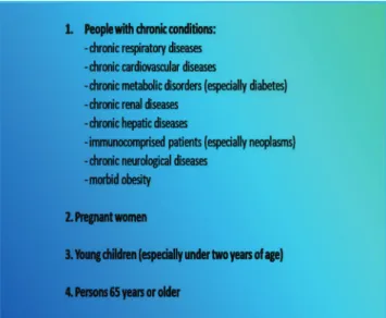
The reported cases reveal that about 70% of patients who die or present severe forms of the disease have underlying conditions.22,23,24,25,26,27 Risk groups for hospitalization and
severe disease due to S-OIV infection are:
a. Individuals with underlying chronic diseases: data show that most patients hospitalized due to the need for care present co-morbidities, such as: asthma, chronic obstruc-tive airways disease, diabetes, immunocompromise, chronic cardiovascular disease, chronic renal failure, epilepsy, obesity, and cancer.22,23,24
b. Pregnant women: studies report that pregnant women infected with S-OIV have a four to ive times greater chance of being hospitalized than healthy pregnant women and present more severe forms of the disease.25,26
c. Young children: children under two years of age present high rates of hospitalization and death.27
d. Older individuals: persons 65 years and older have a high likelihood of needing hospital care and exhibit a high case fatality rate.23,24
From these analyses we can describe a list of risk groups, i.e., groups that experience more severe infections than the general population (Figure 2).
Pregnant women have a potential for complicated disease. For that, in Hospital das Clínicas, there is a speciic protocol of care for this group, which include hospital
admission in suspected or conirmed cases with clinical complications and a careful vigilance system for mild forms of disease with home treatment.
5.1 Hospital admission
To date, most human cases of new inluenza A (H1N1) virus infection have exhibited an uncomplicated illness of limited duration. Hospitalization or antiviral therapy is therefore not likely to be required for most patients. Supportive care includes antipyretics, such as dypirone or acetaminophen for fever or pain, and luid rehydration that can be provided as needed.22
The speciic risk factors that predict an increased risk of progressive disease are incompletely understood. Clinicians and caregivers should watch for signs of possible clinical deterioration (for example, dificulty breathing, chest pain, coughing up colored sputum, dyspnea, altered level of consciousness, and confusion) and refer immediately such patients to the hospital. Clinicians should also take into account any underlying co-morbidities, the already described risk groups (such as immunocompromising conditions, pre-existing chronic lung or cardiovascular disease, diabetes, pregnancy, and young age).
Patients who present one or more of the following signs and symptoms must be hospitalized in an intensive care unit: a) Hemodynamic instability
b) Acute respiratory failure
c) Extensive lung compromise on chest X-ray d) Severe hypoxemia
e) An PO2/FiO2 < 300, characteristic of acute lung injury f) Compromise of other organs: acute renal failure,
myosi-tis, encephalimyosi-tis, and other g) Organ dysfunction
5.2 Intensive care:
5.2.1 Ventilatory support: until now, there has been no unique, deinite pattern of lung disease. Patients who present serious hypoxemia may present different forms of lung damage.23,24 Most patients present diffuse alveolar damage,
but localized disease, bronchiolitis, lobar pneumonia, and pleural effusion may be present. Ventilator support indications will then depend on the clinical condition of the patients, evaluated through signs, symptoms, and laboratory analysis (including arterial gas).
are the same as those for patients with respiratory failure due to other etiologies.28 NIV should be used in patients
without hemodynamic instability or consciousness altera-tions.28,29 The preferred modality is the BiPAP, with
in-spiratory pressure (IPAP) and expiratory pressure (EPAP) adjusted independently. After selecting and itting the mask, the recommended initial settings are IPAP = 8-12 cmH2O and EPAP = 3-5 cmH2O. The IPAP is increased gradually as tolerated, with the therapeutics goals of dyspnea relief, good patient-ventilator synchrony, and improved gas exchange. The EPAP may be increased as needed for alveolar recruitment.
- Invasive ventilation:
- Treatment of ARDS associated with new inluenza A (H1N1) virus infection should be based on published ev-idence-based guidelines for sepsis-associated ARDS.29,30
Lung-protective mechanical ventilation strategies should be used. The rationale is not to cause damage of non-affected areas. We recommend the following therapy:
a) Ventilatory mode: most ARDS patients are ven-tilated using conventional volume-cycled positive-pressure ventilators. Most studies using low tidal volume for ARDS employed this mode.29,30 However,
no differences in outcomes are detected if pressure-controlled ventilation is chosen, since the low tidal volume strategy is reached.
b) Oxygen: treatment of hypoxemia in ARDS induced by H1N1 virus is almost always initiated using 100% oxygen (FIO2 = 1.0), and the concentration of O2 is reduced with the goal of maintaining a PaO2 greater than 60 mmHg (arterial O2 saturation of about 90%). The FIO2 should be lowered to less than 0.5 as soon as possible to reduce the risk of lung damage due to oxygen toxicity.
c) Positive End-Expiratory Pressure (PEEP): Positive end-expiratory pressure works by counteracting the tendency toward alveoli collapse during pulmonary edema, low lung volume, and loss of surfactant.31
In ARDS, the majority of the lung is atelectatic.29
PEEP recruits partially collapsed areas, improving gas exchange.31 However, in some cases it is
associ-ated with barotrauma, pneumothorax, lung injury, inlammation, a cardiac output decrease, and shock.32
Consequently, we recommend a PEEP level between 5 and 12 cm H2O in most patients, and we can adjust the PEEP levels using a combination of the PV curve, the response of arterial blood gases, the hypothetical maximum and minimum PEEP values, and the hemo-dynamic response.
d) Low-tidal-volume strategy: the most important development in the management of ARDS is that
mechanical ventilation with a lower tidal volume than previously used is associated with an improved clinical outcome.29,30 This type of ventilation has been
termed as the low-tidal volume or lung-protective strategy. The best risk:beneit ratio would be gained with a tidal volume of 6 ml/Kg or less, to reach a target plateau pressure of less than 30 cm H2O.30
e) Recruitment maneuvers and the prone position:
recruitment maneuvers and the prone position must be reserved for refractory hypoxemia, as in other cases of ARDS.33,34
5.2.2 Hemodynamic support: Patients admitted to the intensive care unit with S-OIV infection and ARDS may present shock. It is of main importance to maintain hemodynamic goals in these patients to avoid organ failure. Fluid management in this population should be performed carefully. Current evidence indicates that a net negative balance is desirable in ARDS.35 However, in these
patients, hypovolemia must be avoided. Therefore, we recommend dynamic evaluation of the luid status and a more conservative strategy for luid replacement. In some cases, albumin associated with furosemide may offer beneit in the net balance.35,36
Hemodynamic goals:
- Mean arterial pressure > 65 mmHg - SVO2 > 70%
- Lactate < 2 mmol/L
- Diuresis higher than 1ml/Kg/h
For optimal tissue perfusion and to obtain adequate arterial pressure, it is often necessary to use vasopressors and inotropics. We suggest norepinephrine as the irst-line vasopressor and dobutamine as the inotropic. Vasopressin should be reserved for refractory shock.37
5.2.3 Renal support: acute renal failure has been observed in patients with S-OIV infection, especially in cases presenting shock.22 The etiology of acute renal failure
is usually acute tubular necrosis, and in such cases renal replacement therapy may be needed.
5.2.4 Corticosteroids: controversy exists as to whether we should use high-dose corticosteroids in ARDS due to S-OIV infection.38 The positive effect would be a reduction
of the inlammatory lung injury, potentially resulting in a better PO2/FIO2 ratio and less intubation, as in ARDS of other etiologies.39 The adverse events in inluenza
do not have pathology data for H1N1 infection to guide the choice of therapy. So, we recommend the use of methylprednisolone (2 mg/Kg per day) just in cases of ARDS that do not response to initial measures.
Low doses of corticosteroids (hydrocortisone 50 mg IV four times per day) may be considered for patients in septic shock who require vasopressors and have suspected adrenal insuficiency.
5.2.5 Antibiotics: antibiotic chemoprophylaxis should not be used. When pneumonia is present, treatment with antibiotics should follow the recommendations from published guidelines. However, seasonal inluenza and past inluenza pandemics have been associated with an increased risk of secondary Staphylococcus aureus infections, which may be severe, progress rapidly, have necrotizing effects, and, in some areas, may be caused by methicillin-resistant strains.40 The results of microbiological studies,
wherever possible, should be used to guide antibiotic usage for suspected bacterial coinfection in patients infected with the new inluenza A (H1N1) virus. Several patients worldwide have developed community pneumoniae due
to Streptococcus pneumoniae and Haemophilus influenza
and ventilator-associated pneumonia or hospital-acquired pneumonia caused by typical nosocomial pathogens.41
5.2.6 Glycemic control: tight glucose control (< 140 mg/ dL) must be achieved in critically ill patients, according to the local protocol. This measure is associated with a decrease of morbidity and mortality in critical patients.42
5.2.7 Thromboembolic prophylaxis: patients infected with S-OIV should receive mechanical thromboprophylaxis and pharmacological if possible because, as critically ill patients with co-morbidities, they present a high risk for thromboembolic events. In a series of cases from Michigan, of 10 patients with S-OIV infection, ive had a pulmonary embolism, although the higher incidence of thrombosis could be explained because seven were extremely obese (body mass index > 40).43
6. DIAGNOSIS
Laboratory confirmation of the novel influenza A (H1N1) virus, especially at the beginning of a new community outbreak, or for unusual cases, has important implications for case management, consideration of antiviral treatment options, and avoidance of the inappropriate use of antibiotics.44
Reverse transcriptase-polymerase chain reaction (RT-PCR) will provide the most timely and sensitive evidence
of infection with the novel inluenza A (H1N1) virus.44,45
Real-time RT-PCR is available in reference centers around the world.46 Samples for laboratory tests should be obtained
from the deep nasal passages (nasal swab), nasopharynx (nasopharyngeal swab), or the bronchial aspirate if available.47 Upper respiratory tract sampling using a
combination of nasal or nasopharyngeal and a throat swab is advised and may facilitate virus detection. It is not yet known which clinical specimen provides the best diagnostic yield for this speciic infection.48 Specimen collection should
be carried out with precautions since the procedure may expose the collector to respiratory secretions from infected patients.
The real-time test through polymerase chain reaction, includes the use of speciic primers and probes to diagnose S-OIV infection.46 The set of inluenza primers and probes
was created to detect:
(a) Seasonal inluenza A virus (b) Swine-origin inluenza A virus (c) Novel virus inluenza A (H1N1)
Currently, the data show that this method has a sensitivity of 99.3% and a speciicity of 92.3% for the diagnosis of infection due to novel virus influenza A (H1N1).46 We
recommend the following types of cases to be submitted for the test:
a) patients who require hospitalization
b) patients with risk factors for severe forms of disease c) patients in an individualized protocol – according to the
clinical judgment
No validated rapid bedside diagnostic test is presently available to detect novel inluenza A (H1N1) virus infection. As part of the HC “bundles” for control of novel H1N1 infection, a real-time RT-PCR is available for patients and workers who ill criteria to be tested.
7. TREATMENT AND PROPHYLAXIS
The novel influenza A (H1N1) virus is currently susceptible to the antiviral medications known as neuraminidase inhibitors (NAIs), speciically oseltamivir and zanamivir.49,50 The virus is resistant to the adamantane
medications amantadine or rimantadine.49 Clinical eficacy
data on antiviral treatment are not yet available. Based on its
in vitro susceptibility patterns and the clinical experiences derived from seasonal and avian inluenza infection, early administration of NAIs might reduce the severity and duration of illness caused by the novel H1N1 virus infection and might also help to prevent progression to severe disease and death.51 Only sporadic cases of oseltamivir-resistant
worldwide, including nine cases in the United States.52 Eight
of nine patients had a documented exposure to oseltamivir through either treatment or prophylaxis.52
Clinical judgment is an important factor in the treatment decision. People with suspected novel H1N1 inluenza who present with an uncomplicated febrile illness typically do not require treatment unless they are at higher risk for inluenza complications, and in areas with limited antiviral availability, local public health authorities might provide guidance about prioritizing treatment within groups at higher risk.53
Antiviral therapy may be beneicial, especially for the following groups:54
1. All hospitalized patients with conirmed, probable, or suspected novel inluenza (H1N1).
2. Patients who are at a higher risk for complications (listed above).
If used, antiviral treatment should ideally be initiated early, as soon as possible, but it may also be used at any stage of the active disease when ongoing viral replication is anticipated or documented. Evidence for the beneits of antiviral treatment in studies of seasonal inluenza is highest when treatment is started within 48 hours of the onset of illness.55 However, studies investigating oseltamivir
treatment of hospitalized patients have indicated a beneit, including reductions of mortality or duration of hospital stay, even for patients whose treatment was initiated after 48 hours.56
There are important pharmacological differences to consider when choosing NAIs for treatment. Oseltamivir is administered orally and provides a higher systemic level.57
Zanamivir is delivered by oral inhalation, with low systemic absorption.57 Oseltamivir is the recommended treatment for
lower respiratory tract complications.57 The recommended
treatment duration is ive days. In Brazil, oseltamivir is used for the treatment and prophylaxis of S-OIV infection (Tables 1, 2 and 3).
Rare neuropsychiatric symptoms, such as confusion or abnormal behavior, have occurred after beginning treatment for seasonal influenza with oseltamivir, particularly in
children, but the contribution of oseltamivir to these events remains unknown.58
Post-exposure antiviral chemoprophylaxis with either oseltamivir or zanamivir for 10 days can be considered for the following situations:59
1. Close contact of cases (conirmed, probable, or suspect-ed) who are at a high risk for inluenza complications 2. Health care personnel, public health workers, or irst
responders who have had a recognized, unprotected close contact exposure to a person with novel (H1N1) inluenza virus infection during the patient’s infectious period.
In patients presenting renal failure, with a creatinine clearance between 10 and 30 ml/min/m2, a 50% dosage reduction of oseltamivir is recommended.60 There are no
data available on the ideal dosage during renal replacement therapy. Patients with hepatic failure do not require dosage correction.60
Zanamivir is indicated for the treatment of inluenza in adults and children (>5 years).57 The recommended dose
for treatment of adults and children older than 5 years of age is two inhalations (2 x 5mg) twice daily for 5 days.
Table 1 - Recommended antiviral treatment and prophylaxis for novel inluenza A (H1N1) infection
Treatment Chemoprophylaxis
Oseltamivir Oseltamivir
Adults 75-mg capsule twice per day for 5 days 75-mg capsule once per day for 10 days
Children ≥ 12 months 15 kg or less 60 mg per day divided into 2 doses 30 mg once per day 16-23 kg 90 mg per day divided into 2 doses 45 mg once per day 24-40 kg 120 mg per day divided into 2 doses 60 mg once per day >40 kg 150 mg per day divided into 2 doses 75 mg once per day
Table 2 - Recommended antiviral treatment for children younger than 1 year using oseltamivir
Age Recommended treatment for 5 days
<3 months 12 mg twice daily 3-5 months 20 mg twice daily 6-11 months 25 mg twice daily
Table 3 - Recommended antiviral chemoprophylaxis for children younger than 1 year using oseltamivir
Age Recommended prophylaxis for 10 days
<3 months Not recommended unless the situation is judged as critical
Inhaled zanamivir has been temporally associated with bronchospasm, and patients with preexisting airway disease appear to be at an increased risk for this severe adverse reaction.57
Available products in Brazil:
(a) Tamilu® Roche – 75-mg capsule and 52-ml oral suspen-sion (12 mg/ml)
(b) Oseltamivir Farmanguinhos – 75-mg capsule
(c) Oseltamivir HC – 50-ml oral suspension (15 mg/ml)
8. PREVENTION
Until now, no vaccine has been commercially available to protect against novel H1N1 virus, although there are everyday actions known to prevent the spread of infection, such as:61
• Wash your hands often with soap and water. Alcohol-based hand cleaners are also effective.
• Try to avoid close contact with sick people.
• Cover your nose and mouth with a tissue when you cough or sneeze.
• Stay home if you are sick for 7 days after your symptoms begin or until you have been symptom-free for 24 hours.
• Follow public health advice regarding school closures, avoid crowds.
9. CARE OF PATIENTS WITH CONFIRMED, SUS-PECTED, OR PROBABLE INFECTION WITH THE NOVEL VIRUS INFLUENZA A (H1/N1)
Health care professionals must use the following personal protective equipment62:
a) A surgical mask when the professional is working at a distance of less than1 meter from the patients, in proce-dures without aerosol production.
b) Special clothes to avoid contact with blood and luids. c) A N95 mask, protective glasses, and gloves in procedures
with aerosol production, such as intubation, secretion manipulation, and autopsies.
10. HC “BUNDLES” FOR NOVEL INFLUENZA A (H1N1) INFECTION
As already mentioned, as a reference center for H1N1 infection, the Hospital das Clínicas da Universidade de São Paulo created the “Cabinet Crisis” with the objectives of implementing “bundles” for control of S-OIV infection, a group of interventions to obtain better outcomes in the management of this disease. We adopted some measures to obtain continuous and everyday information on the H1N1
infection, to implement actions to control the pandemics and to evaluate the impact of these interventions such as: 1. Periodical meetings among healthcare professionals from
all institutions – directors, professors, epidemiologists, infectologists, intensive care physicians – to discuss new data and information about the disease
2. Continuous local Epidemiologic Surveillance to collect and analyze the data to assess the impact of the virus and determine the groups at an increased risk of complica-tions.
3. Information for all professionals and patients about the infection, symptoms, diagnosis, and prevention.
4. Availability of a real time RT-PCR for novel inluenza A (H1N1) virus in admitted patients.
5. Reduction of daily visits of families to the hospital. 6. Implementation of hygienic measures: alcohol-based
hand cleaner, gloves, masks.
7. Internet published recommendations on the management of infection (www.hcnet.usp.br)
8. Speciic units for the care of patients with suspected or conirmed S-OIV infection including emergency room attending, regular ward and specialized intensive care units.
9. Guidance for the staff and coworkers to limit contact with other people when symptoms are present.
10. Risk groups specialized protocols of care: pregnant women, children, immunocompromised patients, and chronic diseases.
11. Immediate availability of antiviral therapy.
In the last weeks, we could observe a significant reduction in the incidence and mortality of novel inluenza A (H1N1) infection in São Paulo and in Hospital das Clínicas. We suppose that an association of factors contribute to these numbers including a global actions plan, the continuous epidemiologic monitoring and the acquired expertise to diagnosis and adequate treatment of patients. We wish to maintain all the adopted measures in Hospital das Clínicas with the ongoing objective of obtain better outcomes in the management of novel inluenza A (H1N1) infection. With the accumulating experience and groups of study, we hope to contribute continuously with information regarding epidemiology, pathogenesis and clinical aspects of this pandemics.
11. CONCLUSIONS AND PERSPECTIVES
200 days of known disease in humans, we learned that the replication competence and virulence of the novel H1N1 virus enable it to cause severe clinical presentations and death. As a consequence of this outbreak, the world learned the value of nonpharmacologic interventions, which can save many lives. A vaccine is expected as a potential control measure for the pandemic.63 In the Hospital das Clínicas da
Universidade de São Paulo, a “Cabinet Crisis” was created to implement a group of interventions to obtain better outcomes in the management of H1N1 infection.
In our opinion, to control this pandemic, global actions are required without a geographic barrier. More studies are
needed in zoonotic virology to allow a better understanding concerning the pathogenesis and epidemiologic aspects of the new viruses to prevent emergent diseases.
ACKNOWLEDGEMENTS
Adriana Sayuri Hirota, Werther Brunow de Carvalho, Alberto José da Silva Duarte, Felipe Silva Fittipaldi, Ho Yeh Li, Marcelo Magri, Marcelo Park, Marcelo Zugaib, Pedro Paulo Pereira, Rossana Pulcineli Vieira Francisco, Ruy Pires Neto, Sonia Lucena Cipriano, Vanusa Barbosa Pinto, and Clarisse Machado, and Renata Lobo.
REFERENCES
1. Update: swine inluenza A (H1N1) infections--California and Texas, April 2009. MMWR Morb Mortal Wkly Rep. 2009;58:435-7. 2. Update: novel inluenza A (H1N1) virus infection - Mexico, March-May,
2009. MMWR Morb Mortal Wkly Rep. 2009;58:585-9.
3. Swine influenza A (H1N1) infection in two children--Southern California, March-April 2009. MMWR Morb Mortal Wkly Rep. 2009;58:400-2.
4. Dawood FS, Jain S, Finelli L, Shaw MW, Lindstrom S, Garten RJ et al. Emergence of a novel swine-origin inluenza A (H1N1) virus in humans. N Engl J Med. 2009;360:2605-15.
5. Pandemic (H1N1) 2009 - ECDC Daily Update. 2009. (Accessed August 24, 2009, at http://www.ecdc.europa.eu/en/healthtopics/ Documents/090821_Inluenza_AH1N1_Situation_Report_1700hrs. pdf.)
6. Woolhouse ME, Gowtage-Sequeria S. Host range and emerging and reemerging pathogens. Emerg Infect Dis. 2005;11:1842-47.
7. Peiris JSM, Poon LLM, Guan Yi. Emergence of a novel swine-origin inluenza A virus (S-OIV) H1N1 virus in humans. Journal of Clinical Virology. 2009;45:169-73.
8. Morens DM, Taubenberger JK, Fauci AS. The persistent legacy of the 1918 inluenza virus. N Engl J Med. 2009;361(3):225-29.
9. Sandrock C, Kelly T. Clinical review: update of avian inluenza A infections in humans. Crit Care. 2007;11:209-.
10. Kendal AP. Epidemiologic implications of changes in the inluenza virus genome. Am J Med. 1987;82:4-14.
11. Brazilian Epidemiologic Report - Inluenza A (H1N1). 2009. (Accessed August 24, 2009, at http://portal.saude.gov.br/portal/arquivos/pdf/ informe_inluenza_se_32_publicacao_18ago2009.pdf.)
12. Zimmer SM, Burke DS. Historical perspective--Emergence of inluenza A (H1N1) viruses. N Engl J Med 2009;361:279-85.
13. Rambaut A, Pybus OG, Nelson MI, Viboud C, Taubenberger JK, Holmes EC. The genomic and epidemiological dynamics of human inluenza A virus. Nature. 2008;453:615-19.
14. Garten RJ, Davis CT, Russel CA, Shu B, Lindstrom S, Balish A, et al. Antigenic and genetic characteristics of swine-origin 2009 A(H1N1) inluenza viruses circulating in humans. Science. 2009; 10;325(5937):197-201.
15. Webster RG, Bean WJ, Gorman OT, Chambers TM, Kawaoka Y. Evolution and ecology of influenza A viruses. Microbiol Rev. 1992;56:152-79.
16. Taubenberger JK, Morens DM. The pathology of influenza virus infections. Annu Rev Pathol. 2008;3:499-522.
17. Heltzer ML, Cofin SE, Maurer K, et al. Immune dysregulation in severe inluenza. J Leukoc Biol. 2009;85:1036-43.
18. Morens DM, Taubenberger JK, Fauci AS. Predominant role of bacterial pneumonia as a cause of death in pandemic inluenza: implications for pandemic inluenza preparedness. J Infect Dis. 2008;198:962-70.
19. Centers for Disease Control and Prevention. Update: novel inluenza A (H1N1) virus infection -Worldwide May 6, 2009. MMWR. 2009;58:453-8. 20. Centers for Disease Control and Prevention. Update: novel inluenza A
(H1N1) virus infection - Worldwide. MMWR. 2009;58:453-8. 21. Centers for Disease Control and Prevention. Outbreak of swine-origin
inluenza A (H1N1) virus infection - Mexico, March--April 2009. MMWR 2009;58:467-70.
22. ECDC working group on inluenza A(H1N1)v. Preliminary analysis of inluenza A(H1N1)v individual and aggregated case reports from EU and EFTA countries. Eurosurveillance 2009, 14(23). Available from: http://www.eurosurveillance.org/ViewArticle.aspx?ArticleId=19238 23. Perez-Padilla R, de la Rosa-Zamboni D, Ponce de Leon S, Hernandez
M, Quiñones-Falconi F, Bautista E, et al. Pneumonia and respiratory failure from swine-origin inluenza A (H1N1) in Mexico. N Engl J Med. 2009;361:680-89.
24. Chowell G, Bertozzi SM, Colchero MA, Lopez-Gatell H, Alpuche-Aranda C, Hernandez M, Miller MA. Severe respiratory disease concurrent with the circulation of H1N1 inluenza. N Engl J Med. 2009;361:674-79.
25. Jamieson DJ, Honein MA, Rasmussen SA, Williams JL, Swerdlow DL, Biggerstaff MS, Lindstrom S, et al. H1N1 2009 inluenza virus infection during pregnancy in the USA. Lancet. 2009;374:451-58.
26. Centers for Disease Control and Prevention. Novel inluenza A (H1N1) virus infections in three pregnant women—United States, April–May, 2009. MMWR Morb Mortal Wkly Rep. 2009;58: 497–500.
27. Uyeki TM. Inluenza diagnosis and treatment in children: a review of studies on clinically useful tests and antiviral treatment for inluenza. Pediatr Infect Dis J. 2003;22:164-77.
28. Antonelli M, Conti G, Moro ML, Esquinas A, Gonzalez-Diaz G, Confalonieri M, et al. A comparison of noninvasive positive-pressure ventilation and conventional mechanical ventilation in patients with acute respiratory failure. N Engl J Med. 1998; 339:429–35.
29. Ferguson ND, Frutos-Vivar F, Esteban A, Anzueto A, Alía I, Brower RG, et al. Airway pressures, tidal volumes, and mortality in patients with acute respiratory distress syndrome. Crit Care Med. 2005; 33:21–30. 30. The Acute Respiratory Distress Syndrome Network: Ventilation with
lower tidal volumes as compared with traditional tidal volumes for acute lung injury and the acute respiratory distress syndrome. N Engl J Med. 2000; 342:1301–08.
31. Villar J, Kacmarek RM, Pérez-Méndez L, Aguirre-Jaime A, for the ARIES Network: A high PEEP-low tidal volume ventilatory strategy improves outcome in persistent ARDS: A randomized controlled trial. Crit Care Med. 2006; 34:1311–18.
33. Gattinoni L, Caironi P, Cressoni M, Chiumello D, Ranieri VM, Quintel M, et al. Lung recruitment in patients with acute respiratory distress syndrome. N Engl J Med. 2006;354:1775–86.
34. Stocker R, Neff T, Stein S, Ecknauer E, Trentz O, Russi E. Prone positioning and low-volume pressure limited ventilation improve survival in patients with severe ARDS. Chest. 1997; 111:1008–17. 35. Dubois MJ, Orellana-Jimenez C, Melot C, De Backer D, Berre J, Leeman
M, et al. Albumin administration improves organ function in critically ill hypoalbuminemic patients: A prospective, randomized, controlled, pilot study. Crit Care Med. 2006; 34: 2536–40.
36. Wiedemann HP, Wheeler AP, Bernard GR, Thompson BT, Hayden D, deBoisblanc B, et al. Comparison of two luid-management strategies in acute lung injury. N Engl J Med. 2006; 354:2564–75.
37. Russell JA, Walley KR, Singer J, Gordon, AC, Hébert PC, Cooper J, et al. Vasopressin versus Norepinephrine Infusion in Patients with Septic Shock. N Engl J Med. 2008; 358(9):877-87.
38. Marik PE, Pastores SM, Annane D, Meduri GU, Sprung CL, Wiebke A, et al. Recommendations for the diagnosis and management of corticosteroid insuficiency in critically ill adult patients: consensus statements from an international task force by the American College of Critical Care Medicine. Crit Care Med. 2008;36:1937-49. 39. Meduri GU, Marik PE, Chrousos GP, Pastores SM, Arlt W, Beishuizen
A, et al. Steroid treatment in ARDS: A critical appraisal of the ARDS network trial and the recent literature. Intensive Care Med. 2008; 34:61–9.
40. Hers JF, Masurel N, Mulder J. Bacteriology and histopathology of the respiratory tract and lungs in fatal Asian inluenza. Lancet. 1958;2:1141-3.
41. Louria DB, Blumenfeld HL, Ellis JT, Kilbourne ED, Rogers DE. Studies on inluenza in the pandemic of 1957-1958. II. Pulmonary complications of inluenza. J Clin Invest. 1959;38:213-65
42. Fahy BG, Sheehy AM and Coursin DB. Glycemic control. Crit Care Med. 2009;37:1769-76.
43. Centers for Disease Control and Prevention. Intensive-Care Patients with severe novel inluenza A (H1N1) virus infections – Michigan, June 2009. MMWR 2009;58(27):749-52.
44. Ginocchio CC, Zhang F, Manji R, Arora S, Bornfreund M, Falk L, et al. Evaluation of multiple test methods for the detection of the novel 2009 inluenza A (H1N1) during the New York City outbreak. J Clin Virol 2009;45:191-95.
45. Faix DJ, Sherman SS, Waterman SH. Rapid-test sensitivity for novel swine-origin inluenza A (H1N1) virus in humans. N Engl J Med. 2009; 13;361(7):728-9.
46. Carr MJ, Gunson R, Maclean A, Coughlan S, Fitzgerald M, Scully M, et al. Journal of Clinical Virology. 2009;45:196-99.
47. Chan KH, Lai ST, Poon LL, Guan Y, Yuen KY, Peiris JS. Analytical sensitivity of rapid inluenza antigen detection tests for swine-origin inluenza virus (H1N1). J Clin Virol. 2009;45:205-7.
48. Uyeki TM, Prasad R, Vukotich C, Stebbins S, Rinaldo CR, Ferng YH, et al. Low sensitivity of rapid diagnostic test for inluenza. Clin Infect Dis. 2009;48(9):e88-e92.
49. Centers for Diseases Control. Interim guidance on antiviral recommendations for patients with novel inluenza A (H1N1) virus infection and their close contacts. Atlanta, GA: US Department of Health and Human Services, CDC; 2009. Available at http://www.cdc. gov/h1n1lu/recommendations.htm.
50. Ellis C, McEwen R. Who should receive Tamilu for swine lu ? BMJ. 2009; 6;339:b2698.
51. Tanaka T, Nakajima K, Murashima A, Garcia-Bournissen F, Koren G, Ito S. Safety of neuraminidase inhibitors against novel inluenza A (H1N1) in pregnant and breastfeeding women. CMAJ. 2009;7;181(1-2):55-8. 52. Dharan NJ, Gubareva LV, Meyer JJ, Okomo-Adhiambo M, McClinton
RC, Marshall SA, et al. Infections with oseltamivir-resistant inluenza A(H1N1) virus in the United States. JAMA. 2009;301:1034--41. 53. Wallensten A, Oliver I, Lewis D, Harrison S. Compliance and side
effects of prophylactic oseltamivir treatment in a school in South West England. Euro Surveill. 2009;14(30):19285.
54. Rungrotmongkol T, Intharathep P, Malaisree M, Nunthaboot N, Kaiyawet N, Sompornpisut P, et al. Susceptibility of antiviral drugs against 2009 inluenza A (H1N1) virus.Biochem Biophys Res Commun. 2009; 31;385(3):390-4.
55. Couzin-Frankel J. Swine lu outbreak. What role for antiviral drugs? Science. 2009; 8;324(5928):705.
56. Poland GA, Jacobson RM, Ovsyannikova IG. Influenza virus resistance to antiviral agents: a plea for rational use.Clin Infect Dis. 2009;48(9):1254-6.
57. CDC. Updated interim recommendations for the use of antiviral medications in the treatment and prevention of inluenza for the 2009--2010 season. Atlanta, GA: CDC; September 8, 2009. Available at http:// www.cdc.gov/h1n1lu/recommendations.htm.
58. Uyeki TM. Inluenza diagnosis and treatment in children: a review of studies on clinically useful tests and antiviral treatment for inluenza. Pediatr Infect Dis J. 2003;22:164--77.
59. Khazeni N, Bravata DM, Holty JE, Uyeki TM, Stave CD, Gould MK. Safety and Eficacy of Extended-Duration Antiviral Chemoprophylaxis Against Pandemic and Seasonal Inluenza. Ann Intern Med. 2009 Aug 3. [Epub ahead of print].
60. Robson R, Buttimore A, Lynn K, Brewster M, Ward P. The pharmacokinetics and tolerability of oseltamivir suspension in patients on haemodialysis and continuous ambulatory peritoneal dialysis. Nephrol Dial Transplant. 2006;21(9):2556-62.
61. Lurie N. H1N1 inluenza, public health preparedness, and health care reform. N Engl J Med. 2009; 27;361(9):843-5.