Dement Neuropsychol 2015 December;9(4):413-421
413
Prado et al. Neuroimaging in C9ORF72 mutations
Original Article
C9ORF72
and the FTD-ALS spectrum
A systematic review of neuroimaging studies
Laura de Godoy Rousseff Prado1,2, Isabella Carolina Santos Bicalho1,2, Daiane Magalhães3,
Paulo Caramelli1,4,5, Antônio Lúcio Teixeira1,2,4,5, Leonardo Cruz de Souza1,2,4,5
ABSTRACT. Objective: To perform a systematic review of the literature on the neuroimaging investigation of frontotemporal dementia (FTD) and amyotrophic lateral sclerosis (ALS) associated with C9ORF72 mutation. Methods: The search was performed on PubMed and LILACS with the following terms: C9ORF72, MRI, SPECT, PET, ALS, FTD. No filters were added.
Results: Twenty articles were selected. Most studies found consistent involvement of frontotemporal regions in C9ORF72 carriers, including prefrontal cortex, and also cingulate, subcortical regions, especially the thalami, and posterior regions such as the parietal and occipital lobes. Functional connectivity was also explored and impaired sensorimotor connectivity in striatum and thalami was found in behavioral variant FTD C9ORF72 carriers. Some papers have reported an absence of significant abnormalities on brain imaging. Conclusion: The inclusion of patients at different stages of the disease, differences in neuroimaging methods across studies, and distinct clinical phenotypes associated with C9ORF72 may account for the heterogeneity of results.
Key words: amyotrophic lateral sclerosis, frontotemporal dementia, C9ORF72 repeat expansion, neuroimaging.
MUTAÇÕES NO CROMOSSOMA 9 E O ESPECTRO DFT-ELA: REVISÃO SISTEMÁTICA DE ESTUDOS DE NEUROIMAGEM RESUMO. Objetivo: Realizar uma revisão sistemática da literatura sobre os estudos de neuroimagem da demência frontotemporal (DFT) e esclerose lateral amiotrófica (ELA), associadas à mutação C9ORF72. Métodos: A pesquisa foi realizada nas bases PubMed e LILACS com os seguintes termos: C9ORF72, MRI, SPECT, PET, ALS, FTD. Nenhum filtro foi utilizado. Resultados: Vinte artigos foram incluídos. A maioria dos estudos encontrou, nos portadores da expansão C9ORF72, envolvimento significativo das regiões frontotemporais, incluindo o córtex pré-frontal e também o cíngulo,
regiões subcorticais (especialmente o tálamo) e regiões posteriores, como os lobos parietal e occipital. A conectividade funcional também foi investigada e disfunção sensório-motora foi demonstrada no estriado e no tálamo em pacientes com a variante comportamental da DFT associada à expansão C9ORF72. Alguns trabalhos não evidenciaram alterações significativas na neuroimagem. Conclusão: A inclusão de pacientes em diferentes estágios da doença, a variabilidade dos métodos de neuroimagem utilizados nos estudos e os distintos fenótipos de C9ORF72 podem contribuir para a heterogeneidade dos resultados.
Palavras-chave: esclerose lateral amiotrófica, demência frontotemporal, expansão C9ORF72, neuroimagem.
INTRODUCTION
F
rontotemporal dementia (FTD) and amyotrophic lateral sclerosis (ALS) share common clinical, pathological and genetic features. FTD encompasses a heterogeneous group of clinical presentations, with variable phenotypes including behavioral changes and deicits in language and other cognitivefunc-tions.1,2 On the other hand, besides motor
symptoms, ALS is also characterized by cog-nitive impairment and behavioral disorders, overlapping with the cognitive proile of FTD.3
Indeed, the association between dementia and ALS has been recognized since the nineteenth century and almost 50% of ALS patients are
This study was conducted at the Universidade Federal de Minas Gerais (UFMG), Belo Horizonte, MG, Brazil.
1Postgraduate Program of Neuroscience, Universidade Federal de Minas Gerais (UFMG), Belo Horizonte, MG, Brazil. 2Neuromuscular Diseases Center, Department of Neurology, University Hospital, UFMG. 3Universidade José do Rosário Vellano – UNIFENAS, Belo Horizonte, MG, Brazil. 4Internal Medicine Department, Medical School, UFMG. 5Department of Neurology - University Hospital, UFMG.
Leonardo Cruz de Souza. Avenida Professor Alfredo Balena, 190 / sl 281 – 30130-100 Belo Horizonte MG – Brasil. E-mail: [email protected]
Disclosure: The authors report no conflits of interest.
Received August 01, 2015. Accepted in final form October 10, 2015.
Dement Neuropsychol 2015 December;9(4):413-421
414 Neuroimaging in C9ORF72 mutations Prado et al.
believed to have cognitive impairment and up to 15% of these fulill criteria for FTD.3,4 Conversely, motor neuron
disease can appear during the course of FTD in up to 15% of patients.5 herefore there is a clinical and
patho-physiological continuum between FTD and ALS. he recent discovery that an expanded hexanucleo-tide (GGGGCC) repeat insertion in a noncoding promoter region of open-reading frame 72 (C9ORF72) is a cause of familial FTD and ALS opened a promising window for the understanding of the FTD-ALS spectrum.6,7 he
neu-robiological functions of C9ORF72 and the pathophysi-ological mechanisms by which it participates in neuro-degenerative processes are unknown.8 he C9ORF72
genotype may account for 10-50% of familial cases of behavioral variant FTD (bvFTD).1,8 Conversely, up to 41%
of familial ALS and 5% of sporadic ALS cases may have C9ORF72 mutation.9 Co-morbid FTD is more common
in ALS patients with the C9ORF72 genotype, and these patients may have faster disease progression and more pronounced cognitive and behavioral disorders.9,10
Since its discovery, there has been an intense research efort to investigate the clinical phenotypes associated with C9ORF72 repeat expansion. More speciically, neu-roimaging methods have been employed to investigate neuroanatomical features of FTD and/or ALS patients with C9ORF72 mutation. Brain imaging may provide clinical markers for both the diagnosis and/or the fol-low-up of these patients, and may also shed light on the pathophysiological mechanisms of neurodegeneration associated with C9ORF72 repeat expansion. In the cur-rent paper, we aimed to review the literature on neuroim-aging studies of FTD and/or ALS patients with C9ORF72 mutation.
METHODS
We conducted a systematic review of the literature according to a predetermined protocol as described elsewhere.11 he search aimed to identify original
papers reporting neuroimaging data in FTD and/or ALS patients with C9ORF72 repeat expansion.
he search was performed in July 26th 2015 on two
electronic databases: PubMed and LILACS. he following terms (alone and in combination) were employed for the search on PubMed: C9ORF72, MRI, SPECT, PET, ALS, FTD. he same keywords were entered for the search on the LILACS database. We did not employ language or chronological ilters in the search.
Titles and abstracts of the papers retrieved in the ini-tial search were screened according to the following eligi-bility criteria: [1] original research, [2] case series, cohort or cross-sectional design, and [3] imaging methods
(MRI, PET and/or SPECT). Abstracts with insuicient information, individual case reports and review articles were not included in the inal selection. Disagreements on eligibility were resolved through discussion among the authors.
RESULTS
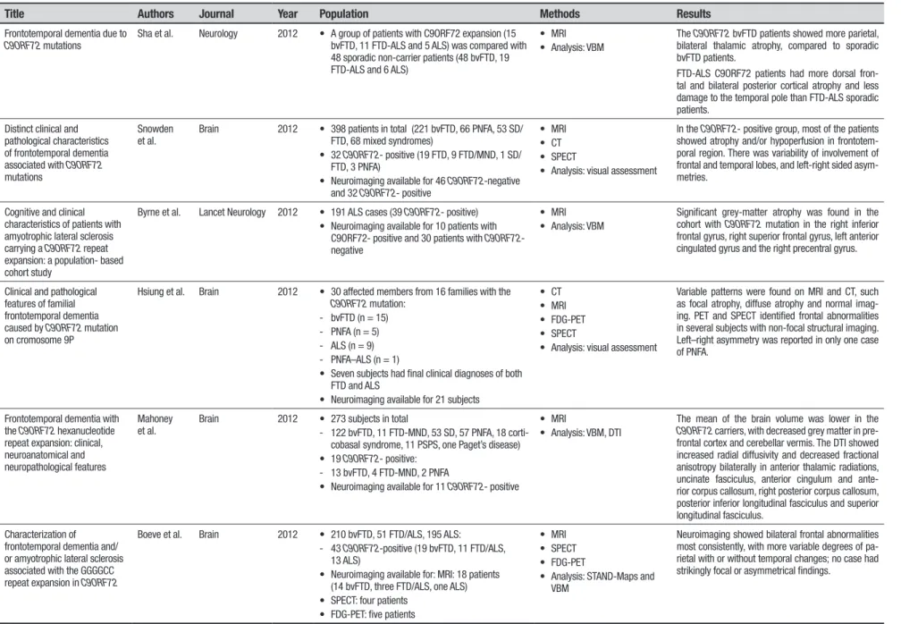
Table 1 presents indings reported in the selected studies, including the number of patients, neuroim-aging technique, and main results.
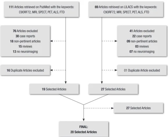
he initial search resulted in 110 and 69 papers retrieved on PubMed and LILACS, respectively. After this initial screening, papers were selected according to the aforementioned inclusion criteria and duplicate articles removed. he inal selection comprised twenty articles (Figure 1).
Selected publications are presented below in three parts: Part I, comprising studies which included FTD patients only; Part II, which describes studies limited to ALS patients; and Part III, which presents studies that included ALS, FTD and FTD-ALS patients.
Part I: FTD Patients. A series of studies assessed the pattern of brain atrophy in FTD patients with C9ORF7210,12-21
using mainly MRI volumetric analysis.
A widespread, symmetrical pattern of brain atrophy was reported in FTD-C9ORF72 patients compared with healthy controls.12,13 he more atrophic compromised
areas were the anterior brain regions, including temporal lobes and all the main subregions of the prefrontal cortex (dorsolateral, orbitofrontal and medial regions). Atrophy in posterior regions (parietal and occipital regions) was also observed in C9ORF72 carriers.13,14 However, these
indings were not replicated in a series of C9ORF72 FTD patients in which brain atrophy was assessed using a visual rating scale, and which failed to ind signiicant diferences in atrophy patterns between carriers and healthy controls in prefrontal regions (orbitofrontal cortex, anterior cingulate) or temporal regions.16 A
recent study reported that carriers of C9ORF72 repeat expansion exhibited signiicant atrophy in speciic brain regions in the pre-symptomatic phase of FTD (before the onset of clinical symptoms).19 Compared to healthy
controls, C9ORF72 carriers had marked atrophy in sub-cortical (thalamus, e.g.) and sub-cortical regions (including frontal, temporal and parietal regions) 20-25 years prior to expected disease onset.19
Investigating white matter tract changes in difer-ent genetic groups of bvFTD21 compared with healthy
Dement Neuropsyc
hol 2015 December;9(4):413-421
415
Prado
et
al.
Neuroimaging in
C9ORF72 mutations
Table 1. Synthesis of articles included in the present review.
Title Authors Journal Year Population Methods Results
Frontotemporal dementia due to
C9ORF72 mutations
Sha et al. Neurology 2012 • A group of patients with C9ORF72 expansion (15 bvFTD, 11 FTD-ALS and 5 ALS) was compared with 48 sporadic non-carrier patients (48 bvFTD, 19 FTD-ALS and 6 ALS)
• MRI • Analysis: VBM
The C9ORF72 bvFTD patients showed more parietal, bilateral thalamic atrophy, compared to sporadic bvFTD patients.
FTD-ALS C9ORF72 patients had more dorsal fron-tal and bilateral posterior cortical atrophy and less damage to the temporal pole than FTD-ALS sporadic patients.
Distinct clinical and pathological characteristics of frontotemporal dementia associated with C9ORF72
mutations
Snowden et al.
Brain 2012 • 398 patients in total (221 bvFTD, 66 PNFA, 53 SD/ FTD, 68 mixed syndromes)
• 32 C9ORF72- positive (19 FTD, 9 FTD/MND, 1 SD/ FTD, 3 PNFA)
• Neuroimaging available for 46 C9ORF72-negative and 32 C9ORF72- positive
• MRI • CT • SPECT
• Analysis: visual assessment
In the C9ORF72- positive group, most of the patients showed atrophy and/or hypoperfusion in frontotem-poral region. There was variability of involvement of frontal and temporal lobes, and left-right sided asym-metries.
Cognitive and clinical characteristics of patients with amyotrophic lateral sclerosis carrying a C9ORF72 repeat expansion: a population- based cohort study
Byrne et al. Lancet Neurology 2012 • 191 ALS cases (39 C9ORF72- positive) • Neuroimaging available for 10 patients with
C9ORF72- positive and 30 patients with C9ORF72 -negative
• MRI • Analysis: VBM
Significant grey-matter atrophy was found in the cohort with C9ORF72 mutation in the right inferior frontal gyrus, right superior frontal gyrus, left anterior cingulated gyrus and the right precentral gyrus.
Clinical and pathological features of familial frontotemporal dementia caused by C9ORF72 mutation on cromosome 9P
Hsiung et al. Brain 2012 • 30 affected members from 16 families with the
C9ORF72 mutation: - bvFTD (n = 15) - PNFA (n = 5) - ALS (n = 9) - PNFA–ALS (n = 1)
• Seven subjects had final clinical diagnoses of both FTD and ALS
• Neuroimaging available for 21 subjects
• CT • MRI • FDG-PET • SPECT
• Analysis: visual assessment
Variable patterns were found on MRI and CT, such as focal atrophy, diffuse atrophy and normal imag-ing. PET and SPECT identified frontal abnormalities in several subjects with non-focal structural imaging. Left–right asymmetry was reported in only one case of PNFA.
Frontotemporal dementia with the C9ORF72 hexanucleotide repeat expansion: clinical, neuroanatomical and neuropathological features
Mahoney et al.
Brain 2012 • 273 subjects in total
- 122 bvFTD, 11 FTD-MND, 53 SD, 57 PNFA, 18 corti-cobasalsyndrome, 11 PSPS, one Paget’s disease) • 19 C9ORF72- positive:
- 13 bvFTD, 4 FTD-MND, 2 PNFA
• Neuroimaging available for 11 C9ORF72- positive
• MRI
• Analysis: VBM, DTI
The mean of the brain volume was lower in the
C9ORF72 carriers, with decreased grey matter in pre-frontal cortex and cerebellar vermis. The DTI showed increased radial diffusivity and decreased fractional anisotropy bilaterally in anterior thalamic radiations, uncinate fasciculus, anterior cingulum and ante-rior corpus callosum, right posteante-rior corpus callosum, posterior inferior longitudinal fasciculus and superior longitudinal fasciculus.
Characterization of frontotemporal dementia and/ or amyotrophic lateral sclerosis associated with the GGGGCC repeat expansion in C9ORF72
Boeve et al. Brain 2012 • 210 bvFTD, 51 FTD/ALS, 195 ALS:
- 43 C9ORF72-positive (19 bvFTD, 11 FTD/ALS, 13 ALS)
• Neuroimaging available for: MRI: 18 patients (14 bvFTD, three FTD/ALS, one ALS) • SPECT: four patients
• FDG-PET: five patients
• MRI • SPECT • FDG-PET
• Analysis: STAND-Maps and VBM
Neuroimaging showed bilateral frontal abnormalities most consistently, with more variable degrees of pa-rietal with or without temporal changes; no case had strikingly focal or asymmetrical findings.
Dement Neuropsyc
hol 2015 December;9(4):413-421
416
Neuroimaging in
C9ORF72 mutations
Prado et al.
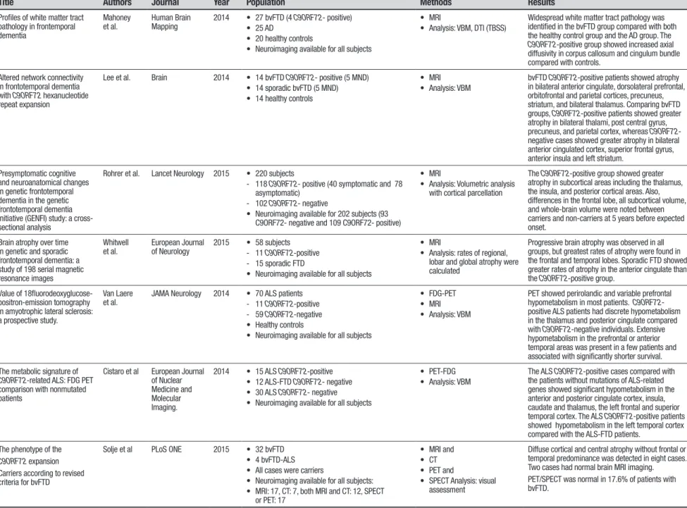
Table 1. Continuation.
Title Authors Journal Year Population Methods Results
Neuroimaging signatures of frontotemporal dementia genetics: C9ORF72, TAU, progranulin and sporadics
Whitwell et al.
Brain 2012 • 76 FTD; imaging available for: - 19 bvFTD C9ORF72- positive - 20 sporadic bvFTD (10 ALS)
• MRI • Analysis: VBM
The C9ORF72 group showed symmetrical atrophy involving dorsolateral, medial and orbitofrontal lobes, and even more loss in anterior temporal lobes, pari-etal lobes, occipital lobes and cerebellum
Frontal asymmetry in behavioral variant frontotemporal dementia: clinicoimaging and pathogenetic correlates
Whitwell et al.
Neurobiology of Aging
2013 • 97 bvFTD • 11 C9ORF72- positive
• MRI • Analysis: VBM
Almost all of the C9ORF72- positive patients had symmetrical frontal atrophy predominantly in the temporofrontoparietal lobes.
Longitudinal neuroimaging and neuropsychological profiles of frontotemporal dementia with
C9ORF72 expansions
Mahoney et al.
Alzheimer’s Research and Therapy
2012 • 20 C9ORF72- positive
• Neuroimaging available for 6 C9ORF72- positive
• MRI
• Analysis: volumetric measures of cortical and subcortical regions; rates of hemispheric and whole brain atrophy were calculated
Carriers exhibited a higher rate of ventricular enlargement, significant atrophy in thalamus and cerebellum and symmetrical atrophy between the cerebral hemispheres.
Cognitive decline and reduced survival in C9ORF72 expansion frontotemporal degeneration and amyotrophic lateral sclerosis
Irwin et al. Neurology Neurosurgery Psychiatry Journal
2013 • 64 C9ORF72-positive (31 ALS, 33 FTLD) • 79 C9ORF72-negative (36 ALS, 43 FTLD) • Neuroimaging available for 41 FTLD patients (14
C9ORF72- positive and 27 C9ORF72- negative)
• MRI • Analysis: VBM
C9ORF72 positive group had greater atrophy in the right fronto-insular, thalamus, cerebellum and bilateral parietal regions compared to C9ORF72
negative group.
Multiparametric MRI study of als stratified for the C9ORF72
genotype
Bede et al. Neurology 2013 • 39 ALS subjects:
- 9 C9ORF72-positive (6 with evidence of FTD) - 30 C9ORF72- negative (3 with evidence of FTD) • 44 healthy controls
• Neuroimaging available for all subjects
• MRI
• Analysis: VMB, DTI (TBSS)
Cortical and subcortical involvement was identified in C9ORF72 carriers, affecting fusiform, thalamic, su-pramarginal, and orbitofrontal regions. White matter abnormalities in the sporadic group were restricted to corticospinal and cerebellar pathways. The body of the corpus callosum and superior motor tracts were affected in both groups.
Basal ganglia involvement in amyotrophic lateral sclerosis
Bede et al. Neurology 2013 • 39 ALS
- 9 C9ORF72- positive - 30 C9ORF72- negative • 44 healthy controls
• Neuroimaging available for all subjects
• MRI
• Analysis: VBM, measures of cortical thickness, DTI (TBSS)
Compared with controls, C9ORF72-negative subjects had significant volume reductions in the left caudate nucleus, left hippocampus, and right accumbens nucleus. In the same comparison of groups, vertex-wise shape analyses revealed changes affecting the superior and inferior aspects of the bilateral thalami, the lateral and inferior portion of the left hippocampus, and the medial and superior aspect of the left caudate. Basal ganglia pathology was more extensive in ALS carriers.
Frontotemporal dementia associated with the C9ORF72
mutation: a unique clinical profile
Devenney et al.
JAMA Neurology 2014 • 114 subjects (84 FTD, 23 FTD/ALS, 7 corticobasal syndrome)
• Neuroimaging • MRI:
- 10 C9ORF72-positive cases with bvFTD - 19 C9ORF72-negative cases with bvFTD - 35 healthy controls
• FDG-PET:
- 6 C9ORF72- positive cases with normal findings on MRI
• MRI • FDG-PET
• Analysis: MRI visual rating scale
A comparison of the C9ORF72 carriers and noncar-riers confirmed no significant difference in the pre-cuneus region, but significant differences were found in the orbitofrontal cortex, anterior temporal lobe, in-sula, and anterior cingulate, with noncarriers showing greater atrophy across these regions. For 3 out 6 pa-tients, the FDG-PET showed hypometabolism in the frontal and/or temporal regions, but FDG-PET showed atypical findings for the other 3 cases.
Dement Neuropsyc
hol 2015 December;9(4):413-421
417
Prado
et
al.
Neuroimaging in
C9ORF72 mutations
Table 1. Continuation.
Title Authors Journal Year Population Methods Results
Profiles of white matter tract pathology in frontemporal dementia
Mahoney et al.
Human Brain Mapping
2014 • 27 bvFTD (4 C9ORF72- positive) • 25 AD
• 20 healthy controls
• Neuroimaging available for all subjects
• MRI
• Analysis: VBM, DTI (TBSS)
Widespread white matter tract pathology was identified in the bvFTD group compared with both the healthy control group and the AD group. The
C9ORF72-positive group showed increased axial diffusivity in corpus callosum and cingulum bundle compared with controls.
Altered network connectivity in frontotemporal dementia with C9ORF72 hexanucleotide repeat expansion
Lee et al. Brain 2014 • 14 bvFTD C9ORF72- positive (5 MND) • 14 sporadic bvFTD (5 MND) • 14 healthy controls
• MRI • Analysis: VBM
bvFTD C9ORF72-positive patients showed atrophy in bilateral anterior cingulate, dorsolateral prefrontal, orbitofrontal and parietal cortices, precuneus, striatum, and bilateral thalamus. Comparing bvFTD groups, C9ORF72-positive patients showed greater atrophy in bilateral thalami, post central gyrus, precuneus, and parietal cortex, whereas C9ORF72- negative cases showed greater atrophy in bilateral anterior cingulated cortex, superior frontal gyrus, anterior insula and left striatum.
Presymptomatic cognitive and neuroanatomical changes in genetic frontotemporal dementia in the genetic frontotemporal dementia initiative (GENFI) study: a cross-sectional analysis
Rohrer et al. Lancet Neurology 2015 • 220 subjects
- 118 C9ORF72- positive (40 symptomatic and 78 asymptomatic)
- 102 C9ORF72- negative
• Neuroimaging available for 202 subjects (93 C9ORF72- negative and 109 C9ORF72- positive)
• MRI
• Analysis: Volumetric analysis with cortical parcellation
The C9ORF72-positive group showed greater atrophy in subcortical areas including the thalamus, the insula, and posterior cortical areas. Also, differences in the frontal lobe, all subcortical volume, and whole-brain volume were noted between carriers and non-carriers at 5 years before expected onset.
Brain atrophy over time in genetic and sporadic frontotemporal dementia: a study of 198 serial magnetic resonance images
Whitwell et al.
European Journal of Neurology
2015 • 58 subjects - 11 C9ORF72-positive - 15 sporadic FTD
• Neuroimaging available for all subjects
• MRI
• Analysis: rates of regional, lobar and global atrophy were calculated
Progressive brain atrophy was observed in all groups, but greatest rates of atrophy were found in the frontal and temporal lobes. Sporadic FTD showed greater rates of atrophy in the anterior cingulate than the C9ORF72-positive group.
Value of 18fluorodeoxyglucose-positron-emission tomography in amyotrophic lateral sclerosis: a prospective study.
Van Laere et al.
JAMA Neurology 2014 • 70 ALS patients - 11 C9ORF72-positive - 59 C9ORF72-negative • Healthy controls
• Neuroimaging available for all subjects
• FDG-PET • MRI • Analysis: VBM
PET showed perirolandic and variable prefrontal hypometabolism in most patients. C9ORF72 -positive ALS patients had discrete hypometabolism in the thalamus and posterior cingulate compared with C9ORF72-negative individuals. Extensive hypometabolism in the prefrontal or anterior temporal areas was present in a few patients and associated with significantly shorter survival.
The metabolic signature of
C9ORF72-related ALS: FDG PET comparison with nonmutated patients
Cistaro et al European Journal of Nuclear Medicine and Molecular Imaging.
2014 • 15 ALS C9ORF72-positive • 12 ALS-FTD C9ORF72- negative • 30 ALS C9ORF72- negative • Neuroimaging available for all subjects
• PET-FDG • Analysis: VBM
The ALS C9ORF72-positive cases compared with the patients without mutations of ALS-related genes showed significant hypometabolism in the anterior and posterior cingulate cortex, insula, caudate and thalamus, the left frontal and superior temporal cortex. The ALS C9ORF72-positive patients showed hypometabolism in the left temporal cortex compared with the ALS-FTD patients.
The phenotype of the
C9ORF72 expansion
Carriers according to revised criteria for bvFTD
Solje et al PLoS ONE 2015 • 32 bvFTD • 4 bvFTD-ALS • All cases were carriers
• Neuroimaging available for all subjects: • MRI: 17, CT: 7, both MRI and CT: 12, SPECT
or PET: 17
• MRI and • CT • PET and
• SPECT Analysis: visual assessment
Diffuse cortical and central atrophy without frontal or temporal predominance was detected in eight cases. Two cases had normal brain MRI imaging. PET/SPECT was normal in 17.6% of patients with bvFTD.
Dement Neuropsychol 2015 December;9(4):413-421
418 Neuroimaging in C9ORF72 mutations Prado et al. 76 Articles excluded
30 case reports 18 non-pertinent articles
15 reviews 13 no neuroimaging
41 Articles excluded 22 case reports 09 non-pertinent articles
03 reviews 07 no neuroimaging
16 Duplicate Articles excluded 01 Duplicate Article excluded
19 Selected Articles 27 Selected Articles
27 Selected Articles
FINAL: 20 Selected Articles 111 Articles retrieved on PubMed with the keywords:
C9ORF72, MRI, SPECT, PET, ALS, FTD
69 Articles retrieved on LILACS with the keywords: C9ORF72, MRI, SPECT, PET, ALS, FTD
Figure 1. Flowchart depicting selection of items for systematic review on PubMed and Lilacs databases using the terms
C9ORF72, ALS, FTD, MRI, SPECT and PET.
data are limited by the small size of the sample (only four bvFTD carriers).
Some studies compared neuroimaging features of C9ORF72-bvFTD with sporadic bvFTD and other muta-tions. C9ORF72-bvFTD patients had less gray matter loss than sporadic bvFTD in the anterior cingulate, orbi-tofrontal cortex, anterior temporal lobe and insula.16
Another study reported that the majority of subjects with mutation in the microtubule associated protein tau gene (MAPT) and C9ORF72 subjects had symmetric frontal atrophy, while most subjects with mutation in the progranulin gene (GRN) had asymmetric atrophy.13
C9ORF72 carriers had greater atrophy in posterior (pari-etal and occipital) lobes in comparison with MAPT and sporadic bvFTD groups, while patients with MAPT muta-tions had greater impairment in temporal poles, com-pared with the C9ORF72 group.13 Patients with GRN mutation had more loss in parietal lobes than C9ORF72 carriers.13 In the same study, by applying a multinomial
logistic regression model based on atrophic patterns, it was possible to classify FTD patients with diferent geno-types with 93% accuracy, suggesting that neuroimaging
may be useful to distinguish C9ORF72-FTD patients from patients with other mutations at a single-subject level.13
Only one study investigated white matter patterns across bvFTD patients grouped according to genetic sta-tus.21 here were no diferences between C9ORF72 and
sporadic bvFTD cases, but MAPT patients had abnormal fractional anisotropy in the anterior region of the left temporal lobe, compared with the C9ORF72 group.
he integrity of the intrinsic connectivity network in bvFTD was explored in a group of 14 bvFTD C9ORF72 carriers and 14 bvFTD non-carriers.12 hese groups
were compared against healthy controls. Patients with C9ORF72 did not exhibit diferences in the default mode network compared to controls. Conversely, bvFTD non-carriers exhibited a diferent pattern, presenting both impaired (in striatum and thalamus) and enhanced (in precuneus and posterior cingulate) connectivity com-pared with controls.12 In the same study, it was reported
Dement Neuropsychol 2015 December;9(4):413-421
419
Prado et al. Neuroimaging in C9ORF72 mutations
salience network connectivity between carriers and non-carriers.12
he progression of brain atrophy in bvFTD patients with diferent genetic status was assessed in a longitu-dinal study.18GRN patients had greater rates of atrophy
than sporadic, MAPT and C9ORF72 groups. Sporadic bvFTD patients had greater rates of gray matter loss in anterior cingulate than C9ORF72 carriers, while the lat-ter had grealat-ter rates of atrophy in cerebellum and occipi-tal lobes, compared with MAPT carriers.18 Another study
found that C9ORF72-bvFTD patients had increased rates of brain atrophy and ventricular expansion compared with healthy controls.14
In summary, widespread brain atrophy was reported in FTD C9ORF72 patients, mostly in anterior brain regions, but also with possible damage in posterior cor-tical areas. Brain atrophy may be identiied before dis-ease onset. However, the absence of signiicant changes in FTD C9ORF72 carriers has also been reported.
Part II: ALS Patients. Five articles investigated neuroim-aging features of ALS patients with C9ORF72 expan-sion. Four of these studies employed the MRI tech-nique9,22-24 while the remainder used FDG-PET.25
ALS patients with C9ORF72 mutation had greater atrophy in prefrontal regions, including frontal gyri and the anterior cingulate, compared to those with sporadic ALS.9,22 he right precentral gyrus was also afected in
one study.9 Mild hypometabolism in the thalamus and
posterior cingulate was found on PET-FDG in ALS car-riers compared with non-carcar-riers.25 Compared with
ALS non-carriers, C9ORF72 carriers had more cal and subcortical involvement, afecting both corti-cal (fusiform, supramarginal, and orbitofrontal cortex and Broca’s area) and subcortical regions (thalamus).23
Interestingly, in the same study, white matter abnor-malities in ALS non-carriers were relatively limited to corticospinal and cerebellar pathways, while carriers had more widespread involvement. hese data suggested that non-motor changes (e.g. cognitive impairment) in ALS could be largely driven by C9ORF72 repeat expan-sion.23 Basal ganglia involvement was also more
exten-sive in ALS patients with C9ORF72 mutation than in non-carriers.24
In short, ALS C9ORF72 carriers had greater atrophy, with predominance in prefrontal regions, compared to sporadic ALS patients. Mild hypometabolism in the thalamus and posterior cingulate, more widespread abnormalities of white matter, and greater basal ganglia involvement has also been demonstrated in ALS carriers compared with non-carriers.
Part III: FTD, ALS and FTD-ALS patients. he imaging patterns of patients with ALS, FTD or FTD-ALS according to their genetic status were compared in a series of studies.5,10,20,22,26,27 Most of the papers employed
structural brain MRI.
In a group of eighteen patients with C9ORF72 repeat expansion (fourteen bvFTD, three with FTD/ALS and one with ALS), gray matter loss was found in cortical areas including frontotemporal regions,26 in a similar
pattern to that reported by others.10,20,22 Most studies
reported symmetrical patterns of brain atrophy, except for patients presenting with predominant language dei-cit. Some patients may have parietal cortical atrophy and thalamic involvement.10,26 hese studies are limited by
the absence of direct comparisons between bvFTD and FTD-ALS.
A group of patients with C9ORF72 expansion (15 bvFTD, 11 FTD-ALS and 5 ALS) was compared against 48 sporadic non-carrier patients (48 bvFTD, 19 FTD-ALS and 6 ALS).27 he authors found that bvFTD-C9 patients
had more parietal and bilateral thalamic atrophy and less medial frontal atrophy compared to sporadic bvFTD patients. FTD-ALS C9ORF72 patients had more dorsal frontal and bilateral posterior cortical atrophy and less damage to the temporal pole than sporadic FTD-ALS patients.27
Conversely, some studies reported that C9ORF72 car-riers may not have brain atrophy.22,26 hese indings were
expanded by a recent study, which demonstrated that almost 18% of bvFTD cases with C9ORF72 mutation had no abnormalities on PET/SPECT.5
In a study that investigated the metabolic patterns of C9ORF72 carriers on PET-FDG, ALS carriers of C9ORF72 had more pronounced hypometabolism in cortical (cin-gulate cortex, and frontotemporal regions) and subcor-tical structures (caudate and thalami) compared with sporadic ALS patients.28 In the same study, ALS patients
with C9ORF72 expansion had impaired metabolism in the left temporal cortex, compared with the ALS-FTD group.28 Accordingly, ALS C9ORF72 patients may have a more severe clinical picture and more widespread central nervous system involvement than sporadic ALS patients, regardless of the association with bvFTD.
Dement Neuropsychol 2015 December;9(4):413-421
420 Neuroimaging in C9ORF72 mutations Prado et al.
DISCUSSION
For many years, neuroimaging was of limited applica-bility in the everyday evaluation of neurodegenerative disorders. For instance, the exclusion of focal lesions or hydrocephalus as causes of cognitive deicits was the main utility of imaging exploration in patients sufering from cognitive disorders. his picture has changed, with modern imaging techniques which provide useful and speciic markers for the diagnosis and the follow-up of neurodegenerative diseases, such as ALS and FTD.29 In
this paper we systematically reviewed neuroimaging data in FTD and/or ALS patients with C9ORF72 repeat expansion.
Most studies that investigated the neuroimaging features of C9ORF72 carriers found consistent involve-ment of frontotemporal regions, including prefrontal cortex, (dorsolateral, orbitofrontal and medial regions), and also cingulate and posterior regions such as the parietal and occipital lobes.10,12-15 Subcortical regions,
especially thalami, may also be afected in C9ORF72 carriers.10,19,22 It is of note that some studies reported
that patients with C9ORF72 mutation may not have abnormalities on structural and functional brain
imag-ing.5,16,22,26 hese disparate patterns may be due to a
number of diferent reasons. he inclusion of patients at diferent stages of disease and diferences in neuroim-aging methods across studies may account for the vari-ability of results. One factor that may partially account for these disparate indings is that diferent phenotypes are associated with C9ORF72 and heterogeneity may occur even among patients with the same clinical phe-notype.22 Besides ALS, bvFTD and ALS/FTD, C9ORF72 mutation has also been associated with primary progres-sive aphasia, Huntington’s disease-like syndrome, and atypical parkinsonism syndromes, such as corticobasal degeneration and progressive supranuclear palsy.22,30-33
Repeated expansion in C9ORF72 may also contribute to Alzheimer’s disease.33,34 In summary, although FTD and/
or ALS are the most common phenotypes of C9ORF72 repeat expansion, other clinical presentations may occur, with diferent neuroimaging patterns. It remains unclear why some patients with the C9ORF72 expansion have minimal atrophy on neuroimaging studies. he possible pathways by which C9ORF72 mutation participates in the pathophysiological process associated with diferent neurodegenerative diseases also remain elusive.
From a clinical perspective, the variability of clinical indings associated with C9ORF72 limits the interpre-tation of neuroimaging features at an individual level. A single-center study reported the utility of a multino-mial regression model to accurately identify C9ORF72
patients based on patterns of brain atrophy13 at
single-subject level. However, this strategy seems limited to research centers with advanced expertise in neuroimag-ing techniques. Moreover, C9ORF72 carriers may have no structural abnormalities on brain MRI.16,22,26
here-fore, atrophic features in brain MRI are of limited value for identiication of C9ORF72 carriers in clinical practice. On the other hand, neuroimaging assessment may be useful for the follow-up of patients with C9ORF72 repeat expansion and for suggesting prognostic aspects. FTD and/or ALS patients with C9ORF72 mutation may have faster disease progression and shorter survival than non-carriers,10,16 even though this is not consistent across
studies.27 In this scenario, neuroimaging can identify
markers of disease progression, such as the rate of brain atrophy and ventricular expansion.14 hese markers
could help track disease changes and guide clinical man-agement, especially in the prospect of disease-modifying drugs that will target the pathophysiological process of neurodegenerative disorders.
New modern neuroimaging techniques may provide useful biomarkers for the diagnosis and follow-up of C9ORF72 carriers. Disruption of functional connectivity may be seen in the absence of brain atrophy and could be regarded as an early marker of disease.12 Only one study
to date explored functional connectivity in C9ORF72 car-riers, and found that there is a convergent, large-scale, disrupted network among diferent patterns of brain atrophy.12 he investigation of functional connectivity
may enhance our understanding about the neural net-works compromised by C9ORF72 mutation, thus provid-ing valuable information for the comprehension of the pathophysiology of the FTD-ALS spectrum.
Techniques exploring the integrity of the white mat-ter tract may also be of clinical value in the assessment of patients with C9ORF72 repeat expansion. Degenera-tion of the corticospinal tract is a hallmark of ALS, and disruption of this tract can diferentiate ALS patients from bvFTD and ALS-FTD patients.23,35 Further studies
are needed to describe putative white matter changes associated withC9ORF72 mutation.
Besides its value for diagnostic purposes, neuroimag-ing is also important for the understandneuroimag-ing of the neural basis of cognitive and behavioral disorders observed in the FTD-ALS spectrum. ALS and bvFTD patients with C9ORF72 mutation have a greater frequency of psychi-atric disorders, especially psychotic symptoms, such as delusions, paranoid ideation and hallucinations.5,16,22,36
Dement Neuropsychol 2015 December;9(4):413-421
421
Prado et al. Neuroimaging in C9ORF72 mutations
Paranoid or irrational thinking were also frequent in the same study.22 he neuropsychological proile of bvFTD
patients with C9ORF72 expansion is similar to non-car-rier bvFTD patients,16 with comparable performance in
memory, language and executive skills. Deicits in execu-tive functions are the most common observed feature in ALS-FTD patients3 and can also be associated with
prefrontal dysfunction.
Taken together, these data emphasize the complex interaction between C9ORF72 mutation and clinical presentations of neurodegenerative diseases, especially the FTD-ALS spectrum. he discovery of the C9ORF72
repeat expansion has opened a window for the under-standing of the continuum between FTD and ALS. he next advances in neuroimaging investigation may pro-vide valuable markers for the diagnosis and follow-up of these patients, and may also clarify the common patho-physiological pathways between ALS and FTD, with pos-sible clinical outcomes.
Acknowledgments: his study was partly funded by CNPq. We thank the reviewers for their valuable comments on the paper.
REFERENCES
1. Pressman PS, Miller BL. Diagnosis and management of behavioral variant frontotemporal dementia. Biol Psychiatry. 2014;75:574-581. 2. Piguet O, Hornberger M, Mioshi E, Hodges JR. Behavioural-variant
frontotemporal dementia: diagnosis, clinical staging, and management. Lancet Neurol 2011;10:162-172.
3. Goldstein LH, Abrahams S. Changes in cognition and behaviour in amyotrophic lateral sclerosis: nature of impairment and implications for assessment. Lancet Neurol 2013;12:368-380.
4. Lomen-Hoerth C, Murphy J, Langmore S, Kramer JH, Olney RK, Miller B. Are amyotrophic lateral sclerosis patients cognitively normal? Neurology 2003;60:1094-1097.
5. Solje E, Aaltokallio H, Koivumaa-Honkanen H, et al. The Phenotype of the C9ORF72 Expansion Carriers According to Revised Criteria for bvFTD. PLoS One. 2015;10:e0131817.
6. Renton AE, Majounie E, Waite A, et al. A hexanucleotide repeat expan-sion in C9ORF72 is the cause of chromosome 9p21-linked ALS-FTD. Neuron 2011;72:257-268.
7. DeJesus-Hernandez M, Mackenzie IR, Boeve BF, et al. Expanded GGGGCC hexanucleotide repeat in noncoding region of C9ORF72 causes chromosome 9p-linked FTD and ALS. Neuron. 2011;72:245-256. 8. Takada LT. The Genetics of Monogenic Frontotemporal Dementia.
Dement Neuropsychol 2015;9:219-229.
9. Byrne S, Elamin M, Bede P, et al. Cognitive and clinical characteris-tics of patients with amyotrophic lateral sclerosis carrying a C9orf72 repeat expansion: a population-based cohort study. Lancet Neurol 2012; 11:232-240.
10. Irwin DJ, McMillan CT, Brettschneider J, et al. Cognitive decline and reduced survival in C9orf72 expansion frontotemporal degeneration and amyotrophic lateral sclerosis. J Neurol Neurosurg Psychiatry 2013; 84:163-169.
11. Vargas C, Lopez-Jaramillo C, Vieta E. A systematic literature review of resting state network--functional MRI in bipolar disorder. J Affect Disord 2013;150:727-735.
12. Lee SE, Khazenzon AM, Trujillo AJ, et al. Altered network connectivity in frontotemporal dementia with C9orf72 hexanucleotide repeat expansion. Brain 2014;137:3047-3060.
13. Whitwell JL, Weigand SD, Boeve BF, et al. Neuroimaging signatures of frontotemporal dementia genetics: C9ORF72, tau, progranulin and sporadics. Brain 2012;135:794-806.
14. Mahoney CJ, Downey LE, Ridgway GR, et al. Longitudinal neuroim-aging and neuropsychological profiles of frontotemporal dementia with C9ORF72 expansions. Alzheimers Res Ther 2012;4:41.
15. Mahoney CJ, Beck J, Rohrer JD, et al. Frontotemporal dementia with the C9ORF72 hexanucleotide repeat expansion: clinical, neuroanatomical and neuropathological features. Brain 2012;135:736-750.
16. Devenney E, Hornberger M, Irish M, et al. JAMA Neurology. 2014;71: 331-339.
17. Whitwell JL, Xu J, Mandrekar J, et al. Frontal asymmetry in behavioral variant frontotemporal dementia: clinicoimaging and pathogenetic corre-lates. Neurobiol Aging 2013;34:636-639.
18. Whitwell JL, Boeve BF, Weigand SD, et al. Brain atrophy over time in genetic and sporadic frontotemporal dementia: a study of 198 serial magnetic resonance images. Eur J Neurol 2015;22:745-752.
19. Rohrer JD, Nicholas JM, Cash DM, et al. Presymptomatic cognitive and neuroanatomical changes in genetic frontotemporal dementia in the Genetic Frontotemporal dementia Initiative (GENFI) study: a cross-sectional analysis. Lancet Neurol 2015;14:253-262.
20. Hsiung GY, DeJesus-Hernandez M, Feldman HH, et al. Clinical and path-ological features of familial frontotemporal dementia caused by C9ORF72 mutation on chromosome 9p. Brain 2012;135:709-722.
21. Mahoney CJ, Ridgway GR, Malone IB, et al. Profiles of white matter tract pathology in frontotemporal dementia. Hum Brain Mapp 2014; 35:4163-4179.
22. Snowden JS, Rollinson S, Thompson JC, et al. Distinct clinical and pathological characteristics of frontotemporal dementia associated with C9ORF72 mutations. Brain 2012;135:693-708.
23. Bede P, Bokde AL, Byrne S, et al. Multiparametric MRI study of ALS stratified for the C9orf72 genotype. Neurology 2013;81:361-369. 24. Bede P, Elamin M, Byrne S, et al. Basal ganglia involvement in
amyo-trophic lateral sclerosis. Neurology 2013;81:2107-2115.
25. Van Laere K, Vanhee A, Verschueren J, et al. Value of 18fluorodeoxyglu-cose-positron-emission tomography in amyotrophic lateral sclerosis: a prospective study. JAMA Neurology 2014;71:553-561.
26. Boeve BF, Boylan KB, Graff-Radford NR, et al. Characterization of fronto-temporal dementia and/or amyotrophic lateral sclerosis associated with the GGGGCC repeat expansion in C9ORF72. Brain 2012;13:765-783. 27. Sha SJ, Takada LT, Rankin KP, et al. Frontotemporal dementia due to
C9ORF72 mutations: clinical and imaging features. Neurology 2012;79: 1002-1011.
28. Cistaro A, Pagani M, Montuschi A, et al. The metabolic signature of C9ORF72-related ALS: FDG PET comparison with nonmutated patients. Eur J Nucl Med Mol Imaging 2014;41:844-852.
29. de Souza LC, Lehericy S, Dubois B, Stella F, Sarazin M. Neuroimaging in dementias. Curr Opin Psychiatry 2012;25:473-479.
30. Simon-Sanchez J, Dopper EG, Cohn-Hokke PE, et al. The clinical and pathological phenotype of C9ORF72 hexanucleotide repeat expansions. Brain 2012;135:723-735.
31. Le Ber I, Camuzat A, Guillot-Noel L, et al. C9ORF72 repeat expansions in the frontotemporal dementias spectrum of diseases: a flow-chart for genetic testing. J Alzheimers Dis 2013;34:485-499.
32. Hensman Moss DJ, Poulter M, Beck J, et al. C9orf72 expansions are the most common genetic cause of Huntington disease phenocopies. Neurology 2014;82:292-299.
33. Beck J, Poulter M, Hensman D, et al. Large C9orf72 hexanucleotide repeat expansions are seen in multiple neurodegenerative syndromes and are more frequent than expected in the UK population. Am J Hum Genet 2013;92:345-353.
34. Kohli MA, John-Williams K, Rajbhandary R, et al. Repeat expansions in the C9ORF72 gene contribute to Alzheimer’s disease in Caucasians. Neurobiol Aging 2013;34:1519 e1515-1512.
35. Lillo P, Mioshi E, Burrell JR, Kiernan MC, Hodges JR, Hornberger M. Grey and white matter changes across the amyotrophic lateral sclerosis-frontotemporal dementia continuum. PLoS One 2012;7:e43993. 36. Dobson-Stone C, Hallupp M, Bartley L, et al. C9ORF72 repeat