Neuroimaging in normal pressure hydrocephalus
Benito Pereira Damasceno1
ABSTRACT. Normal pressure hydrocephalus (NPH) is a syndrome characterized by the triad of gait disturbance, mental deterioration and urinary incontinence, associated with ventriculomegaly and normal cerebrospinal fluid (CSF) pressure. The clinical presentation (triad) may be atypical or incomplete, or mimicked by other diseases, hence the need for supplementary tests, particularly to predict postsurgical outcome, such as CSF tap-tests and computed tomography (CT) or magnetic resonance imaging (MRI). The CSF tap-test, especially the 3 to 5 days continuous external lumbar drainage of at least 150 ml/day, is the only procedure that simulates the effect of definitive shunt surgery, with high sensitivity (50-100%) and high positive predictive value (80-100%). According to international guidelines, the following are CT or MRI signs decisive for NPH diagnosis and selection of shunt-responsive patients: ventricular enlargement disproportionate to cerebral atrophy (Evans index >0.3), and associated ballooning of frontal horns; periventricular hyperintensities; corpus callosum thinning and elevation, with callosal angle between 40º and 90º; widening of temporal horns not fully explained by hippocampal atrophy; and aqueductal or fourth ventricular flow void; enlarged Sylvian fissures and basal cistern, and narrowing of sulci and subarachnoid spaces over the high convexity and midline surface of the brain. On the other hand, other imaging methods such as radionuclide cisternography, SPECT, PET, and also DTI or resting-state functional MRI, although suitable for NPH diagnosis, do not yet provide improved accuracy for identifying shunt-responsive cases. Key words: normal pressure hydrocephalus, neuroimaging, magnetic resonance, cerebrospinal fluid tap test, shunt surgery.
NEUROIMAGEM NA HIDROCEFALIA DE PRESSÃO NORMAL
RESUMO. A hidrocefalia de pressão normal (HPN) é uma síndrome caracterizada por alteração da marcha, transtorno mental-cognitivo e incontinência urinária, associados a ventriculomegalia e pressão liquórica normal. A apresentação clínica (tríade) pode ser atípica ou incompleta, ou pode ser mimetizada por outras doenças, daí a necessidade de testes suplementares, principalmente para predição do resultado cirúrgico, tais como teste da punção lombar e tomografia computadorizada (TC) ou ressonância magnética (MR) de crânio. O teste da punção liquórica lombar, especialmente a drenagem externa contínua (≥150 ml/dia, por 3 a 5 dias), é o único método que simula o efeito da cirurgia, com alta sensibilidade (50-100%) e alto valor preditivo positivo (80-100%). Consensos internacionais consideram os seguintes achados da TC ou RM como decisivos para o diagnóstico de HPN e a seleção de pacientes bons respondedores à cirurgia: dilatação ventricular desproporcional em relação ao grau de atrofia cerebral (índice de Evans >0.3), associada a arredondamento dos cornos frontais; hipersinal difuso periventricular; adelgaçamento e elevação do corpo caloso, com ângulo do corpo caloso entre 40º e 90º; dilatação dos cornos temporais não explicada por atrofia hipocampal; sinal do fluxo vazio no aqueduto e quarto ventrículo; dilatação das fissuras Sylvianas e cisterna basal, e estreitamento ou apagamento dos sulcos e espaços subaracnoides nas superfícies cerebrais da convexidade alta e linha média. Por outro lado, a cisternografia isotópica, SPECT, PET, e mesmo técnicas mais modernas de RM funcional e tensor de difusão, embora compatíveis com o diagnóstico de HPN, não melhoram a acurácia na identificação de casos responsivos à cirurgia. Palavras-chave: hidrocefalia de pressão normal, neuroimagem, ressonância magnética, teste da punção lombar, cirurgia de derivação liquórica.
INTRODUCTION
N
ormal pressure hydrocephalus (NPH) is a syndrome characterized by the triad of gait disturbance, mental deterioration andurinary incontinence, which are associated with enlargement of the ventricular system and normal cerebrospinal luid (CSF) pres-sure. In NPH, CSF pressure may be normal
This study was conducted at the Department of Neurology, Medical School, University of Campinas (UNICAMP), Campinas SP, Brazil.
1MD, PhD, Department of Neurology, Medical School, University of Campinas (UNICAMP), Campinas SP, Brazil.
Benito Pereira Damasceno. Rua Maria Monteiro 1710 / apt 24 – 13025-1252 Campinas SP – Brazil. E-mail: [email protected] Disclosure: The authors report no conflicts of interest.
Received August 03, 2015. Accepted in final form October 20, 2015.
at one spinal tap, but episodes of increased CSF pres-sure can occur, and hence NPH is also termed “intermit-tent pressure hydrocephalus”. It is caused by excessive accumulation of CSF in the ventricular system due to an impairment of its low distally to the fourth ventricle (“communicating” hydrocephalus). About 50% of cases with communicating NPH have a known cause (second-ary or symptomatic NPH, or SNPH), such as meningitis, subarachnoid hemorrhage, or cranial trauma, while the remaining 50% of cases are idiopathic (INPH), usually presenting in the 7th decade of life. Epidemiological data on NPH incidence and prevalence are scarce, but surveys in Germany, Norway, Sweden and Japan have estimated the annual incidence of INPH to be between 1.8/100,000 and 5.5/100,000 inhabitants, with a prevalence ranging from 0.2% to 2.9% among individuals aged 65 years or older,1-4 and that it is the cause of dementia in up to 6%
of all dementia cases. his review was based on a PubMed literature search from 1996 to date.
DIAGNOSIS
he diagnosis of NPH is based on the following criteria: [1] a history of gait disturbance, progressive mental deterioration, and urinary urgency or incontinence; [2] hydrocephalus, deined as Evans’ ratio >0.30 on computed tomography (CT) or magnetic resonance (MR) imaging; and [3] a CSF opening pressure (appro-priately measured) of <24 cm of water.
Diferential diagnostic diiculties may arise when the clinical manifestations of the triad are atypical or incomplete, or when they are mimicked by other dis-eases. Indeed, many other conditions may cause the complete triad, such as vascular dementia, parkinsonism, Lewy body disease, corticobasal degeneration, progres-sive supranuclear palsy, multiple system atrophy, neu-rosyphilis, medication side efects; or in combination with other diseases, particularly cerebrovascular and Alzheimer’s disease, which are present in up to 75% of patients with INPH.5
NEUROIMAGING AND COMPLEMENTARY
PROGNOSTIC TESTS
Shunt surgery can improve all NPH symptoms and quality of life in up to 80% of properly selected cases, but has complications rates (35-52%) that dissuade us from shunting every suspected case. Surgical decision may be diicult and yield varying results, particularly in the elderly with comorbidities or vascular or degen-erative brain diseases, which can mimic or worsen NPH symptoms. herefore, in order to improve diag-nosis and management of NPH cases, complementary
tests have to be used, especially neuroimaging and CSF tap tests.
Radionuclide cisternography (RC), intracranial pres-sure monitoring (ICP) and lumbar infusion tests can show CSF dynamics malfunction, but none are able to conirm whether the patient will beneit from surgery.6,7
A ‘positive’ RC (with ventricular relux and convexity block) can be seen in other dementia disorders and even in healthy subjects, thus having questionable predictive value. Most clinicians suggest it should no longer be per-formed,8 and the International INPH Guidelines9 have
not included it as an option, since it does not improve the diagnostic accuracy of identifying shunt-responsive cases.
he CSF tap test (CSF-TT) consists of quantitative testing of gait and cognitive functions before and after the drainage of 40-50 ml lumbar CSF. It is the only proce-dure that can temporarily simulate the efect of a deini-tive shunt, and can predict not only the outcome of sur-gery but also the degree of improvement.10,11 Since the
one tap CSF-TT has low sensitivity (26-61%), a “negative” result cannot be used to exclude patients from surgery. In such cases, the alternative is a repeated CSF-TT (RTT) performed on three consecutive days with a minimum of 30 to 40 ml CSF removed; or continuous external lumbar drainage (ELD) for 3 to 5 days, with a minimum of 150 ml CSF drained daily. Due to ELD’s high sensitivity (50-100%) and high positive predictive value (80-(50-100%), it is considered by the 2005 International INPH Guidelines the most efective test for identifying shunt-responsive cases, even though it requires hospital admission and is associated with higher complication rates (meningitis, subdural hematoma, nerve root inlammation).
In NPH, computed tomography (CT) and magnetic resonance imaging (MRI) show ventricular enlargement disproportionate to cerebral atrophy, with associated bal-looning of frontal horns, periventricular hyperintensi-ties, thinning and elevation of the corpus callosum, and widening of temporal horns without evidence of hip-pocampal atrophy. Although diagnosis can be made on the basis of CT indings alone, MRI is more accurate for disclosing associated pathologies (such as cerebrovascu-lar disease) and also for detecting NPH typical signs of prognostic value, besides avoiding exposure to ionizing radiation.
he International Guidelines have recommended the following key imaging features for diagnosis of INPH and selection of shunt-responsive patients:
1. Ventricular enlargement not entirely attributable to cerebral atrophy or congenital enlargement (Evans index >0.3).
3. At least one of the following supportive features: a) Enlargement of the temporal horns of the lateral
ventricles not entirely attributable to hippocampus atrophy;
b) Callosal angle of 40° or greater;
c) Evidence of altered brain water content, including periventricular signal changes on CT and MRI not attributable to microvascular ischemic changes or demyelination;
d) An aqueductal or fourth ventricular low void on MRI. he Japanese INPH Guidelines12 also include the
fol-lowing key imaging features: (1) enlarged Sylvian issures and basal cistern; and (2) narrowing of the sulci and sub-arachnoid spaces over the high convexity and midline surface of the brain. Unlike the International Guidelines, periventricular changes are not considered essential.
Other neuroimaging methods have also been used in NPH. Single photon emission computed tomogra-phy (SPECT) and positron emission tomogratomogra-phy (PET) can show reduction of cerebral blood low and metabo-lism, mainly in frontobasal and anterior periventricular regions, even with improvement of regional cerebral metabolic rate of glucose in shunt responsive patients.13
Newer MRI techniques such as DTI14 and resting-state
functional MRI15 have also been studied in NPH allowing
the possibility of detecting biomarkers and improving NPH diagnosis. However, the diagnostic and prognostic value of all these neuroimaging techniques is not well established and they are not part of the routine selection procedures for shunt surgery.
Ventricular enlargement can be measured by the Evans índex,16 which is the ratio between the maximal
width of the frontal horns and the maximal width of the inner table of the cranium at the level of the frontal horns; or by an equivalent measure, such as by dividing the diameter of the frontal horns by the widest brain diameter (Figure 1).
he reliability of the Evans index has been questioned by some studies using modern brain imaging techniques (FreeSurfer) for ventricular volumetric analysis.17
How-ever, a more recent study18 has addressed this issue and
showed that the Evans index and other linear measure-ments (frontal-occipital horn ratio, third ventricular width, and callosal angle at the level of the posterior commissure) reliably determine ventricular enlarge-ment, without the need for expensive, time-intensive and technically challenging computer software, unavail-able in many healthcare services.
Periventricular signal changes may be associated with subcortical vascular encephalopathy (also with lacunar infarctions) in NPH, but this does not predict poor
sur-gical outcome and should not exclude patients from shunting.5,19
In typical NPH cases, the ventricles are dispropor-tionately more dilated than the cortical sulci, which are narrow or obliterated at the high convexity and midline, with local narrowing of the subarachnoid space sur-rounding the outer brain surface, as can be seen in a MRI coronal section at the level of the posterior commissure (Figure 2 and 3). In this context, the presence of enlarged ventricles associated with large basal cisterns and Syl-vian issures, and also focally dilated sulci, should not be misinterpreted as cerebral atrophy. On the contrary, these indings tend to support rather than exclude the diagnosis of shunt-responsive NPH.20,21 In normal aging
and degenerative diseases (Alzheimer’s, Pick’s), the thin-ning of the gyri and the corresponding dilation of the sulci are more generalized, occurring to a similar degree in the afected brain regions.22
Callosal angle (CA) as well as temporal horns and hip-pocampus are best evaluated with coronal MRI. CA is the angle between the lateral ventricles (Figure 2), and is typical of NPH when between 40° and 90°. CA greater than 90º suggests brain atrophy,5 as occurs in
degen-erative diseases such as Alzheimer’s and Lewy body dementia.23 In a recent study, shunt-responders had a
signiicantly smaller mean preoperative CA compared
Figure 1. Axial CT slice of the brain in a patient with NPH. The Evans
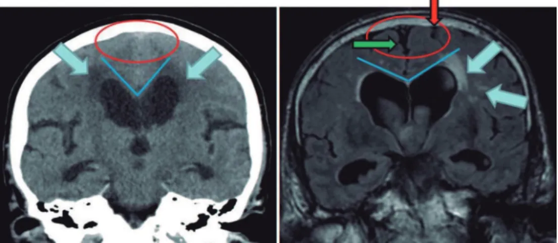
Figure 2. Coronal head CT (left) and MRI (right) at the level of the posterior commissure: in the left image, the CSF spaces over the convexity near the vertex are narrowed (“tight convexity”, red circle), as are the medial cisterns (red circle) - these are typical findings of NPH. On the right image, however, the CSF spaces over the convexity near the vertex (red arrow) and the medial cisterns (green arrow) are widened, a finding consistent with brain atrophy. The blue lines in both images indicate the callosal angle: an angle less than 90º is typical of NPH (left image), while an angle greater than 90º is typical of brain atrophy (right image). The blue arrows indicate periventricular signal alterations. The unilateral occurrence of these alterations (right image) suggests they are probably due to vascular encephalopathy. The abnormalities seen in the left image may well represent transependymal CSF diapedesis due to NPH. (From Kiefer & Unterberg, Dtsch Arztebl Int, 2012, with permission).
A
B
C
Figure 3. Coronal head CT of a 73-year-old man with idiopathic NPH. [A, B and C] show disproportionately enlarged ventricles with periventricular
with non-responders (59% versus 68%), with a cut-of value of 63% providing the best prognostic accuracy.24
Another consistent inding in NPH, best recognized with mid-sagittal MRI, is callosal upward elevation, stretching and thinning, usually showing total or partial recovery after shunting.25
Wide temporal horns are typical of NPH and repre-sent signiicant predictors of a positive shunt outcome,26
although narrow temporal horns are also compatible with NPH diagnosis.27 Evaluation of temporal horns
and hippocampal/perihippocampal structures may dis-tinguish hydrocephalic enlargement of the ventricle, as occurs in NPH (with obliteration of perihippocampal sulcal markings) from ventriculomegaly secondary to cerebral atrophy (which may expose sulcal markings in this region).28 Measurement of the hippocampus volume
may be of diagnostic and prognostic value in cases of sus-pected NPH versus Alzheimer’s disease (AD), in which the hippocampus is signiicantly smaller.29 Difusion
tensor imaging (DTI) of the fornix may also diferenti-ate NPH from AD. Fornix volume, cross-sectional area, and fractional anisotropy are smaller both in NPH and in AD relative to controls. Fornix length, however, is sig-niicantly greater in NPH than in controls yet not altered in AD, probably explained by mechanical stretching due to lateral ventricular dilation and corpus callosum defor-mation in NPH and by degeneration secondary to hip-pocampal atrophy in AD.30
Aqueductal low void is a loss (increased hypointen-sity) of signal seen within the aqueduct and neighbouring third and fourth ventricles, particularly on T2-weighted MRI images. he signal loss is usually increased in states of hyperdynamic CSF motion, with higher velocities, tur-bulent and accelerated low, which occur mainly in pas-sages with smaller cross-sectional areas such as the aque-duct. In NPH there is greater outlow of CSF through the aqueduct with subsequent increase in signal loss (void sign). he irst studies31,32 showed correlation between
void sign in the aqueduct and shunt results, while oth-ers33 did not, with the same frequency of void sign
occur-ring in NPH patients and healthy controls.
Further advances in CSF low imaging have enabled quantiication by means of cine phase-contrast MRI (PC-MRI) throughout the cardiac cycle. With this technique,
the slice is positioned on an angled axial plane perpen-dicular to the aqueduct using higher spatial resolution MRI because of the small size of this structure, as well as short TR to achieve adequate temporal resolution.34,35
he aqueductal CSF stroke volume (ACSV, deined as the average of the volume lowing down during cardiac systole and up during diastole) is then calculated. Ini-tial studies36 found that NPH patients who responded
to shunting had aqueductal stroke volume ≥ 42 µL or at least twice the ACSV of healthy elderly subjects, while others did not.37,38 For this reason, a single PC-MRI
mea-surement of stroke volume cannot reliably predict which patients will improve after shunting.35 In one study, 14%
of patients who did not improve after a high-volume lumbar tap test had signiicantly higher aqueductal CSF low rates than patients who improved.38
On the other hand, the combination of PC-MRI with the tap test by measuring the peak CSF low velocity at the level of the aqueduct, before and after lumbar CSF drainage, has been shown to be a sensitive method to support the diagnosis of NPH and select patients who are likely to beneit (or not) from shunt surgery.34,39
hus, the indings of aqueductal CSF low void alone, or of a single PC-MRI measurement, have long been observed even in healthy persons, and cannot safely sup-port NPH diagnosis or postsurgical prognosis.
More recent MRI techniques for CSF low study, such as time-spatial labeling inversion pulse (Time-SLIP),40
have been introduced, but their predictive value for selecting shunt-responsive NPH patients needs to be further investigated with more extensive studies.35
In conclusion, NPH is a treatable cause of dementia, with the best shunting results occurring when the CSF tap test is positive and CT or MRI show signs of high diagnostic and predictive value, such as: ventricular enlargement disproportionate to cerebral atrophy (Evans index > 0.3) with ballooning of frontal horns; periven-tricular hyperintensities; corpus callosum thinning and elevation, callosal angle between 40º and 90º; widening of temporal horns not entirely explained by hippocam-pal atrophy; aqueductal or fourth ventricular low void; enlarged Sylvian issures and basal cistern, and narrow-ing of sulci and subarachnoid spaces over the high con-vexity and midline surface of the brain.
REFERENCES
1. Krauss JK, Halve B. Normal pressure hydrocephalus: survey on contem-porary diagnostic algorithms and therapeutic decision-making in clinical practice. Acta Neurochir (Wien) 2004;146:379-388.
2. Brean A, Eide PK. Prevalence of probable idiopathic normal pressure hydrocephalus (iNPH) in a Norwegian population. Acta Neurol Scand 2008;118:48-53.
3. Tanaka N, Yamaguchi S, Ishikawa H, Ishii H, Meguro K. Prevalence of possible idiopathic normal pressure hydrocephalus in Japan: the Osaki-Tajiri project. Neuroepidemiology 2009;32:171-175.
5. Kiefer M, Unterberg A. The differential diagnosis and treatment of normal-pressure hydrocephalus. Dtsch Arztebl Int 2012;109:15-26.
6. Damasceno BP, Carelli EF, Honorato DC, Facure JJ. The predictive value of cerebrospinal fluid tap-test in normal pressure hydrocephalus. Arq Neuropsiquiatr 1997;55:179-185.
7. Damasceno BP. The predictive value of the tap-test in normal pressure hydrocephalus. Postdoctorate Thesis, Medical School, State University of Campinas (UNICAMP), Brazil, 2000. Abstract in Arq Neuropsiquiatr 2000;58:1155-1157.
8. Vanneste J. Diagnosis and management of normal-pressure hydroceph-alus. J Neurol 2000;247:5-14.
9. Marmarou A, Bergsneider M, Klinge P, Relkin N, Black PM. The value of supplemental prognostic tests for the preoperative assessment of idiopathic normal-pressure hydrocephalus: INPH Guidelines, part III. Neurosurgery 2005;57:S17-S28.
10. Wikkelso C, Andersson H, Blomstrand C, Lindqvist G, Svendsen P. Normal pressure hydrocephalus: predictive value of the cerebrospinal fluid tap-test. Acta NeurolScand 1986;73:566-573.
11. Damasceno BP. Normal pressure hydrocephalus: diagnostic and predic-tive evaluation. Dement Neuropsychol 2009;3:8-15.
12. Mori E, Ishikawa M, Kato T, et al. Guidelines for management of idio-pathic normal pressure hydrocephalus: second edition. Neurol Med Chir 2012;52:775-809.
13. Calcagni ML, Taralli S, Mangiola A, et al. Regional cerebral metabolic rate of glucose evaluation and clinical assessment in patients with idiopathic normal-pressure hydrocephalus before and after ventricular shunt place-ment: a prospective analysis. Clin Nucl Med 2013;38:426-431. 14. Hoza D, Vlasák A, Horinek D, Sames M, Alfieri A. DTI-MRI biomarkers
in the search for normal pressure hydrocephalus aetiology: a review. Neurosurg Rev 2015;38:239-244.
15. Khoo HM, Kishima H, Tani N, et al. Default mode network connectivity in patients with idiopathic normal pressure hydrocephalus. J Neurosurg 2015;21:1-9.
16. Evans WA. An encephalographic ratio for estimating ventricular and cere-bral atrophy. Arch Neurol Psychiatry 1942;47:931-937.
17. Toma AK, Holl E, Kitchen ND, Watkins LD. Evans’ index revisited: the need for an alternative in normal pressure hydrocephalus. Neurosurgery 2011;68:939-944.
18. Reinard K, Basheer A, Phillips S, et al. Simple and reproducible linear measurements to determine ventricular enlargement in adults. Surg Neurol Int 2015;6:59.
19. Tullberg M, Jensen C, Ekholm S, Wikkelso C. Normal pressure hydro-cephalus: vascular white matter changes on MRI must not exclude patients from shunt surgery. Am J Neuroradiol 2001;22:1665-1673. 20. Kitagaki H, Mori E, Ishii K, et al. CSF spaces in idiopathic normal
pres-sure hydrocephalus: morphology and volumetry. Am J Neuroradiol 1998; 19:1277-1284.
21. Hashimoto M, Ishikawa M, Mori E, Kuwana N. Study of INPH on neuro-logical improvement (SINPHONI): diagnosis of idiopathic normal pressure hydrocephalus is supported by MRI-based scheme: a prospective cohort study. Cerebrospinal Fluid Res 2010;7:18.
22. Holodny AI, George AE, De Leon MJ, et al. Focal dilation and paradoxical collapse of cortical fissures and sulci in patients with normal-pressure hydrocephalus. J Neurosurg 998;89:742-747.
23. Cagnin A, Simioni M, Tagliapietra M, et al. A simplified callosal angle measure best differentiates idiopathic normal pressure hydrocephalus from neurodegenerative dementia. J Alzhemers Dis 2015;46:1033-1038.
24. Virhammar J, Laurell K, Cesarini KG, Larsson EM. The callosal angle measured on MRI as a predictor of outcome in idiopathic normal pres-sure hydrocephalus. J Neurosurg 2014a;120:178-184.
25. Mataró M, Matarín M, Poca MA, et al. Functional and magnetic reso-nace imaging correlates of corpus callosum in normal pressure hydro-cephalus before and after shunting. J Neurol Neurosurg Psychiatry 2007;78:395-398.
26. Virhammar J, Laurell K, Cesarini KG, Larsson EM. Preoperative prog-nostic value of MRI findings in 108 patients with idiopathic normal pres-sure hydrocephalus. Am J Neuroradiol 2014b;35:2311-2318. 27. Kojoukhova M, Koivisto AM, Korhonen R, et al. Feasibility of radiological
markers in idiopathic normal pressure hydrocephalus. Acta Neurochir (Wien) 2015;157:1709-1719.
28. Relkin N, Marmarou A, Klinge P, Bergsneider M, Black PM. Diagnosing idiopathic normal-pressure hydrocephalus: INPH Guidelines, part II. Neurosurgery 2005;57:S4-S16.
29. Savolainen S, Laakso MP, Paljarvi L, et al. MR imaging of the hippo-campus in normal presure hydrocephalus: correlations with cortical Alzheimer’s disease confirmed by pathologic analysis. Am J Neuroradiol 2000;21:409-414.
30. Hattori T, Sato R, Aoki S, et al. Different patterns of fornix damage in idiopathic normal pressure hydrocephalus and Alzheimer disease. Am J Neuroradiol 2012;33:274-279.
31. Jack CR, Mokri B, Laws ER, et al. MR findings in normal-pressure hydro-cephalus: significance and comparison with other forms of dementia. J Comput Assist Tomogr 1987;6:923-931.
32. Bradley WG, Whittemore AR, Watanabe AS, et al. Association of deep matter infarction with chronic communicating hydrocephalus: implica-tions regarding the possible origin of normal-pressure hydrocephalus. Am J Neuroradiol 1991;12:31-39.
33. Krauss JK, Regel JP, Vach W, et al. Flow void of cerebrospinal fluid in idiopathic normal pressure hydrocephalus of the elderly: can it predict outcome after shunting? Neurosurgery 1997;40:67-73.
34. Bradley WG. CSF flow in the brain in the context of normal pressure hydrocephalus. Am J Neuroradiol 2015;36:831-838.
35. Yamada S, Tsuchiya K, Bradley WG, et al. Current and emerging MR imaging techniques for the diagnosis and management of CSF flow disorders: a review of phase-contrast and time-spatial labeling inversion pulse. Am J Neuroradiol 2015;36:623-630.
36. Bradley WG, Scalzo D, Queralt J, et al. Normal-pressure hydrocephalus: evaluation with cerebrospinal fluid flow measurements at MR imaging. Radiology 1996;198:523-529.
37. Kahlon B, Annertz M, Stahlberg F, et al. Is aqueductal stroke volume, measured with cine phase-contrast magnetic resonance imaging scans useful in predicting outcome of shunt surgery in suspected normal pres-sure hydrocephalus? Neurosurgery 2007;60:124-129.
38. Dixon GR, Friedman JA, Luetmer PH, et al. Use of cerebrospinal fluid flow rates measured by phase-contrast MR to predict outcome of ventriculoperitoneal shunting for idiopathic normal-pressure hydro-cephalus. Mayo Clin Proc 2002;77:509-514.
39. Sharma AK, Gaikwad S, Gupta V, et al. Measurement of peak CSF flow velocity at cerebral aqueduct, before and after lumbar CSF drainage, by use of phase-contrast MRI: utility in the management of idiopathic normal pressure hydrocephalus. Clin Neurol Neurosurg 2008;110:363-368. 40. Yamada S, Miyazaki M, Kanazawa H, et al. Visualization of cerebrospinal