76
Ximenes-Netto, Manoel, et al.
Paraganglioma de mediastino com metástases pulmonares
Case Report
Pulmonary metastasis of mediastinal paraganglioma
MANOEL XIMENES NETTO(TE SBCT), PEDRO R. PANIÁGUA, MARCOS A. PIAUILINO(TE SBCT),
HUMBERTO ALVES DE OLIVEIRA(TE SBCT), LUCI ISHII
J Bras Pneumol 2005; 31(1): 76-9.
* Study conducted in the Chest Clinic, Cardiovascular Surgery Department and Oncology Sector of the Hospital Santa Lúcia, Brasília, DF
Correspondence to: Manoel Ximenes Netto. Clínica do Tórax - Hospital Santa Lúcia - W3 Sul Quadra 716 Bloco C - CEP 70390-000 Brasília, DF . Tel: 55-61 245 7341 - E. mail. [email protected]
Submitted: 22 March 2004. Accepted, after review: 13 April 2004.
Herein, we describe the case of a 27-year-old female presenting with paraganglioma of the anterior and middle mediastinum and bilateral pulmonary nodules. Treatment consisted of pulmonary resection by anterior bilateral thoracotomy and transverse sternotomy, in which the paraganglioma was excised with the aid of extracorporeal circulation. As neoadjuvant treatments, radiotherapy and chemotherapy were applied. Postoperative evolution was uneventful, and the patient was classified as asymptomatic after 14 months.
Key words: Mediastinal tumor. Paraganglioma. Lung metastasis
INTRODUCTION
Pheochromocytomas are tumors derived from chromaffin cells. They are highly vascularized and are categorized as 10% tumors because 10% are bilateral, 10% are malignant or multiple, 10% are familiar and 10% are extra-adrenal. Therefore, tumors of this type are called paragangliomas. Paraganglioma was first described in 1950 by Lattes(1). Both functioning and nonfunctioning tumors might originate in the proximity of any area along the sympathetic neural chain, and less than 2% of the tumors that secrete catecholamine originate in the chest. Less than 150 cases of
77
Jornal Brasileiro de Pneumologia 31(1) - Jan/Fev de 2005
cases of partial resection or biopsy(2). These tumors can be divided into four types, according to their location: branchiomeric (related to arterial vessels and cranial nerves); vagal; aortic sympathetic; and autonomous visceral.
It has been reported that paragangliomas are multicentric in 43% of cases and there seems to be no specific distribution(3). Carney’s Triad, a nonfamilial syndrome, has been described in 14% of the cases and involves young women who present gastric epithelioid leiomyosarcoma and pulmonary chondromas, as well as functioning extra-adrenal paraganglioma(4).
The main objective of this study was to present the case of a 27-year-old female with p u l m o n a r y m e t a s t a s i s o f a m e d i a s t i n a l paraganglioma. The surgical option was bilateral, simultaneous resection of the pulmonary nodules, and excision of the paraganglioma with the aid of extracorporeal circulation.
CASE REPORT
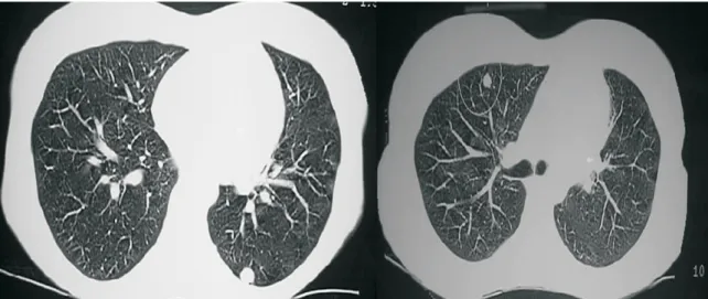
A 27-year-old patient was referred for evaluation of a tumor in the anterior and middle mediastinum and of pulmonary nodules in the lower right lobe and upper left lobe (Figure 1).
The patient was submitted to biopsy through parasternal incision with pericardial opening. A diagnosis of paraganglioma, with positivity for
synaptophysin, chromogranin and S100 protein
and negativity for pancreatin, was established.
Scintigraphy withmetaiodobenzylguanidine was
negative. Arterial pressure was normal. Magnetic resonance angiography revealed the details of the paraganglioma, which was positioned amidst the aortic arch, the pulmonary artery and the right atrium (Figure 2).
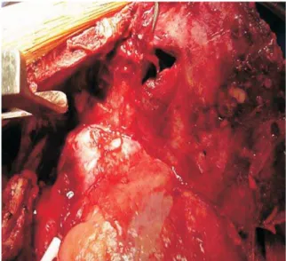
The patient was submitted to resection of the pulmonary metastases through anterior bilateral transsternal thoracotomy, followed by initiation of extracorporeal circulation and resection of the paraganglioma, positioned amidst the aorta, the trunk of the pulmonary artery and right atrium, involving the adventitia of the blood vessels and the pericardium (Figure 3).
Histology and immunohistochemistry confirmed the diagnosis of paraganglioma, including in the pulmonary lesions and pericardium (Figure 4).
Metallic clips were placed in the area of the resection. In the postoperative period, the patient was submitted to radiotherapy, guided by the clips, in the resected areas, as well as to systemic c h e m o t h e r a p y i n v o l v i n g t h e u s e o f cyclophosphamide, vincristine, doxorubicin and dacarbazine. The evolution of the patient after the surgery was extremely favorable, and she was discharged on the fifth postoperative day. Fifteen months later, she was asymptomatic.
78
Ximenes-Netto, Manoel, et al.
Paraganglioma de mediastino com metástases pulmonares
DISCUSSION
Among the various imaging modalities used to locate intra-thoracic paragangliomas, magnetic resonance imaging, especially magnetic resonance angiography, is the most (100%) efficient method ( a t 1 0 0 % e f f i c i e n c y ) , a n d s c a n n i n g w i t h metaiodobenzylguanidine can also be very efficient if a high resolution scanner is used. In a study involving 236 patients with paraganglioma evaluated at the Mayo Clinic, false-negative results were obtained in the following proportions: 0% for magnetic resonance imaging, 5.8% for computed tomography, 3.4% for angiography, 10.7% for ultrasound and 39% for scintigraphy with metaiodobenzylguanidine(5).
When located in the posterior mediastinum, most of these tumors present little technical difficulty in the surgical approach, in contrast to that which occurs when they are located in the anterior and middle mediastinum. Among 104 patients submitted to resection of a mediastinal paraganglioma, 3.8% required the aid of extracorporeal circulation in order to effect the safe removal of the tumor mass(2-6).
Radiotherapy has been considered partially efficient in the treatment of paraganglioma. It can diminish its growth or induce partial regression, but a cure should not be expected. Despite the fact that our patient had been submitted to radical surgery, we believed that the tumor mass had not
Figure 3 – Paraganglioma located at the level of the aortic arch
Figure 2 – Magnetic resonance angiography showing paraganglioma
79
Jornal Brasileiro de Pneumologia 31(1) - Jan/Fev de 2005
been completely excised. Therefore, we opted for
postoperative irradiation of the tumor bed, in
addition to chemotherapy. The chemotherapy regimen used with our patient consisted of cyclophosphamide, vincristine, doxorubicin and dacarbazine(7).
Because of the extreme vascularization of these tumors, prior embolization was occasionally necessary and was carried out according to the technique described by Rakovich et al.(8)
In conclusion, paragangliomas are very rare tumors that present a complex pathology, requiring multidisciplinary treatment and, in treatment centers possessing adequate facilities, the use of all available resources for its handling.
ACKNOWLEDGMENTS
We would like to thank Professor Carlos E. Bachi, pathologist at the Faculdade de Medicina da Universidade Estadual Paulista (Paulista State University School of Medicine) and Professor Saul Suster, chief of the Surgical Pathology Department at the Ohio State University School of Medicine, Columbus, OH, USA.
REFERENCES
1. Lattes R. Nonchromaffin paraganglioma of ganglion nodosum, carotid body and aortic-arch bodies. Cancer 1950; 3: 667-94
2. Lamy AL, Fradet GJ, Luoma A, Nelems B. Anterior and m i d d l e m e d i a s t i n u m p a r a g a n g l i o m a : C o m p l e t e resection is the treatment of choice. Ann Thorac Surg 1994; 57: 249-52
3. Herrera MF, van Heerden JA, Puga FJ. Mediastinal paraganglioma: a surgical experience. Ann Thorac Surg 1993; 56: 1096-1100
4. C a r n e y J A . T h e t r i a d o f g a s t r i c e p i t h e l i o i d l e i o m y o s a r c o m a , p u l m o n a r y c h o n d r o m a a n d functioning extra-adrenal paraganglioma: a five year review. Medicine 1983; 62: 159-9
5. Erikson D, Kudva Y, Ebersold M. Benign paragangliomas: Clinical presentation and treatment outcomes in 236 patients. J Clin Endocrinol Metabol 2001; 86:5210-6 6. Andrade CF, Camargo S, Zanchet M, Felicetti JC, Cardoso PF. Nonfunctioning paraganglioma of the a o r t o p u l m o n a r y w i n d o w. A n n T h o ra c S u rg 2003;75:1950-1
7. Patel SR, Winchester DJ, Benjamin RS. A 15 year e x p e r i e n c e w i t h c h e m o t h e r a p y o f p a t i e n t s w i t h paraganglioma. Cancer 1995; 76: 1476-80