Key words:
Hematuria; Urinary Bladder Neoplasms; Radiotherapy
Int Braz J Urol. 2013; 39: 808-16
__________________
Submitted for publication: July 07, 2013
__________________
Accepted after revision: September 09, 2013 Objective: The aim of our study was to assess short and mid-term clinical efficacy of
external beam radiation therapy to achieve hemostasis in patients with bladder-cancer related gross hematuria who were unfit for surgery. We also assessed hypofractionation as a possible alternative option for more severe patients.
Patients and Methods: Thirty-two patients were included for hemostatic radiation thera-py, with two schedules based on Eastern Cooperative Oncology Group performance sta-tus. The standard treatment was 30 Gy in 10 fractions over 2 weeks. More severe patients underwent a hypofractionated regimen, with 20 Gy in 5 fractions over a one week period. Clinical evaluation was performed at 2 weeks and 6 months.
Results: At 2 weeks, 69% of patients were hematuria-free. Subgroup analysis showed that 79% of patients undergoing hypofractionated regimen were hematuria-free. A total of 54% were hematuria-free with the standard regimen. Based on tumor stage, hematuria was controlled at 2 weeks for 57% of non-muscle invasive tumors and 72% of muscle--invasive tumors. After 6 months, 69% of patients had relapsed, regardless of tumor stage or therapy schedules.
Conclusions: Hemostatic radiotherapy is an effective option for palliative-care hematuria related to bladder cancer in patients unfit for surgery. Although it appears to be rapidly effective, its effect is of limited duration. Hypofractionation also seems to be an effective option; however larger cohorts and prospective trials are needed to evaluate its efficacy compared to standard schedules.
The efficacy of hemostatic radiotherapy for bladder
cancer-related hematuria in patients unfit for surgery
_______________________________________________
E. Lacarrière, C. Smaali, A. Benyoucef, C. Pfister, P. Grise
Department of Urology, Rouen University Hospital (EL, CS, CP, PG) and Department of Radiation Oncology and Medical Physics, Centre Henri Becquerel (AB), Rouen, France
ABSTRACT
ARTICLE
INFO
_________________________________________________________ ___________________
INTRODUCTION
The incidence of bladder tumors has been reported to be 10.1 per 100000 male patients and 2.5 per 100000 female patients (1). Urothelial car-cinomas are primarily found in elderly patients. Two distinct groups of tumors occur: muscle sive bladder tumors (MIBT) and non-muscle inva-sive bladder tumors (NMIBT). The management of these patients is very different, and the prognosis involves a minimum amount of risk of local
pro-gression and metastasis for more advanced tumors (2). Surgery remains a key element to successful management for both types of tumors.
Elderly patients can become unfit for sur-gery. These patients must be included in a com-prehensive, global care network which attempts to alleviate the patient’s symptoms. This remains the main objective of support and palliative care (3).
he-morrhage or pain. Unfortunately, the optimal schedule for palliative EBRT in bladder tumors unfit for surgery remains a matter of debate (4).
The aim of our study was to evaluate the efficacy of palliative EBRT for a distressing symp-tom for patients and their caregivers, i.e. gross he-maturia. Two different schedules were used. This provided the more affected population with a hy-pofractionated regimen, in order to preserve their quality of life.
MATERIALS AND METHODS
This retrospective, bi-center study was conducted on thirty-two patients treated between January 1993 and January 2009 in the Depart-ment of Urology at Rouen University Hospital, Rouen, France and the Becquerel Cancer Center in Rouen, France.
Forty consecutive patients were considered for enrollment. Eight patients were excluded from the study (six prior pelvic radiation therapy, one had Crohn’s disease, one had von Willebrand disease). Thirty two patients (Table-1) with bladder cancer were enrolled for palliative pelvic external beam ra-diation therapy from bladder cancer-related hema-turia. All patients were considered unfit for surgical treatment due to age or medical comorbidities. The comorbidities involved are listed in Table-2.
Inclusion in this study was based on the following criteria:
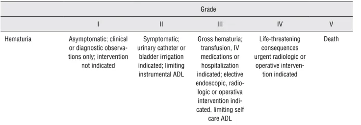
Gross hematuria from advanced bladder cancer. All patients were considered grade 3 ac-cording to the Common Terminology Criteria for Adverse Events (CTCAE) 4.02 classification.
Histological proof of bladder cancer (NMI-BT or MI(NMI-BT);
1. Failure of local treatment (emboliza-tion, electrocoagulation and intravesi-cal instillation of hemostatic agents); 2. Unfit for radical surgical treatment
(cystectomy).
Exclusion criteria of this study were: • Contraindication to perform
radiothe-rapy (i.e. digestive disease);
• Patients submitted to prior radiothera-py in the pelvic area;
• Patients with a coagulation disorder.
All patients were evaluated by an uro-logist and a radiation oncouro-logist, and had pre-viously undergone transurethral resection of the bladder (TURB). Bladder cancer was confirmed by pathological tests, based on the 2004 WHO clas-sification, and classified into two groups: muscle invasive tumors (MIBT) and non-muscle invasive tumors (NMIBT).
Pathological tumor stage was assessed according to the 2002 modification of the TNM (UICC) classification.
The general health conditions before and after treatment were assessed based on the Eastern Cooperative Oncology Group performance status (ECOG PS Table-3).
External radiotherapy was performed using high energy photon therapy, with four or-thogonal beams. The more recent patients un-derwent a computer tomography (CT) dosimetry scan. The clinical target volume (CTV) was the bladder. Lymph nodes were not considered for tre-atment, in this palliative setting. Beam definition was performed using a simulator or via a bone localization, with no contrast agent. Additional areas were assessed to take into account bladder filling and patient movements.
Two protocols A and B were used, depen-ding on the general health conditions of the pa-tient as follows: Protocol A (13 papa-tients) delivered 30 Gy in 10 fractions for 2 weeks, if the ECOG PS was less than or equal to 2. Patients in group B (19 patients) underwent a hypofractionated regi-men of 20 Gy in 5 fractions for 1 week if they had a ECOG PS of more than 2.
The patients were evaluated 2 weeks and 6 months after the beginning of radiotherapy. The common terminology criteria for adverse events CTCAE version 4.02 (2009 version) (5) published by the US Department of Health and Human Ser-vice was used to estimate the intensity of the he-maturia (Table-4).
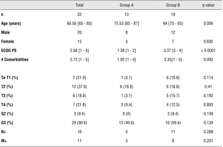
Table 1 - Patients’ characteristics.
Total Group A Group B p value
n 32 13 19
Age (years) 80.56 [65 - 93] 75.53 [65 - 87] 84 [70 - 93] 0.006
Male 20 8 12
Female 12 5 7 0.692
ECOG PS 2.56 [1 - 4] 1.38 [1 - 2] 3.37 [3 - 4] < 0.0001
# Comorbidities 2.72 [1 - 5] 1.92 [1 - 4] 3.35[1 - 5] 0.092
Ta-T1 (%) 7 (21.9) 1 (3.1) 6 (18.8) 0.114
T2 (%) 12 (37.6) 6 (18.8) 6 (18.8) 0.41
T3 (%) 6 (18.8) 1 (3.1) 5 (15.7) 0.192
T4 (%) 7 (21.8) 3 (9.4) 4 (12.5) 0.893
G2 (%) 3 (9.4) 0 (0) 3 (9.4) 0.139
G3 (%) 29 (90.6) 13 (40.6) 16 (59.4) 0.139
N+ 16 5 11 0.288
M+ 11 3 8 0.201
Table 2 - Major comorbidities encountered during patien-tenrollment.
Comorbidities Times mentioned
Chronic bronchitis (COPD) 23
Ischemic heart disease 11
Chronic kidney disease 10
Morbid obesity 9
Chronic heart failure 7
Sleep apnea requiring aid 7
CKD requiring dialysis 3
Cirrhosis (Child-Pugh A) 2
Non-bladder metastatic cancer 1
Blood transfusion, in the absence of gross hema-turia, was not an exclusion criteria for our study.
Statistical analysis was performed using XLSTAT V 7.5 software (Addinsoft, 2004). Two-sided p-values were obtained using Mann-Whitney non--parametric bilateral tests. Results were considered significant when the p-value was below 0.05.
RESULTS
Thirty-two patients were evaluated. The main results are shown in Table-5.
The mean patient age was 81 years (ran-ge 65-93). Mean follow-up length was 25 months (range 7-42). 11 patients (35%) were alive two ye-ars after radiation therapy (RT). Mean length be-fore relapse in the entire cohort was 3.6 months (0-42). Mean global survival was 7 months (3-42).
status (PS) based on ECOG criteria was 2.56 for the general cohort. Group A had a lower PS than group B (1.38 vs. 3.37; p < 0.0001). The mean number of major comorbidities was 2.72 in the general cohort, with less co-morbidities in group A than group B (1.92 vs. 3.26, p = 0.092).
Twenty-five patients had MIBT (78.2%) and 7 patients had NMIBT (21.8%). The majority of patients had tumor stage T2 (37.6%), and the most frequent tumor grade was grade 3 (90.6%). Table 3 - ECOG – PS Eastern Cooperative Oncology Group performance status.
ECOG Description
0 Fully active, able to carry on all pre-disease performance without restriction
1 Restricted in physically strenuous activity but ambulatory and able to carry out work a light se-dentary nature, e.g., light house work, office work
2 Ambulatory and capable of all self care but unable to carry out any work activities. Up and
about more than 50% of waking hours
3 Capable of only limited self care, confined to bed or chair more than 50% of waking hours
4 Completely disabled. Cannot on self care. Totally confined to bed or chair
Table 4 - CTCAE – Common Terminology Criteria for Adverse Events grading for hematuria.
Grade
I II III IV V
Hematuria Asymptomatic; clinical or diagnostic observa-tions only; intervention
not indicated
Symptomatic; urinary catheter or
bladder irrigation indicated; limiting instrumental ADL
Gross hematuria; transfusion, IV medications or hospitalization indicated; elective endoscopic, radio-logic or operativa intervention indi-cated. limiting self
care ADL
Life-threatening consequences urgent radiologic or
operative interven-tion indicated
Death
Definition = A disorder characterized by laboratory test results that indicate blood in the urine
No significant differences between treatment groups were found concerning tumor stage or grade.
Malignancy grade 3 was found in 29 pa-tients (90.6%), 13 group A papa-tients had a grade 3 tumor, while 16 group B patients had a grade 3 tumor (p = 0.139).
Sixteen patients (50%) had lymph node involvement. More patients in group B than group A had lymph node involvement (LNI) (5 vs. 11, p = 0.288). Eleven patients had metastasis at inclusion (34%), which was more frequent in group B than in group A (8 vs. 3, p = 0.201).
Thirteen patients (41%) underwent the standard EBRT regimen based on ECOG PS test results. Nineteen patients (59%) had more severe health conditions and were selected to undergo a hypofractionated EBRT regimen.
After two weeks of EBRT, twenty-two pa-tients (69%) presented no hematuria. There appe-ared to be a better response to EBRT in the muscle invasive group than in the non-muscle invasive group (Figure-1) although no statistical signifi-cance was reached (72% hematuria-free vs. 57% p = 0.469).
After 6 months, both groups showed high relapse rates: 69% of all patients had relapsed, with no difference in tumor subgroups (62% vs. 71%) or ECOG PS subgroups (77% vs. 63%).
There was no statistical difference in the length of the period to relapse found between the subgroups: In tumor stage subgroups (3.1 months vs. 3.3 p > 0.05) or in PS subgroups (3.4 vs. 3.5 months p > 0.05). After 2 years, 7 patients (23%) had no hematuria.
Table 5 - Results at 2 weeks and 6 months.
n Hematuria at 2 weeks (%) Hematuria at 6 months (%)
Ta-T1 7 3 (43) 5 (71)
T2-T4 25 7 (28) 17 (68)
p value 0.469 ns
ECOG PS 0-2 13 6 (46) 10 (77)
ECOG PS 3-4 19 4 (21) 12 (63)
p value 0.139 ns
TOTAL 32 10 (31) 22 (69)
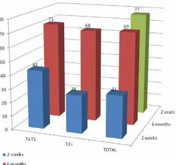
Figure 1 - Results based on tumor stage (initial staging). The rate of relapse is shown in blue, green and red.
Figure 2 - Results based on ECOG – PS Eastern Cooperative Oncology Group performance status (therefore the External beam radiotherapy scheme was used). The rate of relapse is shown in blue, green and red.
DISCUSSION
Bladder tumor management remains a rou-tine procedure in clinical urology practice. The stan-dard treatment for non-metastatic bladder tumors remains surgery (1,3,6).
unfit for surgery, radiotherapy associated with che-motherapy remains a possible alternative (3,6,7).
However, insufficient local tumor con-trol has been previously reported using this ma-nagement approach (8). Many patients unfit for surgery will suffer from ongoing tumor-related symptoms (pain, urgency, hematuria). Life expec-tancy is short (under 9 months) in this population (2,9). Some cases of severe hematuria, especially in patients with cardiovascular history, can be life-threatening (10). Non-surgical hematuria ma-nagement involves bladder irrigation, coagulation correction, and sometimes intravesical procoagu-lant agent use or hypogastric arterial emboliza-tion. Other treatments have been reported, such as formalin, F2 prostaglandin or alum intravesical instillation, or hyperbaric oxygen therapy. Howe-ver, results have been disappointing and their use cannot be widely encouraged (11).
Patients with bladder cancer and no surgi-cal option should benefit from support and pallia-tive care, which is defined by the alleviation of symptoms without extending the life expectancy of the patient (12,13). Palliative treatment must strike a balance between efficacy, convenien-ce, toxicity and duration. The main objective of palliative irradiation remains to provide adequate symptomatic relief throughout a patient’s antici-pated life span while limiting the risk of both acu-te and laacu-te treatment-relaacu-ted complications (14).
Radiation therapy has a double effect on urothelial tumors: Shortly after RT, endothelial cell damage occurs, leading to small and medium vessel rupture or thrombosis. Longer term effects include the disappearance of the microvascular network (15). Other effects that could lead to he-mostasis can include: vasoconstriction and plate-let aggregation through a decrease in endothelial nitric oxide synthase, an increase in interleukin 1 and TNF alpha through endothelial cell damage, and fibrinolysis inhibition due to the inhibition of plasminogen 1 activator. (16-18).
Hypofractionation involves delivering the same dose as the standard radiation schedule, in a lower number of sessions using a larger dai-ly fraction. This translates into higher long-term tissue toxicity. However, the population submit-ted to support and palliative care usually has a
short life expectancy and bothersome symptoms. Therefore, short-term relief is a more pressing concern than late toxicity. The ease of execution, reduction of patient transportation and rapid efficacy justify the use of hypofractionated radio-therapy for patients in palliative care (19). These facts suggest that more severe patients should be good candidates to hypofractionated EBRT since the impact on their quality of life would be less important.
Therefore, this therapeutic management should be proposed for populations with short--term limited life expectancy. In fact, Scholten et al. reported high rates of 5-year overall complica-tions (33%) and severe complicacomplica-tions (9%) (20).
According to guidelines, EBRT remains an option to reduce bleeding from various cancers. This palliative and hemostatic approach is main-ly used due to its antitumoral effects and easy administration. Moreover, its cytotoxic effects allow for limitation or regression of an irradiated tumor, which can provide a decrease in tumor--specific symptoms (14,21).
The hemostatic effects of EBRT have been previously reported in patients affected by lung carcinomas. Brundage et al. have shown an 80% reduction in bleeding in non small cell lung can-cers. Hypofractionation has also been reported to be as effective as the standard procedure (22,23).
The efficacy of palliative EBRT has also been observed in gynecological cancers. Biswal et al. (24) reported 100% of bleeding control within three fractions of EBRT. Kraiphibul et al. (25) also observed EBRT efficacy in uterine cervix cancers: 62.9% of cancer-related bleeding was controlled within three fractions and 97% within five frac-tions of radiotherapy.
Duchesne et al. (4) compared two hypo-fractionated EBRT schedules in a prospective trial, with no difference between a 35 Gy-10 fraction and 21 Gy-3 fraction concerning hematuria im-provement (55% imim-provement vs. 50%, p = 0.198). Clinical evaluation was performed at 3 months. Symptoms such as nocturia, dysuria, and urgency were also improved after EBRT, with no signifi-cant difference between the two schedules.
urinary symptoms were not taken into account in our data analysis. Limitations include the re-trospective nature of this study and the limited patient cohort.
Two EBRT schedules were proposed: 30Gy in 10 fractions (Group A - 2 weeks) or 20Gy in 5 fractions (Group B - 1 week). ECOG PS defined which schedule would be assigned: More severe patients (ECOG > 2) underwent the hypofractiona-ted group B procedure. Group B patients were the-refore less subjected to transportation, with less impact on their quality of life, and alleged non--inferior efficacy (4,19,26).
As reported in the Duchesne et al. study, long term follow-up in large cohorts was difficult to obtain considering the patient’s short-term life expectancy. Therefore, we considered the objecti-ve of hemostatic EBRT to be rapidly effectiobjecti-ve, and well tolerated. Therefore, an early 2-week medical evaluation was carried-out. Clinical improvement and sustainability was evaluated at the 6-month medical evaluation.
Efficacy following a 2-week evaluation was high: 22 patients (68.75%) showed no sign of gross hematuria (CTACE grade 1). Rapid control of the bleeding, which was a major objective of the patient care was reported. However, efficacy ra-tes dropped at the 6-month follow-up evaluation: 69% of the patients had relapsed. This corrobora-tes the findings of Duchesne et al., who reported approximately 60% of hematuria-free patients 3 months after EBRT with 35 Gy or 21 Gy schedules (4). Two years after EBRT, only 23% of patients were hematuria-free.
Analysis by ECOG performance status and EBRT schemes suggested a benefit in terms of hematuria control in favor of hypofractionation. The results were once again below the significan-ce threshold: Hypofractionation (Group B) had a 21% relapse rate after 2 weeks, whereas standard EBRT scheme (Group A) had a 46% relapse rate (p = 0.139).
The present data suggest that hemostatic EBRT can be an interesting option for patients with hematuria related to bladder cancer unfit for surgery. This therapeutic approach should be used for patients with poor health status, with limited life expectancy, due to the limited sustainability of
its effect. Patients with a life expectancy of more than 3 months should not be candidates for he-mostatic EBRT. Practitioners should strive to im-prove the patient’s general state of health in order to enable more aggressive, but more effective al-ternatives.
A benefit was found in hypofractionation in terms of hematuria control, although its use should be considered only for patients with more severe general conditions. These patients were able to benefit most from rapid bleeding control with limited transportation and did not suffer from long-term tissue toxicity due to limited life expectancy. Maintaining quality of life should re-main the foremost objective for the management of patients with bladder cancer who are not can-didates for surgery.
Srinivasan et al. reported encouraging results with hypofractionated EBRT on advanced bladder cancer (26). Jose et al. reported a positive effect on 62% of the patients who underwent hy-pofractionated palliative EBRT, although signifi-cant bladder and digestive toxicity was observed, one year after EBRT (27).
Further research is required to establish the higher efficacy of hypofractionated EBRT. Lar-ger cohorts are needed to confirm the trend that we have reported in our retrospective series. This would be useful to define the place of hypofrac-tionated EBRT as the standard therapy for bladder cancer-related hematuria in severe patients unfit for surgical management.
CONCLUSIONS
Palliative external beam radiation thera-py is an effective short-term option for bladder cancer-related gross hematuria when patients are unfit for surgery. Hypofractionation appears to be an interesting option. However, this approach should be used only for patients with poor life ex-pectancy, as its long term toxicity is higher than the standard schedules. Its efficacy compared to usual schedules requires further study.
surgical options as a means of bleeding control, and it cannot offer long-term control for patients with a longer life expectancy.
ABBREVIATIONS
CT = Computer tomography
CTCAE = Common Terminology Criteria for
Ad-verse Events
CTV = Clinical target volume
ECOG PS = Eastern Cooperative Oncology Group
performance status
Gy = Grays units of radiation
LNI = Lymph node involvement
MIBT = Muscle invasive bladder tumors
NMIBT = Non-muscle invasive bladder tumors
RT = Radiation therapy
TURB = Transurethral resection of the bladder
ACKNOWLEDGEMENTS
The authors are grateful to Richard Medei-ros - Medical Editor, Medical Editing International for editing the manuscript.
REFERENCES
1. Babjuk M, Oosterlinck W, Sylvester R, Kaasinen E, Böhle A, Palou-Redorta J,et al.: EAU guidelines on non-muscle-inva-sive urothelial carcinoma of the bladder, the 2011 update. Eur Urol. 2011; 59: 997-1008.
2. Duncan W, Quilty PM: The results of a series of 963 patients with transitional cell carcinoma of the urinary bladder primar-ily treated by radical megavoltage X-ray therapy. Radiother Oncol. 1986; 7: 299-310.
3. EAU Guidelines, edition presented at the 25th EAU Annual Congress. 2010.
4. Duchesne GM, Bolger JJ, Griffiths GO, Trevor Roberts J, Gra-ham JD, Hoskin PJ, et al.: A randomized trial of hypofraction-ated schedules of palliative radiotherapy in the management of bladder carcinoma: resultsof medical research council trial BA09. Int J Radiat Oncol Biol Phys. 2000; 47: 379-88. 5. Common Terminology Criteria for Adverse Events (CTCAE)
Version 4.0. May 28, 2009, U.S Department of Health and Hu-man Services; National Institutes of Health; National Cancer Institute.
6. Pfister C, Roupret M, Wallerand H, Davin JL, Quintens H, Guy L, et al.: Recommendations Onco-Urology 2010: Urothelial tu-mors. Prog Urol. 2010; 20(Suppl 4): S255-74.
7. Fletcher A, Choudhury A, Alam N: Metastatic bladder cancer: a review of current management. ISRN Urol. 2011; 2011: 545241.
8. Housset M, Maulard C, Chretien Y, Dufour B, Delanian S, Huart J, et al.: Combined radiation and chemotherapy for invasive transitional-cell carcinoma of the bladder: a prospective study. J Clin Oncol. 1993; 11: 2150-7.
9. McLaren DB, Morrey D, Mason MD: Hypofractionated radio-therapy for muscle invasive bladder cancer in the elderly. Ra-diother Oncol. 1997; 43: 171-4.
10. Pereira J, Phan T: Management of bleeding in patients with advanced cancer. Oncologist. 2004; 9: 561-70.
11. Ghahestani SM, Shakhssalim N: Palliative treatment of intrac-table hematuria in context of advanced bladder cancer: a sys-tematic review.Urol J. 2009; 6: 149-56.
12. National Institute of Clinical Excellence. Guidance on Cancer Services. Improving Outcomes in Urological Cancers. The Manual. 2002.
13. Brierly RD, O’Brien TS: The importance of palliative care in urology. Urol Int. 2008; 80: 13-8.
14. Hoskin PJ: Radiotherapy in symptom management. Oxford Textbook of Palliative Medicine, 2nd edition, ed. H.G. Doyle D, MacDonald N. 1998: Oxford University Press
15. Fajardo LF: The pathology of ionizing radiation as defined by morphologic patterns. Acta Oncol. 2005; 44: 13-22.
16. Hallahan DE, Spriggs DR, Beckett MA, Kufe DW, Weichsel-baum RR: Increased tumor necrosis factor alpha mRNA after cellular exposure to ionizing radiation. Proc Natl Acad Sci U S A. 1989; 86: 10104-7.
17. Woloschak GE, Chang-Liu CM, Jones PS, Jones CA: Modula-tion of gene expression in Syrian hamster embryo cells fol-lowing ionizing radiation. Cancer Res. 1990; 50: 339-44. 18. Kanai T, Konno H, Tanaka T, Baba M, Matsumoto K, Nakamura
S, et al.: Anti-tumor and anti-metastatic effects of human-vascular-endothelial-growth-factor-neutralizing antibody on human colonand gastric carcinoma xenotransplanted ortho-topically into nude mice. Int J Cancer. 1998; 77: 933-6. 19. Lutz ST, Chow EL, Hartsell WF, Konski AA: A review of
hypo-fractionated palliative radiotherapy. Cancer. 2007; 109: 1462-70. 20. Scholten AN, Leer JW, Collins CD, Wondergem J, Hermans J,
Timothy A: Hypofractionated radiotherapy for invasive bladder cancer. Radiother Oncol. 1997; 43: 163-9.
21. Kynaston HG, Keen CW, Matthews PN: Radiotherapy for pal-liation of locally advanced prostatic carcinoma. Br J Urol. 1990; 66: 515-7.
22. Brundage MD, Bezjak A, Dixon P, Grimard L, Larochelle M, Warde P, et al.: The role of palliative thoracic radiotherapy in non-small cell lung cancer. Can J Oncol. 1996; 6(Suppl 1): 25-32. 23. Langendijk JA, Aaronson NK, de Jong JM, ten Velde GP,
24. Biswal BM, Lal P, Rath GK, Mohanti BK: Hemostatic radio-therapy in carcinoma of the uterine cervix. Int J Gynaecol Obstet. 1995; 50: 281-5.
25. Kraiphibul P, Srisupundit S, Kiatgumjaikajorn S, Pairachvet V: The experience in using whole pelvic irradiation in man-agement of massive bleeding from carcinoma of the uterine cervix. J Med Assoc Thai. 1993; 76(Suppl 1): 78-81. 26. Srinivasan V, Brown CH, Turner AG: A comparison of two
radiotherapy regimens for the treatment of symptoms from advanced bladder cancer. Clin Oncol (R Coll Radiol). 1994; 6: 11-3.
27. Jose CC, Price A, Norman A, Jay G, Huddart R, Dearnaley DP, et al.: Hypofractionated radiotherapy for patients with carcino-ma of the bladder. Clin Oncol (R Coll Radiol). 1999; 11: 330-3.