Key words:
Melanoma; Penis; Lymph Nodes; Penile Neoplasms Int Braz J Urol. 2013; 39: 823-31
__________________ Submitted for publication: April 19, 2013
__________________ Accepted after revision: August 15, 2013
Purpose: To describe our experience in treating penile melanoma in 06 patients follo-wed at our institution.
Materials and Methods: Between 2004 and 2012 six consecutive patients with penile melanoma were treated at our Institution. Stage of the disease was classified accor-ding to the 2002 AJCC pathologic system. Melanoma in situ (TIS) was diagnosed in one patient. One patient was staged as T1b, two patients as T2b and two patients as T4b. The clinical and pathological findings were evaluated. Immunohistochemical tests were performed for Melan-A, HNB-45, S-100 and C-KIT. All histological speci-mens were examined by the same pathologist (ABSS). The patients with Cis, stages T1b and one patient T2b underwent only local excision. One patient T2b underwent local excision and sentinel lymph node dissection. Two patients with melanoma stage T4b underwent partial penile amputation. One of these last patients had palpable in-guinal lymph nodes at diagnosis and underwent bilateral inin-guinal lymphadenectomy and received systemic chemotherapy (dacarbazine, 30 cycles).
Results: Mean follow-up was 36.3 months. One patient, with stage T2b, died after 12 months due to disease recurrence with bilateral inguinal involvement. The patient who underwent chemotherapy progressed with lung metastases and died after 14 months of follow up. The disease-free survival at five years was 33.3%.
Conclusion: Penile melanoma is a disease with poor prognosis in most cases. Local excision or partial penile amputation may have effective control for stages T1 and T2 lesions. Patients who have clinically proven metastases died despite surgical and adjuvant chemotherapy.
INTRODUCTION
The first case of penile melanoma was des-cribed by Muchison in 1859 and the first report of melanoma of the urethra was made by Tirell in 1871 (1). Primary penile melanoma and in male urethra are rare malignant neoplasms that mostly affects
el-derly patients, from the sixth and seventh decades of life (2). There are approximately 200 cases described in the literature, representing less than 1.4% of pri-mary carcinomas of the penis (3). Most frequently, the lesion is located on the glans (55%), followed by foreskin (28%), penile shaft (9%) and urethral mea-tus (8%) (4).The involvement of urinary tract mucosa
Penile primary melanoma: analysis of 6 patients treated at
Brazilian national cancer institute in the last eight years
_______________________________________________
Gustavo Ruschi Bechara, Aline Barros de Santos Schwindt, Antonio Augusto Ornellas, Diogo Eugênio
Abreu da Silva, Felipe Monnerat Lott, Franz Santos de Campos
Departments of Urology (GRB,AAO,DEAS,FML,FSC) and Pathology (ABSS), Brazilian National Cancer Institute, Rio de Janeiro, RJ, Brazil
ABSTRACT
ARTICLE
INFO
is more common in females and the explanation is the higher concentration of melanocytes in the mu-cocutaneous border of the vulva (5).
A problem in clinical practice is recognizing a pigmented penile lesion as a melanoma. The use of the dermatoscope may be useful in differential diagnosis with other pigmented skin lesions such as: melanosis, nevus, lentigo, and atypical pigmen-ted macula of penis, however the diagnosis must be made by biopsy of the lesion. Indeed, one of the major mimickers of mucosal melanoma, and thus of penile melanomas, is melanosis. Clinically, despite its benign behavior, melanosis can, at times, share features with malignant melanoma as asymmetry, irregular borders, multifocality, variegated pigmen-tary patterns and large size. Due to late diagnosis and lack of well established treatment protocols, the prognosis is generally poor. However, although it is an aggressive disease, it is possible to maximize cure with treatment in its early stages.
Given the rarity of the disease, we report our experience with the treatment of six patients with penile melanoma between 2004 and 2012.
MATERIALS AND METHODS
We reviewed, after approval by the INCA Ethical Committee with the number 38/05, the charts of six patients who were consecutively ad-mitted to Brazilian National Cancer Institute to treat penile melanoma between 2004 and 2012. After detailed anamnesis, physical examination was performed with careful palpation of the pri-mary lesion and the inguinal region, seeking pal-pable lymph nodes. Following, we performed a biopsy of the lesion. A case of melanoma in the penile glans is shown in Figure-1.
All slides were reviewed by a single patho-logist (ABSS). All tumors were evaluated for major prognostic factors. To determine the real extent and dimension of the injury, the analysis inclu-ded the depth (Breslow) and size of the lesion, the presence or absence of necrosis, ulceration and satellite nodules. We also analyzed the number of mitoses per field, presence or absence of associa-ted in situ melanoma (Tis) and characteristics of
Regression was observed in associated area of lesion “in situ”. Figures of mitoses, ulceration, and vascu-lar or perineural invasion were not observed. In the second case, the slides showed ulcerated tissue frag-ments, and necrotic tumor emboli that populated vessels and corpus cavernosum, constituting some-times, metastatic nodules. Areas of regression were observed. Mitoses were not found and the thickness of the lesion and stage could not be assessed. Viral cytopathic changes consistent with HPV were ob-served even in non-neoplastic skin.
In tumors occurring in the mucosa, one ori-ginated in the glans, and the other into the urethral orifice. The glans had a nodular type melanoma measuring 2 mm thick. It had no mitoses, vascu-lar invasion or regression. But there was perineural invasion and foci of ulceration graded as pT1b. In the second case the injury that began in the mucosa of the urinary meatus, in an average of 7 mm, was
Figure 2 - Slides of malignant melanoma of the penis: A) Malignant melanoma showing spindle cell area, containing atypical melanocytes. (HE - 200x). B) Same case immunostained for HMB45 (400x). C) Immunoreativity for Melan - A (400x), a product of gene MART 1. D) c-KIT (400x), product of c-kit gene expression. It is a transmembrane protein.
An immunohistochemical study from the fragments obtained was performed for Melan-A, HNB 45, c-kit (Figure-2) and S-100. In patient with Tis we were unable to perform immunohisto-chemical examination due to shortage of material to be examined. In the other 5 cases, the exami-nation was done for both Melan-A and S100. The HMB45 was done in 2 cases. The c-kit (CD117) was made in 5 cases and marking was performed on the desmoplastic melanoma.
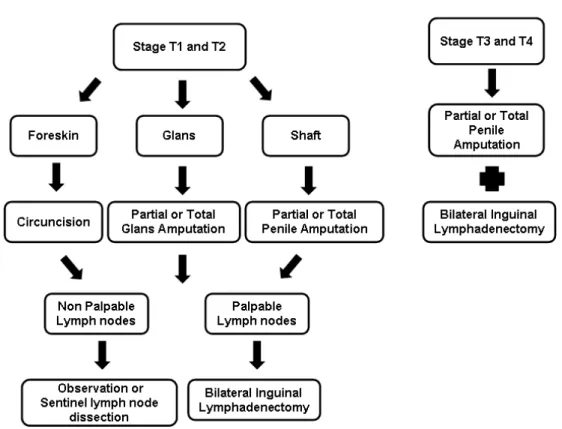
The preoperative staging included chest radiography and abdomen and pelvis CT. The tumor staging was based on the 2002 American Joint Committee on Cancer (AJCC) to classify me-lanoma (Table-1). In this system, pathological tu-mor stage is mainly based on the evaluation of lesion depth (Breslow) and anatomic level of in-vasion (Clark). Our approach for the treatment of melanoma of penis is detailed in Figure-3.
RESULTS
Patient ages ranged from 14 to 78 years with a median age of 72 years. Surgical treatment varied with histopathological staging. Of the 6 patients, 2
(33.3%) had lesions on the glans, 2 (33.3%) in the foreskin, one (16.7%) in the body of the penis and one (16.7%) in the meatus of the urethra (Table-1).
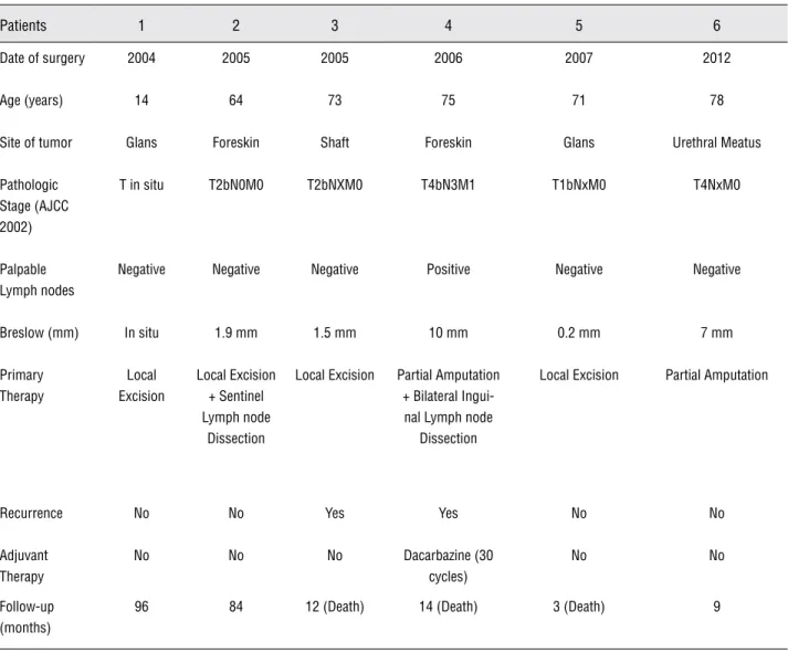
Table 1 - Clinical and pathologic characteristics of the tumors studied.
Patients 1 2 3 4 5 6
Date of surgery 2004 2005 2005 2006 2007 2012
Age (years) 14 64 73 75 71 78
Site of tumor Glans Foreskin Shaft Foreskin Glans Urethral Meatus
Pathologic Stage (AJCC 2002)
T in situ T2bN0M0 T2bNXM0 T4bN3M1 T1bNxM0 T4NxM0
Palpable Lymph nodes
Negative Negative Negative Positive Negative Negative
Breslow (mm) In situ 1.9 mm 1.5 mm 10 mm 0.2 mm 7 mm
Primary Therapy
Local Excision
Local Excision + Sentinel Lymph node
Dissection
Local Excision Partial Amputation + Bilateral Ingui-nal Lymph node
Dissection
Local Excision Partial Amputation
Recurrence No No Yes Yes No No
Adjuvant Therapy
No No No Dacarbazine (30
cycles)
No No
Follow-up (months)
96 84 12 (Death) 14 (Death) 3 (Death) 9
penile amputation. Only one patient had palpable inguinal lymph nodes at diagnosis, and then un-derwent bilateral inguinal lymphadenectomy. The histopathological finding revealed lymph node me-tastases in one of seven lymph nodes on the right and in three of eleven lymph nodes on the left. This patient was then referred to the oncology service, where he underwent chemotherapy with dacarba-zine (30 cycles). The patient developed lung metas-tasis despite systemic chemotherapy and died after 14 months of follow-up. In the other case, we opted for not performing inguinal lymphadenectomy due
one occupying the body of the penis, and 2 the fo-reskin. The tumor in the body of the penis was in the vertical growth phase, had a thickness of 10 mm and pT4bN3M1 stage, contained satellite nodules, ulceration and regression. Mitotic figures reached the highest number (17/mm2) and tumor infiltration
was observed in the dermis and in the corpora ca-vernosa, making neoplastic emboli. Inguinal lymph node metastases were found bilaterally (Table-2).
Figure 3 - An algorithm showing our treatment for patients with melanoma of penis.
Melanoma of the Penis: Management of the Primary Tumor
Table 2 - Histological characteristics of the tumors.
Pts. Invasion Ulcers Necrosis Satellites nodules
Regression Inflammation Mitosis (mm³)
Tis Associated Lesion
1 Dermis present present present absent mild 5 absent Lupus
2 in situ absent absent absent present mild absent absent absent
3 dermis /
epithelium
absent absent absent present mild absent present absent
4 dermis /
epithelium / vascular / sebaceous
present present absent present absent 17 present absent
5 dermis/
epithelium
present present absent present moderate absent present HPV
staged as pT4 and had angiolymphatic invasion. The tumor had 5 mitoses / mm2, ulceration and was
in the vertical growth phase, with peripheral radial growth area represented by melanoma “in situ”. The regression did not follow the development of the lesion. Lupus erythematosus was observed in non--tumor skin area.
The predominant histological pattern was that of melanocytic cells rounded and large, with cytoplasm ranging from clear to eosinophilic, con-taining nuclei presenting gross atypia and evident nucleoli. Multinucleated giant cells were seen in smaller numbers. Melanocytes were isolated from tumor nests or neoplastic mass of cohesiveness va-riable. In case 5, whose tumor was nodular, the pat-tern of rounded melanocyte was mixed with elon-gated fusiform cells, similar to sarcoma. The dermis exhibited varying degrees of stromatous reaction, vascular proliferation and inflammatory infiltrate.
In immunohistochemical examination (Table-3) the staining was strongly positive in 5 cases, both for Melan-A, and for the S100, but the amount of labeled cells was above 50% in 3 ca-ses and 40% in the two other caca-ses. In the HMB45 done in 2 cases at diagnosis, intensity was stron-gly positive, with 30 and 20% of labeled cells in 2 cases respectively. The c-kit (CD117) and marking in the desmoplastic melanoma was strong in 30% of cells balloon-shaped and weak in fusiform cells, over 50% marking in two distinct types of cells. For cases of melanoma in vertical growth, staining
was strong and over 50% of cells, being negative in case 5, the nodular (pT1b).
In our series of penile melanoma patients, disease-free survival was 33.3% at five years. Re-currence occurred in 2 patients after 12 and 14 months respectively, with mean follow-up period of 36.3 months.
DISCUSSION
Penile melanoma is a rare disease that affects mainly elderly patients, from the sixth decade of life, unlike cutaneous melanoma, who-se incidence is higher in young patients (40-50 years) (2). One of our patients, however, with 14 years of age showed an in situ (Tis) melanoma in the glans, successfully treated by local excision and remained disease-free after a follow-up of 96 months after surgery.
Patients are usually asymptomatic, but in advanced stages, may have dysuria, obstructive symptoms, hematuria, urethral discharge and more rarely urinary fistula (6).
Presentation ranges from papule or plaque staining bluish-black or reddish brown with blee-ding ulcer. These lesions are usually benign com-pletely indistinguishable clinically from primary penile melanoma (7). Dermoscopy may prove use-ful for the differential diagnosis between mucosal melanosis, and other mimickers, and early melano-ma. However, its potential role has been limited so
Table 3 - Immunohistochemical characteristics of tumors.
Patients Melan-A HNB 45 S 100 C-kit
1 + 50% Strong x x positive
2 + 50% Strong x x positive
3 + 50% Strong x x positive
4 + 40% Strong x x positive
far because little is known about the dermoscopic features of penile melanoma (8).
Diagnosis is made by biopsy of the lesion. Histopathological examination demonstrates in-creased activity of atypical junctional cells and detachment of pigmented cells in the dermis (8,9).
Microscopic criteria as asymmetry, cell nests confluence, junctional activity, atypia and necrosis of melanocytes are important for a con-clusive diagnosis. In difficult cases we have used immunohistochemistry. Although that is not ne-cessary in cases of well- differentiated tumors, it is indispensable in poorly differentiated tumors. The more specific markers for melanoma are melan--A (MART-1), HMB 45 and S-100 protein. Besides these, information about the aberrations of c-Kit gene in acral melanomas in mucosa or in areas of permanent sun exposure, are targeting therapeutic research, making these aberrations an important marker (10-15). Immunohistochemistry performed in five cases for c-kit was positive in more than 50% of cells with good intensity in 4 cases and negative in one (case 5 with tumor in glans, pT1b).
Thickness of the lesion, presence of ulce-ration, and mitotic figures are considered major predictors of survival. The presence of metastases to lymph nodes and metastatic nests satellites is also valued. This consensus applies to tumors of the skin, and consequently, of the penis, which is covered by skin (16). The thickness of the primary tumor is a predictor of recurrence, especially if as-sociated with ulceration and high mitotic rate (17). Many melanomas begin with the radial growth phase through the stages “in situ” and microinva-sive (18). In the vertical phase, progression occurs through its expansion into the dermis where the measured thickness will vary with the histologi-cal type. For example, nodular melanoma exhibits the largest thickness among the other types (19). The average thickness is increased in cases of fatal melanomas (19). Absence of ulceration is propor-tional to the increased specific survival rate and disease free survival at 5 years (20). As we have seen, the ulceration was present in 4 cases we eva-luated. Two of them reached great depths (7 and 10 mm) and pT4 stage. In a third case, the thick-ness of the lesion could not be measured, probably because of severe necrosis, associated with
ulcera-tion that became the injury brittle, leading to easy fragmentation of the material.
Rate of mitosis is used to stage melanomas and their presence outweighs prognosis necrosis. But the absence of mitosis does not invalidate the diagnosis of malignancy (16). Vascular invasion is another independent factor related to survival and disease-free survival. Presence of intralym-phatic neoplastic cells was criterion for evalua-tion of metastasis to the category N (16). Vascular invasion was present in both melanomas on the skin and on the mucosa, infiltrating vessels and corpus cavernosum. Microscopic metastases found on the reticular dermis or fat, in the same section from the tumor, distant more than 0.05 mm from the lesion, are called satellites (21). Tumor nodu-le with these characteristics was observed in case of melanoma in the penis shaft (pT4N3M1). The behavior of the tumor in the phase of regression has been widely discussed as an indicator of dise-ase progression. Sentinel lymph node metastasis occurs in melanomas with vertical growth and re-gression, but does not occur in cases of radial gro-wth with regression (22). T lymphocytes responsi-ble for autoimmune reaction and mechanisms of programmed cell death are believed to be involved in the mechanism of regression. But it seems that instead of controlling the growth of the tumor, the autoimmune reaction accelerates this growth (23). In this case, the regression would not be the result of immune toxicity, but the rapid differentiation of cells, contributing to a more aggressive behavior (24). In place of regression are found lymphocytes and phagocytic cells loaded with melanin angio-lymphatic vascular reaction and delayed fibrotic scarring permeated with neoplastic cells isolated (25). The lymphocytic infiltrate was present in al-most all our cases, both around the injury and / or in the middle of it, remembering their relationship with immune regression. The regression was pre-sent in over half of our cases, but only in case 2 (pT4N3M1) was accompanied by metastases to lymph nodes.
diagnosed too late, which reduced the cure rates of disease. All patients with lymph node invol-vement or distant metastases at diagnosis died within two years despite surgical treatment. Tu-mor greater than 15 mm, presence of ulceration and tumor depth greater than 3.5 mm had a worse prognosis (26).
Chemotherapy is indicated for disseminated melanoma. The combination chemotherapy consis-ting of six cycles of DTIC, BCNU, cisplatin and ta-moxifen presents the best result. The response rate to therapy varies between 15% and 45% (27).
Given the rarity of the disease, the best treatment for penile melanoma remains uncertain. Stillwell et al. from the Mayo Clinic reported 11 consecutive patients treated over a period of 66 years. All patients underwent conservative treat-ment, including the amputation of the glans in 6 patients, local excision in 3 and partial penile amputation in 2 (3). They believe that conserva-tive surgery such as local excision or partial pe-nile amputation with appropriate safety margin (3-5 cm), can be performed in superficial lesions (less than 1.5 mm) or associated with superficial bilateral inguinal lymphadenectomy in deeper le-sions (greater to 1.5 mm), thus being suitable for melanomas stage T1 or T2 (27-29). However, lack further evidence for defining the optimal surgical margin for lesions with a thickness between 1 and 2 mm, and it is the current trend to practice banks of 2 cm. In addition there are very few studies dealing with more severe injuries, thicker than 4 mm, for example. Melanoma in situ is also poorly evaluated in these aspects and recommendations are very variable ranging from 0.5 to 1 cm of sur-gical margins. In our study, all patients were trea-ted conservatively, including glans amputation in 2, local excision in 2 and distal third penile ampu-tation in 2. Subsequently 4 of the 6 patients (66%) were rendered disease-free.
All patients who persist with palpable in-guinal lymph nodes after antibiotic use shall be submitted to bilateral superficial inguinal lympha-denectomy. The treatment in relation to patients without palpable lymph nodes is controversial. Although the overall incidence of inguinal lymph
early-stage disease is significantly small. Thus, a large proportion of patients clinically negative for the disease can not benefit from prophylactic bilateral inguinal lymphadenectomy. Other au-thors have reported that sentinel lymphadenec-tomy avoids potential morbidity related to ingui-nal lymph node dissection and allows accurate staging for further treatment and assessment of prognosis (27,30). The inguinal lymphadenec-tomy is clearly indicated in patients with stage T3 or T4. Patients with palpable lymph nodes that do not resolve after antibiotic treatment should undergo inguinal lymph node dissection. Chemotherapy and radiation treatments are only palliative or adjuvant. (31).
CONCLUSIONS
Penile melanoma is an uncommon disea-se, in most cases with poor prognosis due to the late diagnosis and lack of treatment protocols well established. Local excision or partial penile am-putation, with appropriate safety margin, can be effective in the control of stages T1 and T2 penile melanomas. Yet patients who had clinically pro-ven metastases died despite surgical procedures and chemotherapy. Although it is an aggressive disease is likely to be cured when in the early sta-ges, making necessary a close cooperation betwe-en urologists and dermatologists to achieve this goal. The treatment of patients without palpable lymph nodes is controversial, however, conside-ring the aggressiveness of the disease and early metastasis, prophylactic inguinal lymphadenec-tomy may be considered in selected patients.
CONFLICT OF INTEREST
None declared.
REFERENCES
1. Gross SD: A System of Surgery, 6th ed. Philadelphia, WB Saunders, 1882.
3. Stillwell TJ, Zincke H, Gaffey TA, Woods JE: Malignant mela-noma of the penis. J Urol. 1988; 140: 72-5.
4. Demitsu T, Nagato H, Nishimaki K, Okada O, Kubota T, Yoneda K, et al.: Melanoma in situ of the penis. J Am Acad Dermatol. 2000; 42: 386-8.
5. Batsakis JG, Suarez P: Mucosal melanomas: a review. Adv Anat Pathol. 2000; 7: 167-80.
6. Oldbring J, Mikulowski P: Malignant melanoma of the penis and male urethra. Report of nine cases and review of the litera-ture. Cancer. 1987; 59: 581-7.
7. Primus G, Soyer HP, Smolle J, Mertl G, Pummer K, Kerl H: Early ‘invasive’ malignant melanoma of the glans penis and the male urethra. Report of a case and review of the literature. Eur Urol. 1990; 18: 156-9.
8. Carli P, De Giorgi V, Soyer HP, Stante M, Mannone F, Giannotti B: Dermatoscopy in the diagnosis of pigmented skin lesions: a new semiology for the dermatologist. J Eur Acad Dermatol Venereol. 2000; 14: 353-69.
9. Soyer HP, Argenziano G, Chimenti S, Ruocco V: Dermoscopy of pigmented skin lesions. Eur J Dermatol. 2001; 11: 270-6. 10. Smalley KS, Sondak VK, Weber JS: c-KIT signaling as the
driv-ing oncogenic event in sub-groups of melanomas. Histol His-topathol. 2009; 24: 643-50.
11. Abu-Abed S, Pennell N, Petrella T, Wright F, Seth A, Hanna W: KIT gene mutations and patterns of protein expression in mucosal and acral melanoma. J Cutan Med Surg. 2012; 16: 135-42.
12. Weider N, Cote RJ, Suster S, Wess LM: Modern Surgical Pa-thology. 2ª edition 2009; cap. 49; page 1936-942. Saunders / Elsevier, Weedon D. Skin Pathology. Churchill Livingstone, second edition. 2002; pp. 821-34.
13. Veronese LA, Marques MEA: Critérios anatomopatológicos para melanoma maligno cutâneo: análise qualitativa de sua eficácia e revisão da literatura. J. Bras. Patol. Med. Lab. 2004; 40: 99-112.
14. Park E, Yang S, Emley A, DeCarlo K, Richards J, Mahalingam M: Lack of correlation between immunohistochemical expres-sion of CKIT and KIT mutations in atypical acral nevi. Am J Dermatopathol. 2012; 34: 41-6.
15. Curtin JA, Busam K, Pinkel D, Bastian BC: Somatic activation of KIT in distinct subtypes of melanoma. J Clin Oncol. 2006; 24: 4340-6.
16. Balch CM, Gershenwald JE, Soong SJ, Thompson JF, Atkins MB, Byrd DR, et al.: Final version of 2009 AJCC melanoma staging and classification. J Clin Oncol. 2009; 27: 6199-206. 17. Francken AB, Accortt NA, Shaw HM, Colman MH, Wiener M,
Soong SJ, et al.: Follow-up schedules after treatment for ma-lignant melanoma. Br J Surg. 2008; 95: 1401-7.
18. Piérard GE: Cell proliferation in cutaneous malignant melano-ma: relationship with neoplastic progression. ISRN Dermatol. 2012; 2012: 828146 [In Press].
19. Criscione VD, Weinstock MA: Melanoma thickness trends in the United States, 1988-2006. J Invest Dermatol. 2010; 130: 793-7.
20. Gajdos C, Griffith KA, Wong SL, Johnson TM, Chang AE, Cim-mino VM, et al.: Is there a benefit to sentinel lymph node biop-sy in patients with T4 melanoma? Cancer. 2009; 115: 5752-60. 21. Rao UN, Ibrahim J, Flaherty LE, Richards J, Kirkwood JM:
Implications of microscopic satellites of the primary and ex-tracapsular lymph node spread in patients with high-risk mela-noma: pathologic corollary of Eastern Cooperative Oncology Group Trial E1690. J Clin Oncol. 2002; 20: 2053-7.
22. Kaur C, Thomas RJ, Desai N, Green MA, Lovell D, Powell BW, et al.: The correlation of regression in primary melanoma with sentinel lymph node status. J Clin Pathol. 2008; 61: 297-300. 23. Lonchay C, van der Bruggen P, Connerotte T, Hanagiri T, Coulie
P, Colau D, et al.: Correlation between tumor regression and T cell responses in melanoma patients vaccinated with a MAGE antigen. Proc Natl Acad Sci U S A. 2004; 101(Suppl 2): 14631-8. 24. Prehn RT: The paradoxical association of regression with a
poor prognosis in melanoma contrasted with a good prognosis in keratoacanthoma. Cancer Res. 1996; 56: 937-940.
25. Attia P, Phan GQ, Maker AV, Robinson MR, Quezado MM, Yang JC, et al.: Autoimmunity correlates with tumor regression in patients with metastatic melanoma treated with anti-cytotoxic T-lymphocyteantigen-4. J Clin Oncol. 2005; 23: 6043-53. 26. van Geel AN, den Bakker MA, Kirkels W, Horenblas S, Kroon
BB, de Wilt JH, et al.: Prognosis of primary mucosal penile melanoma: a series of 19 Dutch patients and 47 patients from the literature. Urology. 2007; 70: 143-7.
27. Zurrida S, Bartoli C, Clemente C, De Palo G: Malignant mela-noma of the penis. A report of four cases. Tumori. 1990; 76: 599-602.
28. Milton GW, Shaw HM: Rare variants of malignant melanoma. World J Surg. 1992; 16: 173-8.
29. Myskow MW, Going JJ, McLaren KM, Inglis JA: Malignant melanoma of penis. J Urol. 1988; 139: 817-8.
30. Han KR, Brogle BN, Goydos J, Perrotti M, Cummings KB, Weiss RE: Lymphatic mapping and intraoperative lymphos-cintigraphy for identifying the sentinel node in penile tumors. Urology. 2000; 55: 582-5.
31. Sánchez-Ortiz R, Huang SF, Tamboli P, Prieto VG, Hester G, Pettaway CA: Melanoma of the penis, scrotum and male ure-thra: a 40-year single institution experience. J Urol. 2005; 173: 1958-65.