Key words:
Muscle Strength; Pelvic Floor; Sexual Behavior; Parity
Int Braz J Urol. 2013; 39: 847-52
__________________
Submitted for publication: March 11, 2013
__________________
Accepted after revision: September 16, 2013 Objective: The aim of this study was to assess pelvic floor muscle (PFM) strength in
diffe-rent body positions in nulliparous healthy women and its correlation with sexual activity. Materials and Methods: Fifty healthy nulliparous women with mean age of 23 years were prospectively studied. Subjective evaluation of PFM was assessed by transvaginal digital palpation (TDP) of anterior and posterior areas regarding the vaginal introitus. A perineometer with inflatable vaginal probe was used to assess the PFM strength in four different positions: supine with extended lower limbs (P1); bent-knee supine (P2); sitting (P3); standing (P4).
Results: Physical activity, 3 times per week, was reported by 58% of volunteers. Sexual activity was observed in 80% of women and 82% of them presented orgasm. The average body mass index (BMI) was 21.76 kg/m2, considered as normal according World Health
Organization (WHO). We observed that 68% of volunteers were conscious about the PFM contraction. TDP showed concordance of 76% when anterior and posterior areas were compared (p = 0.00014). There was not correlation between PFM strength and orgasm in subjective evaluation. The PFM strength was significantly higher in standing position when compared with the other positions (p < 0.000). No statistical difference was obser-ved between orgasm and PFM strength when objective evaluations were performed. Conclusions: There was concordance between anterior and posterior areas in 76% of cases when subjective PFM strength was assessed. In objective evaluation, higher PFM strength was observed when volunteers were standing. No statistical correlation was ob-served between PFM strength and orgasm in nulliparous healthy women.
Pelvic floor muscle strength evaluation in different
body positions in nulliparous healthy women and its
correlation with sexual activity
_______________________________________________
Mônica Orsi Gameiro, Luciana Miraglia, Luiz Felipe Orsi Gameiro, Carlos Roberto Padovani, João
Luiz Amaro
Physiotherapy Service (MOG, LM, LFOG); Department of Biostatistics (CRP) and Department of Urology (JLA) School of Medicine, São Paulo State University, Botucatu, SP, Brazil
ABSTRACT
ARTICLE
INFO
_________________________________________________________ ___________________
INTRODUCTION
The vagina and bladder are correlated to the pelvic floor muscles (PFM), comprising the elevator ani and puborectalis. Likewise, decrease of pelvic floor muscle (PFM) strength in women may cause urinary incontinence (UI) (1)or sexual
848
stronger pelvic floor muscle. Some studies report a relationship between women’s sensation du-ring sexual intercourse as well as the vaginal grip intensity felt by their partner (6,7). Puborectalis muscle may play an important role in the cons-trictor function of PFM. Thus, its assessment, by vaginal pressure measurement, could be a deter-minant factor for PFM evaluation (8,9).
Some authors have advocated different forms of PFM assessment using tools such as ul-trasound (10), electromyography (EMG) (11) and magnetic resonance imaging (MRI) (12). These different approaches may also permit to evaluate other aspects of PFM activity when compared with vaginal squeeze pressure.
Another important point would be women’s body position during pelvic floor evalu-ation. Another study observed that digital muscle testing and vaginal pressures using manometry are reliable tools for measuring maximum volun-tary contraction in supine and upright positions (13). However, there is no consensus on the best form to evaluate the PFM strength as well as its baseline concerning nulliparous healthy women and its relationship with sexual activities. Thus, we propose to assess PFM strength in different body positions and also to evaluate its correlation with sexual activity in this specific population.
MATERIALS AND METHODS
From March to September 2006, fifty he-althy nulliparous volunteers of hehe-althy area were recruited by an invitation letter. Mean age was 23 years old (range 20-30). The group comprised women with higher education level (University). This study was approved by the “Ethical Resear-ch Committee” (protocol n.368/2005). All parti-cipants were informed about its importance and signed the “Free Informed Consent”.
Exclusion criteria were UI or urinary com-plaints, neurological diseases, previous pelvic sur-geries, diabetes, smoking and cognitive problems. Women were evaluated through a clini-cal questionnaire. Sexual activity was assessed by self-applicable anonymous questionnaire, compo-sed of two simples questions: 1 - Have you had sexual intercourse in the last 3 months?Yes/ No;
2 - Did you have orgasm during this intercourse?
Yes/ No. BMI was calculated and classified
accor-ding to the World Health Organization (WHO) (14). Subjective and objective PFM evaluations were performed in all women.
For the subjective evaluation, volunteers were placed in supine position, undressed from waist to feet, covered with a sheet with the lower limbs bent and separated and instructed about the correct PFM contraction. They were evaluated by only one examiner through transvaginal digital palpation (TDP) of anterior and posterior areas regarding the vaginal introitus (Figure-1); they were also required to contract the perineal mus-cles and hold this contraction as long as possible. The classification of the PFM strength contraction
Figure 1 - Bidigital vaginal palpation regarding the vaginal introitus. (A) anterior and (B) posterior areas.
A
was performed according to the description of Ama-ro et al.(4), that it has been tested but not validated. The objective measurement was obtained with a Dynamed portable perineometer (model DM01), in four different patient positions (Figure-2): supine with lower limbs extended (P1); bent-knee lying (P2), sitting (P3), and standing (P4). When the participants were in position, examiner introduced a balloon catheter, sized 11 x 2.6 cm, into the va-gina. The balloon catheter was covered with a non--lubricated condom, and filled with 60 mL of air permitting contact with the vaginal wall. This value was standardized at 60 mL in all participants. The equipment was immediately zeroed, three PFM con-tractions were requested and held as long as possi-ble with nearly 30-second of rest interval between each one. Maximal peak of each contraction was registered in cmH2O. The length of time of these contractions was recorded in seconds with chrono-meter. The average of three measurements was used to avoid biased results.
Statistical analysis
Qualitative variables were analyzed using the proportion test of concordances (15). Mann--Whitney Test was used for comparisons between PFM strength and the presence or not of orgasm. For the comparison of PFM strength in different body positions, the Friedman’s non-parametric test was used and complemented by the Dunn multiple comparison tests. For comparing the time of PFM contractions, the technique of variance analysis for measurements of repeated models was used and complemented by Bonferroni’s test (16). Statistical analysis was performed at 5% of significance level.
RESULTS
The mean age of women’s menarche was 12 years old. Sexual activity was reported by 80% and 82% of these women reported orgasm. Regular physical activity, at least three times a week, was
850
reported by 58% of volunteers and 54% of them presented symptoms of constipation.
Average BMI was 21.76 Kg/m2, considered
as normal according to WHO.
PFM subjective evaluation showed that 68% of women were conscious of musculature contraction. The TDP in anterior position showed that 48% and 52% of women had moderate and normal PFM contractions, respectively. Whereas, in posterior area, we observed that moderate and normal contractions represented 24% and 72% respectively. There was statistical concordance in 76% of cases (p < 0.00014) when both positions of PFM evaluation were compared (Table-1). Ho-wever, there was no statistical correlation between orgasm and subjective PFM evaluation neither in
anterior nor in posterior areas regarding the vagi-nal introitous.
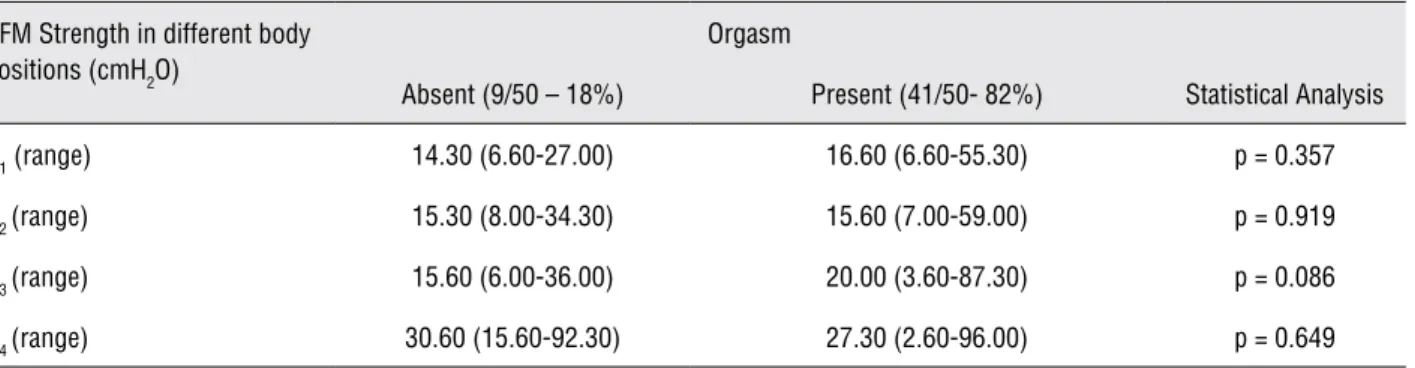
Perineometer evaluation of PFM streng-th was significantly higher in standing position when compared to the others (Table-2). Time of PFM contraction was significantly longer in the standing position (Table-3). There was not statisti-cal difference in the PFM strength, in the different test positions, in women with orgasm compared to those who had not orgasm (Table-3).
DISCUSSION
Mean age of menarche was 12 years old what is in agreement with literature (17). Some au-thors consider menarcheal age important because
Table 1 - Association between the bidigital vaginal palpation in anterior and posterior areas of vaginal introitus according to Amaro’s classification (4).
SE posterior SE anterior
Grade 2 Grade 3 Total
Grade 2 12 (24%)* 0 (0.0 %) 12 (24%)
Grade 3 12 (24%) 26 (52%)* 38 (76%)
Total 24 (48%) 26(52%) 50 (100%)
* Concordance level in moderate grade = 24 % and normal grade = 52%; p < 0.00014 SE = Subjective Evaluation
Table 2 - Maximum amplitude (cmH2O) (median and range) and time (second) (mean ± sd) of PFM contractions in objective evaluation of PFM strength using perineometer (cmH2O) in different positions. Different lower case letters indicate when groups were significantly different at the same moment.
Objective evaluation of PFM
Body Position
P1 P2 P3 P4 Statistical
Analysis
Median and range of PFM strength (cmH2O)
16.30a (6.6 - 55.3)
15.60a (7.0 - 59.0)
19.30a (3.6 - 87.3)
28.65b
(12.6 - 96.3) p < 0.000
Mean and Standard deviation of PFM contraction time (second)
7.26 ± 1.72a 7.16 ± 1.33a 7.68 ± 1.75a 8,45 ± 2.33b p < 0.000
it is influenced by environmental and genetic fac-tors, and also it may be determinant for sexual maturity (17), demonstrating that our population presents these homogenous characteristics.
In our population, BMI was considered normal according to WHO, demonstrating that obesity did not influence outcomes. Some authors consider that this fact could worsen SUI or pelvic disorders (18,19). In our study, there was not in-fluence of this parameter in the results.
Frawley et al. (13), using different metho-ds for PFM evaluation, observed higher reliabi-lity using manometry in comparison to TDP as-sessment. However, other authors (20) reported a strong correlation between EMG and TDP in continent women without PFM disorders, sho-wing that the best methods of PFM assessment are somehow controversial. In our study, we assessed PFM strength through transvaginal digital palpa-tion in anterior and posterior areas and observed statistical concordance between both methods in 76% of cases, demonstrating that this assessment may be used in any of these positions. At the mo-ment, there is no report regarding this subject in literature.
Maximum amplitude and time of PFM con-tractions in objective evaluation of PFM strength using perineometer in standing position were sig-nificantly higher in comparison to the other po-sitions. This fact could be explained because in standing position the pelvic floor muscles suffer of gravity effects and respond with their contrac-tion thereby increasing their strength.
Some authors reported the relation betwe-en orgasm and sexual arousal and PFM strbetwe-eng- streng-th (7). Ostreng-ther austreng-thors observed streng-that PFM function improvement using perineal exercise postpartum (21) or surgical procedure performed to pelvic flo-or dysfunctions can improve the sexual function (22). In our series, there was no statistical correla-tion between PFM strength and orgasm. The fact that we have not used a more specific and com-plete sexual function questionnaire, which could cause bias, suggests that further research in speci-fic population is necessary.
CONCLUSIONS
There was statistical concordance in 76% of cases when PFM strength, in anterior and pos-terior areas, was assessed using transvaginal digi-tal palpation. Objective evaluation of PFM streng-th was significantly higher in standing position when compared to the other positions. Orgasm does not seem to be affected by PFM strength. Further studies should be performed in nullipa-rous continent women to elucidate the effects of different test positions and orgasm in the pelvic floor muscle strength.
ABBREVIATIONS
PFM = pelvic floor muscle)
TDP = transvaginal digital palpation
WHO = World Health Organization
BMI = body mass index
Table 3 - Association between Maximum amplitude (cmH2O) (median and range) of PFM contractions in objective evaluation of PFM strength using perineometer in different positions and presence or not of orgasm.
PFM Strength in different body positions (cmH2O)
Orgasm
Absent (9/50 – 18%) Present (41/50- 82%) Statistical Analysis
P1 (range) 14.30 (6.60-27.00) 16.60 (6.60-55.30) p = 0.357
P2 (range) 15.30 (8.00-34.30) 15.60 (7.00-59.00) p = 0.919
P3 (range) 15.60 (6.00-36.00) 20.00 (3.60-87.30) p = 0.086
P4 (range) 30.60 (15.60-92.30) 27.30 (2.60-96.00) p = 0.649
852
UI = urinary incontinence
EMG = electromyography
MRI = resonance imaging
SUI = stress urinary incontinence
CONFLICT OF INTEREST
None declared.
REFERENCES
1. Amaro JL, Moreira EC, De Oliveira Orsi Gameiro M, Pa-dovani CR: Pelvic floor muscle evaluation in incontinent patients. Int Urogynecol J Pelvic Floor Dysfunct. 2005; 16: 352-4.
2. Shafik A: The role of the levator ani muscle in evacuation, sexual performance and pelvic floor disorders. Int Urogy-necol J Pelvic Floor Dysfunct. 2000; 11: 361-76.
3. Zahariou AG, Karamouti MV, Papaioannou PD: Pelvic floor muscle training improves sexual function of women with stress urinary incontinence. Int Urogynecol J Pelvic Floor Dysfunct. 2008; 19: 401-6.
4. Amaro JL, Oliveira Gameiro MO, Padovani CR: Treatment of urinary stress incontinence by intravaginal electrical stimulation and pelvic floor physiotherapy. Int Urogynecol J Pelvic Floor Dysfunct. 2003; 14: 204-8; discussion 208. 5. Dietz HP, Steensma AB, Vancaillie TG: Levator function in
nulliparous women. Int Urogynecol J Pelvic Floor Dysfunct. 2003; 14: 24-6; discussion 26.
6. Graber B, Kline-Graber G: Female orgasm: role of pubococ-cygeus muscle. J Clin Psychiatry. 1979; 40: 348-51. 7. Lowenstein L, Gruenwald I, Gartman I, Vardi Y: Can
stron-ger pelvic muscle floor improve sexual function? Int Uro-gynecol J. 2010; 21: 553-6.
8. Jung SA, Pretorius DH, Padda BS, Weinstein MM, Nager CW, den Boer DJ, et al.: Vaginal high-pressure zone as-sessed by dynamic 3-dimensional ultrasound images of the pelvic floor. Am J Obstet Gynecol. 2007; 197: 52.e1-7. 9. Brandon CJ, Lewicky-Gaupp C, Larson KA, Delancey JO:
Anatomy of the perineal membrane as seen in magnetic resonance images of nulliparous women. Am J Obstet Gy-necol. 2009 ; 200: 583.e1-6.
10. Santoro GA, Wieczorek AP, Dietz HP, Mellgren A, Sultan AH, Shobeiri SA, et al.: State of the art: an integrated approach to pelvic floor ultrasonography. Ultrasound Obstet Gynecol. 2011; 37: 381-96.
11. Peschers UM, Voduŝek DB, Fanger G, Schaer GN, DeLanc-ey JO, Schuessler B: Pelvic muscle activity in nulliparous volunteers. Neurourol Urodyn. 2001; 20: 269-75.
12. Soljanik I, Janssen U, May F, Fritsch H, Stief CG, Weissen-bacher ER, et al.: Functional interactions between the fossa ischioanalis, levator ani and gluteus maximus muscles of the female pelvic floor: aprospective study in nulliparous women. Arch Gynecol Obstet. 2012; 286: 931-8.
13. Frawley HC, Galea MP, Phillips BA, Sherburn M, Bø K: Re-liability of pelvic floor muscle strength assessment using different test positions and tools. Neurourol Urodyn. 2006; 25: 236-42.
14. World Health Organization [homepage on the Internet] (2006) BMI Classification. Geneva: WHO [cited 2008 nov 1 12]. Available from: www.who.int/bmi.
15. Goodman LA: Simultaneus confidence intervals for multi-nomial proportions. Technometrics. 1965; 7: 247-54. 16. Zar JH: Biostatical analysis. New Jersey, Prentice Hall.
2009; 5th Ed., pp. 1-960.
17. Tavares CH, Haeffner LS, Barbieri MA, Bettiol H, Barbieri MR, Souza L: Age at menarche among schoolgirls from a rural community in Southeast Brazil. Cad Saude Publica. 2000; 16: 709-15.
18. Viktrup L: Female stress and urge incontinence in fam-ily practice: insight into the lower urinary tract. Int J Clin Pract. 2002; 56: 694-700.
19. Kirby M: Managing stress urinary incontinence -- a primary care issue. Int J Clin Pract. 2006; 60: 184-9.
20. Botelho S, Pereira LC, Marques J, Lanza AH, Amorim CF, Palma P, et al.: Is there correlation between electromyog-raphy and digital palpation as means of measuring pelvic floor muscle contractility innulliparous, pregnant, and postpartum women? Neurourol Urodyn. 2013; 32: 420-3. 21. Citak N, Cam C, Arslan H, Karateke A, Tug N, Ayaz R, et
al.: Postpartum sexual function of women and the effects of early pelvic floor muscle exercises. Acta Obstet Gynecol Scand. 2010; 89: 817-22.
22. Kammerer-Doak D: Assessment of sexual function in wom-en with pelvic floor dysfunction. Int Urogynecol J Pelvic Floor Dysfunct. 2009; 20(Suppl 1): S45-50.
_______________________ Correspondence address: