Mônica Nogueira Pigozzo Dalva Cruz Laganá Newton Sesma Glaís Ferrari de Souza Alvaro Luiz Ichi
Departmentof Prosthodontics, School of Dentistry, Universidade de São Paulo - USP, São Paulo, SP, Brazil.
Corresponding Author: Mônica Nogueira Pigozzo E-mail: [email protected]
Photoelastic stress analysis in
mandibular bone surrounding bar-clip
overdenture implants
Abstract: The aim of this in vitro study was to evaluate, using the pho-toelastic analysis method, the stress distribution in mandibular bone sur-rounding a bar-clip overdenture when 2 implant angulations were sim-ulated. Two mandibular photoelastic models were manufactured, with 2 implants embedded in the interforaminal region: model 1 - PAPI, a photoelastic analysis model with parallel implants; and model 2 - PAAI, a photoelastic analysis model with angled implants. A bar-clip retention system and an overdenture were positioned over the implants, and loads of 1.0, 2.0 and 3.0 bars were applied. The resultant stresses that devel-oped in the supporting structure were photoelastically monitored and were recorded photographically. The results showed that there were no similarities in the areas of stress among the photoelastic resin models when the angulation of the implants was evaluated. Model 1 - PAPI pre-sented a higher stress concentration at the implant apex, while in model 2 - PAAI, there were higher stress concentrations on the mesial and distal implant faces. Within the limitations of this study, it was concluded that the PAPI photoelastic model demonstrated better stress transfer com-pared to the PAAI model, since the forces oriented along the axis were better absorbed by the bone.
Descriptors: Dental Prosthesis; Dental Implants; Dentures; Biomechanics.
Introduction
When conventional complete dentures are converted into implant-re-tained overdentures, clear increases in the patient’s masticatory function, denture retention and stability,1 phonetics2-4 and oral hygiene can be
ob-served.5 These advantages are important for explaining the popularity of
overdentures as an alternative to conventional complete dentures.6-8
Bar-clip and O-ring overdenture abutments are the most common types, and they are classiied as resilient systems.9,10 These retentive
mechanisms attached to implants transmit stress to the bone differently than natural teeth supported by periodontal ligaments.5,11,12 If the
inci-dent force on the implant exceeds the physiological limit, it can result in overload, with consequent bone microfractures that can heal with non-mineralized connective tissue or lost tissue, because the implants are not prepared to support excessive force.5,13,14 Therefore, it is essential that
questions be addressed regarding the biomechanical behavior of implants Declaration of Interests: The authors
certify that they have no commercial or associative interest that represents a conflict of interest in connection with the manuscript.
Submitted: Feb 21, 2013
Accepted for publication: Oct 08, 2013 Last revision: Oct 22, 2013
and how they react to the surrounding tissues when submitted to loads, because they are directly related to the preservation of supporting tissues. Moreover, the mandibular nerve and the foramen represent limitations in edentulous patients. An alternative to avoid injury to these structures is to angle the im-plants. Another advantage is a reduction in the pros-thesis cantilever. However, the effects of nonaxial load, caused by implant angulation, on the integrity of osseointegration have rarely been reported in the literature on overdentures. Thus, this study included a photoelastic model with 10° angulated implants, divergent from the midline, because it has been sug-gested that this divergence can usually be tolerated.15
The purpose of this study was to compare the load transfer characteristics of a bar-clip attachment system for 2 implant-retained mandibular overden-ture designs, vertically and 10° inclined implants, using photoelastic analysis.
Methodology
Two photoelastic models of an edentulous man-dible were fabricated with PL-2 photoelastic resin (Vishay Intertechnology, Malvern, USA). These mandibles were obtained from a photoelastic cra-nium (Figure 1).
The photoelastic cranium’s superior teeth and the mandible were reproduced with Flexitime sili-cone impression material (Heraus Kulzer GmbH, Hanau, Germany), resulting in stone casts (Zher-mack SpA, Rovigo, Italy) that were positioned, us-ing a bite record, in a semi-adjustable articulator (Bio-art, São Carlos, Brazil), allowing for artiicial anatomic tooth arrangement (Ivoclar Vivadent, Barueri, Brazil). A silicone mold (Flexitime) facili-tated the duplication of this inferior wax denture to obtain a surgical guide. This guide was placed on a stone mandible to allow the perforations, made with a maxicut drill (KG Sorensen, Cotia, Brazil), to obtain positions with the implant analogs (Con-exão Sistemas de Prótese, Arujá, Brazil). A delinea-tor (Bio-art) was used to enable parallelism between them. These 2 implants analogs, with diameters of 4.1 mm, were embedded in the interforaminal re-gion at a distance of 22 mm.16
Perforations in the stone mandible were created
to allow for the fabrication of 2 photoelastic man-dible models:
• In model I, the implants were vertically oriented, perpendicular to the occlusal plane and parallel to each other;
• in model II, the implants were 10 degrees diver-gent from the midline.12
Flexitime silicone transfer impressions were ob-tained from the stone mandible. After polymeriza-tion, the stone mandible was removed with the im-plant replicas from the impression.
Two Master Screw implants, 3.75 × 13 mm with a 4.1 platform and a hexagonal external connection (Conexão), were screw-retained to the impression’s open coping tray (Conexão), and photoelastic resin was poured into the silicone molds, according to the manufacturer’s recommendations.17 After
Results
All the images obtained of model I, evincing the sequence of load applied and differences among the fringe patterns, are shown in Figure 2. PAPI0, with-out load, produced moderate stress (2 fringes) on the apical region of the right implant and less than 1 fringe order of stress on the body of the left implant; this stress was sparser in its mesial region and more delimited in its distal region. No fringes were ob-served on the right edentulous ridge, and low order stress (less than 1 fringe) was observed on the left.
PAPI1, seen in Figure 2, showed an increase in stress patterns in the previously analyzed areas, which was proportional to the load applied. A load of 1 bar produced a moderate stress (3 fringes) on the apical region of the right implant and 2 fringe orders of stress on the body of the left implant. No fringes were observed on the right edentulous ridge, and a moderate order of stress (2 fringes) was observed on the left. Little or no discernible stress appeared in the mandibular trigon, as indicated by the arrow.
Regarding PAPI2, Figure 2 shows an increase in the intensity of stress proportional to the load applied. High stress (more than 3 fringes) was ob-served on the apical region of the right implant, while moderate stress (2 fringes) was observed on the body of the left. No fringes were observed on the right edentulous ridge and moderate order stress (between 1 and 3 fringes) was observed on the left. Low stress (1 fringe) was noted in the mandibular trigon, as indicated by the arrow.
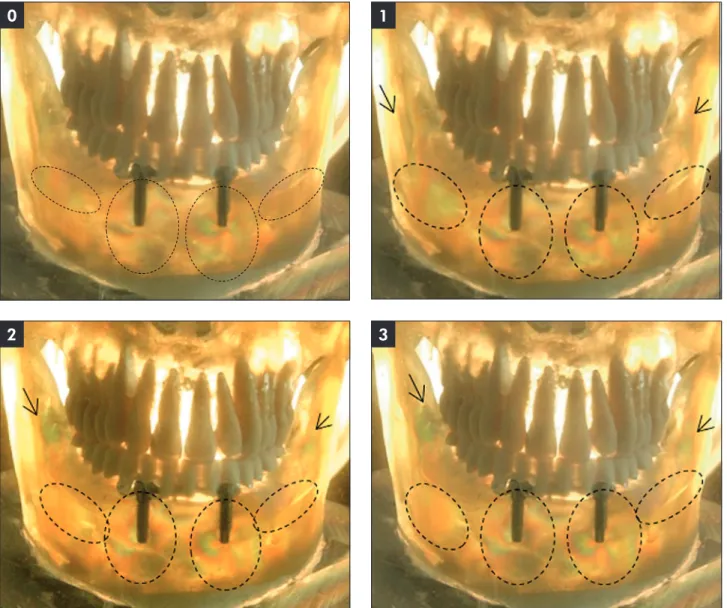
Figure 2 shows that for PAPI3, the load of 3 bars generated similar stress patterns as seen with PAPI2. For Model 2 - PAAI, the angled implant model submitted to photoelastic analysis (Figure 3), the re-gions selected for analysis were the same as those used for Model 1.
In Figure 3, low stress can be observed for PAAI0 (1 fringe or less) on the body of the implants. No discernable stress was noted on either edentu-lous ridge.
In Figure 3, which shows PAAI1 with 1 bar load, an increase was observed in stress pattern when compared with PAAI0. Low stress (1 fringe) was vi-sualized on the apical region, with moderate stress (between 1 and 3 fringes) on the mesial body of the they were invested and cast with Verabond II Ni-Cr
Alloy (Alba Dent Inc., Cordelia, USA).
The overdentures were waxed with the injection lask that was used to make the surgical guide, and they then were fabricated using conventional dental laboratory techniques. The clips (Conexão) were embedded, according to the manufacturer’s recom-mendations.
Light-bodied silicone impression material (Aero-jet Fiberglass, São Paulo, Brazil) was applied to the intaglio surface of the extension base of the over-dentures to mimic the soft tissue.5,12,13,16,18,19
Each photoelastic model was ixed to the photo-elastic cranium. This set was immersed in a tank of mineral oil (Campestre, São Bernardo, Brazil) to fa-cilitate photoelastic observation.
Loads of 1, 2 and 3 bars were applied to the pho-toelastic mandible models from above to the top, as in vivo, during which the mandible moves up to the maxilla.15 The teeth of the cranium were in contact
with the artiicial teeth from the overdenture. The occlusal contact determined the load application. The same overdenture was used in both groups, so in this manner, the points of load application were standardized.
In this study, stresses were observed and record-ed photographically17 using a Fuji S 9500 camera
(Fujiilm Corporation, Tokyo, Japan) in the ields of a circular polariscope. The evaluation was under-taken using the following terminology:
• low stress: 1 fringe or less;
• moderate stress: between 1 and 3 fringes; and
• high stress: more than 3 fringes.20,21
The areas analyzed in both groups were the me-sial, distal and apical regions from each implant and the right and left edentulous ridges from each group.
The photoelastic model images were named ac-cording to the implant angulations, followed by the amount of load applied:
• Model I - PAPI, for the photoelastic analysis model with parallel implants (PAPI0; PAPI1; PAPI2; PAPI3); and
right implant. Moderate stress was observed on the body of the left implant; this stress was more sparse in its mesial region and more delimited and intense in its distal region. The fringes formed by the right and left implants merged in the mental region. Low stress (1 fringe or less) was observed on the right and left edentulous ridges.
When a load of 2 or 3 bars was applied, PAAI2 and PAAI3, as shown in Figure 3, showed low stress (1 fringe) on the apical region, with moderate stress (3 fringes) on the mesial body of the right implant. Moderate stress was observed on the body of the left implant (3 fringes); this stress was more sparse in its mesial region and more delimited and intense in its
distal region. The fringes formed between the im-plants in the mental region were more intense. Low stress (1 fringe or less) was observed on right and left edentulous ridges.
Discussion
The advantages of treatment with overden-tures1,2,4 are important for explaining their
popu-larity, as well as why the authors chose this type of treatment.
Furthermore, this study compared the load transfer of an implant-retained mandibular overden-ture, with vertical and 10° inclined implants, so it was possible to verify the effects of nonaxial load. Figure 2 - PAPI: Bar-clip overdenture on two parallel implants, followed by load application.
0 1
2 3
0 1
The PAPI0 model, shown in Figure 2, presented low to moderate stress concentrated in the regions of the implants. Therefore, the evaluations of stress behaviors were undertaken starting with this initial image. The initial fringes patterns were the result of:
• photoelastic resin component manipulation and homogenization;
• denture adjustments on the photoelastic models; and
• denture ixation on the cranium acrylic base.
These initial fringes appeared although all care was taken to prevent them. Thus, the sequence ad-opted for analyzing the results was planned to eval-uate the intensity of stresses generated during load applications, while always considering the stresses
initially present in the models.
In Model 1 - PAPI, when submitted to loads of 0 to 3 bars, stress concentration was observed at the implant apex and in the edentulous ridge region. The presence of stress at the implant apex is explained by implants having the tendency to intrude when sub-mitted to load. Resin resistance hinders this implant penetration into photoelastic resin due to its hard-ness. Thus, areas of stress are generated at the apex, and as a result, photoelastic fringes occur.22,23
There is a principal difference in the fringe pat-tern along the implant body when the load is applied vertically on the denture occlusal plane. The stress concentration on implants is not reduced; other-wise, there would be a greater stress concentration at the apex.13 This reaction does not occur when the
Figure 3 - PAAI: Bar-clip overdenture on two angled implants, followed by load application.
0 1
2 3
0
2 3
load is applied at a point on the posterior teeth. In this situation, the bar clip retention system tends to rotate, and the stress tends to be transferred per-pendicularly to the posterior region of the ridge.19
Federick and Caputo, in 1996,12 evaluated stress
distribution in overdentures anchored by differ-ent retdiffer-ention systems. The load was applied on the premolar and second molar. The authors concluded that the more posteriorly the load was applied, the greater the stress concentration was on the posterior alveolar ridge, and the lower the stress was on the anterior region of the implants. Thus, some reports in the literature have described greater stress con-centrations on the posterior edentulous ridges and lower stress concentrations on the anterior region, where the implants are placed.17,24 This situation
was not observed in this study.
The difference in stress concentrations, when compared to the posterior edentulous ridges and the anterior region, could be explained by load applica-tion having occurred vertically on the overdenture occlusal plane.21 Kenney and Richards13 concluded
that the greater stress concentration on the anterior region, where the implants were placed, was due to their being joined by a bar-clip retention system, which transmitted a greater stress concentration to the supporting tissue, compared to the posterior alveolar ridge. Ochiai et al.,5 in 2004, compared
stress transmission to the alveolar ridge when over-dentures were retained by bar-clip and O-ring reten-tion systems. The bar-clip system was responsible for a greater stress concentration on the anterior region, where the implants were placed.21 Similar
re-sults were reported by Thayer and Caputo in 1977,22
when they described a stress concentration along the posterior ridges and a higher stress concentration on the root apex because the overdenture was retained using the Dolder bar system.
The literature has also reported that lower stress concentrations on the posterior alveolar ridge might have occurred due to the interposition of a layer of silicone, which was applied to simulate the oral mu-cosa. The capacity of silicone to absorb stress likely reduced the stress concentration on the posterior ridges.16,24
PAAI0, the angled implant model with load
ab-sence submitted to photoelastic analysis, as shown in Figure 3, presented more concentrated fringes in the implant region and no discernable stress on the posterior ridge. The stress pattern observation was undertaken considering the same observations made for the residual stresses in PAPI0.
Model 2 - PAAI, when submitted to loads of 0 to 3 bars, showed an increase in stress concentrations in the implant region and low stress on the posterior ridge. Thayer and Caputo, in 1977,22 afirmed that
after load application, implants tended to intrude into the photoelastic resin. This implant penetra-tion was hindered by the resin resistance, resulting in a photoelastic fringe at the implant apex. When the implant was angled and received perpendicular force on the overdenture occlusal plane, the same tendency of intrusion occurred. However, it tend-ed to descend in an inclintend-ed manner, so the fringe formations occurred not only at the apex but also along the distal face of the implant. This fringe oc-curred because the implant did not tend to intrude vertically, parallel to the load applied.12 Moreover,
Menicucci et al.25 afirmed that stress
concentra-tion occurred in the anterior mandible region when overdentures were anchored by a linked retention system, such as the bar-clip. Mandible deforma-tion during load applicadeforma-tion generated torsion in the mental region, where the implants were placed, which might explain the high stress concentration.
Celik and Uludag21 afirmed that when the
im-plants were angled, their apices were in a closer posi-tion, causing the stresses to aggregate or concentrate in this region, which explains the higher stress con-centration. This inding is in accordance with photo-elastic model 1, with parallel implants, provided that in this model, the stress concentration in the anterior region was less, compared with Model 2.
that all structures are isotropic and homogeneous. In addition, the contact between the implant and the photoelastic resin, which simulates bone tissue, is considered 100% along the entire implant body. In in vivo, it does not occur, because osseointegra-tion is a dynamic process.5,25,26 Furthermore,
three-dimensional photoelastic analysis was undertaken using images that are two-dimensional.13 The
simu-lated loads were applied vertically,5,13 although it is
known that mastication forces occur in many direc-tions. The method to obtain a transfer impression from the stone mandible with the implant replica
was selected because it mimics the real situation.16
Conclusion
Within the limitations of this study, it was con-cluded that PAPI showed better stress transfer com-pared with PAAI, since the forces oriented along the axis were better absorbed by the bone.
Acknowledgments
Fundação de Amparo à Pesquisa do Estado de São Paulo - FAPESP (process number 2007/54281-0) and Conexão Sistemas de Prótese.
References
1. Rutkunas V, Mizutani H, Peciuliene V, Bendinskaite R, Linkevicius T. Maxillary complete denture outcome with two-implant supported mandibular overdentures. A systematic review. Stomatologija. 2008;10(1):10-5.
2. Naert I, Quirynen M, Hooghe M, van Steenberghe D. A comparative prospective study of splinted and unsplinted Brånemark implants in mandibular overdenture therapy: a preliminary report. J Prosthet Dent. 1994 May;71(5):486-92. 3. Spiekermann H, Jansen VK, Richter EJ. A 10-year follow-up
study of IMZ and TPS implants in the edentulous mandible using bar-retained overdentures. Int J Oral Maxillofac Im-plants. 1995 Mar-Apr;10(2):231-43.
4. Naert I, Gizani S, van Steenberghe D. Bone behavior around sleeping and non-sleeping implants retaining a mandibu-lar hinging overdenture. Clin Oral Implants Res. 1999 Apr;10(2):149-54.
5. Ochiai KT, Williams BH, Hojo S, Nishimura R, Caputo AA. Photoelastic analysis of the effect of palatal support on vari-ous implant-supported overdenture designs. J Prosthet Dent. 2004 May;91(5):421-7.
6. Rentsch-Kollar A, Huber S, Mericske-Stern R. Mandibular implant overdentures followed for over 10 years: patient com-pliance and prosthetic maintenance. Int J Prosthodont. 2010 Mar-Apr;23(2):91-8.
7. Payne AG, Solomons YF. The prosthodontic maintenance requirements of mandibular mucosa- and implant-supported overdentures: a review of the literature. Int J Prosthodont. 2000 May-Jun;13(3):238-43.
8. Walton JN, MacEntee MI, Glick N. One-year prosthetic out-comes with implant overdentures: a randomized clinical trial. Int J Oral Maxillofac Implants. 2002 May-Jun;17(3):391-8. 9. Visser A, Raghoebar GM, Meijer HJ, Batenburg RH,
Vis-sink A. Mandibular overdentures supported by two or four endosseous implants. A 5-year prospective study. Clin Oral Implants Res. 2005 Feb;16(1):19-25.
10. Ribeiro PP, Goiato MC, Pellizzer EP, Pesqueira AA, Haddad MF, Dekon SFC, et al. Photoelastic analysis of implant-re-tained and conventional obturator prostheses with different attachment systems and soft relining. J Craniofac Surg. 2011 May;22(3):797-800.
11. Thayer HH, Caputo AA. Photoelastic stress analysis of over-denture attachments. J Prosthet Dent. 1980 Jun;43(6):611-7. 12. Federick DR, Caputo AA. Effects of overdenture retention designs and implant orientations on load transfer character-istics. J Prosthet Dent. 1996 Dec;76(6):624-32.
13. Kenney R, Richards MW. Photoelastic stress patterns pro-duced by implant-retained overdentures. J Prosthet Dent. 1998 Nov;80(5):559-64.
14. Pellizzer EP, Falcón-Antenucci RM, Carvalho PS, Sánchez DM, Rinaldi GA, Aguirre CC, Goiato MC. Influence of im-plant angulation with different crowns on stress distribution. J Craniofac Surg. 2011 Mar;22(2):434-7.
15. Gulizio MP, Agar JR, Kelly JR, Taylor TD. Effect of implant angulation upon retention of overdenture attachments. J Prosthodont. 2005 Mar;14(1):3-11.
16. Sadowsky SJ, Caputo AA. Effect of anchorage systems and extension base contact on load transfer with mandibu-lar implant-retained overdentures. J Prosthet Dent. 2000 Sep;84(3):327-34.
17. Machado AC, Cardoso L, Brandt WC, Henriques GE, Nóbilo MAA. Photoelastic analysis of the distribution of stress in dif-ferent systems of overdentures on osseous-integrated implants. J Craniofac Surg. 2011 Nov;22(6):2332-6.
18. Thayer HH, Caputo AA. Occlusal force transmission by over-denture attachments. J Prosthet Dent. 1979 Mar;41(3):266-71. 19. Porter JA Jr, Petropoulos VC, Brunski JB. Comparison of load
distribution for implant overdenture attachments. Int J Oral Maxillofac Implants. 2002 Sep-Oct;17(5):651-62.
21. Celik G, Uludag B. Photoelastic stress analysis of various retention mechanisms on 3-implant-retained mandibular over-dentures. J Prosthet Dent. 2007 Apr;97(4):229-35.
22. Thayer HH, Caputo AA. Effects of overdentures upon remain-ing oral structures. J Prosthet Dent. 1977 Apr;37(4):374-81. 23. Berg T, Caputo AA. Maxillary distal-extension removable partial denture abutments with reduced periodontal support. J Prosthet Dent. 1993 Sep;70(3):245-50.
24. Ichikawa T, Horiuchi M, Wigianto R, Matsumoto N. In vi-tro study of mandibular implant-retained overdentures: the influence of stud attachments on load transfer to the implant and soft tissue. Int J Prosthodont. 1996 Jul-Aug;9(4):394-9.
25. Menicucci G, Lorenzetti M, Pera P, Preti G. Mandibular implant-retained overdenture: a clinical trial of two anchor-age systems. Int J Oral Maxillofac Implants. 1998 Nov-Dec;13(6):851-6.