1Head of the Pain Center of Hospital das Clinicas, Functional Neuro s u rg e ry Division, Neurology Department, Medical School, University of São Paulo SP, Brazil;2PhD Student, Member of the Orofacial Pain Team, Dentistry Division, Hospital das Clinicas, Medical School, University of São Paulo SP, Brazil; 3Neurology Department, Medical School, University of São Paulo SP, Brazil.
Received 10 April 2006, received in final form 28 June 2006. Accepted 12 August 2006.
Dr. Silvia R.D.T. de Siqueira - Rua Maria Candida 135 - 02071-010 São Paulo SP - Brasil. E-mail: [email protected]
P E R C U TANEOUS RADIOFREQUENCY RHIZOTOMY AND
N E U R O VASCULAR DECOMPRESSION OF THE TRIGEMINAL
N E RVE FOR THE TREATMENT OF FACIAL PA I N
Manoel J. Teixeira
1, Silvia R.D.T. Siqueira
2, Gilberto M. Almeida
3ABSTRACT - Objective:To determine the outcomes of 354 radiofrequency rhizotomies and 21 neuro v a s c u-lar decompressions perf o rmed as treatment for 367 facial pain patients (290 idiopathic trigeminal neural-gia, 52 symptomatic trigeminal neuralneural-gia, 16 atypical facial pain, 9 post-herpetic neuralgia). M e t h o d :
Clinical findings and surg e ry success rate were considered for evaluation. A scale of success rate was deter-mined to classify patients, which considered pain relief and functional/sensorial deficits. Results: Radio-frequency rhizotomy was performed in 273 patients with idiopathic trigeminal neuralgia and in all other patients, except for trigeminal neuropathy; neurovascular decompression was perf o rmed in 18 idiopathic trigeminal neuralgia patients; 100% idiopathic trigeminal neuralgia, 96.2% symptomatic trigeminal neu-ralgia, 37.5% atypical facial pain and 88.9% post-herpetic neuralgia had pain re l i e f . Conclusion:Both t e c h-niques for idiopathic trigeminal neuralgia are usefull. Radiofrequency rhizotomy was also efficient to tre a t symptomatic facial pain, and post-herpetic facial pain, but is not a good technique for atypical facial pain.
KEY WORDS: facial pain, neurosurgery, trigeminal neuralgia, radiofrequency rhizotomy, vascular decom-pression.
Rizotomia percutânea por radiofreqüência e a descompressão neurovascular do nervo trigêmeo no tratamento das algias faciais
RESUMO - Objetivo:D e t e rminar eficácia e achados pós-operatórios após 354 rizotomias por radiofre q ü ê n-cia e 21 descompressões neuro v a s c u l a res como tratamento de 367 pacientes com dor fan-cial (290 neuralgia idiopática do trigêmeo, 52 neuralgia sintomática do trigêmeo, 16 dor facial atípica, 9 neuralgia pós-her-p é t i c a ) . Método:Achados clínicos e taxa de sucesso das ciru rgias foram considerados para a avaliação. Uma escala avaliando alívio da dor e complicações sensoriais e funcionais foi utilizada para classificar os p a c i e n t e s . Resultados:A rizotomia por radiofreqüência foi realizada em 273 pacientes com neuralgia idiopática do trigêmeo e em todos os outros pacientes, exceto neuropatia trigeminal; descompressão neu-rovascular foi realizada em 18 pacientes com neuralgia idiopática do trigêmeo; 100% dos pacientes com neuralgia idiopática do trigêmeo, 96.2% dos pacientes com neuralgia sintomática, 37.5% dos pacientes com dor facial atípica e 88.9% dos doentes com neuralgia pós-herpética tiveram alívio da dor. C o n c l u s ã o :
Ambas as técnicas são úteis para a neuralgia idiopática do trigêmeo. A rizotomia por radiofreqüência foi também eficiente para tratar neuralgia sintomática do trigêmeo e pós-herpética, mas não foi uma boa técnica como tratamento da dor facial atípica.
PALAVRAS-CHAVE: dor facial, neurocirurgia funcional, neuralgia trigeminal, rizotomia por radiofreqüên-cia, descompressão neurovascular.
Idiopathic trigeminal neuralgia (ITN) is a paro x-ysmal shock-like pain restricted to the innerv a t i o n a rea of one or more trigeminal branches, often set o ff by light stimuli in a trigger zone1 , 2. Its clinical tre a t-ment includes anticonvulsants, and carbamazepine is the drug of choice3, whereas surgical treatment is indicated in about 75% of the patients at any mo-ment after diagnosis4. Around 5% of patients pre
with-out any imaging or laboratorial abnorm a l i t i e s6. Post-herpetic neuralgia (PHN) is a complication of Herpes zoster infection in around 4% of patients, character-ized by burning pain at the trigeminal branch involv-e d7 , 8. For both AFP and PHN, treatment with dru g s (eg. antidepressants) is the first choice, but perc u t a-neous neuro s u rg e ry may be a treatment option9. Ra-d i o f requency percutaneous rhizotomy (RPR) is wiRa-de- wide-ly used to treat ITN1 0, and may be a choice for other re c u rrent facial pain1 1. Operated ITN may recur in 18%-20% of the cases in 10 years1 2. It is probable that high-er sensory deficit is an indication of highhigh-er success r a t e1 3. Some complications are: corneal hypoesthesia (16-23%), corneal anesthesia (2-6%), corn e a l - p a l p e-bral reflex deficit (19,7%), keratitis (1.4-4%), corn e a l ulcer (1-2%), paresthesia (8-10.9%), anesthesia dolo-rosa (0-5%), anesthesia of trigeminal branches (17%), Herpes simplex infection (40%), numbness sensation (58-79%), dysesthesia (0.5-18% - 5% need medica-tion), motor masseteric deficit (4-53%), paraliysis of ocular nerv e s1 4. Dysesthesias are usually associated to m o re intense sensory deficit1 5. Severe complications a re rare (intracranial haemorhage, meningitis)1 6.
N e u rovascular decompression (ND) is an open sur-g e ry with the aim of eliminatinsur-g the nerve compre s-sion at the entry zone by a vessel1 7. Most cases pre s-ent immediate relief, with less than 36% of re c u r-rence after 5 years1 8. Up to 26% of patients do not p resent the compression and need section of the trigeminal sensitive root. Complications are: facial hypoesthesia or hypoalgesia, anaesthesia doloro s a , master muscle weakness, paresis of the IV cranial n e rve (4.3%), auditive deficit1 8. Mortality rate is 1-4 . 3 %1 4. It is indicated for young patients intending to pre s e rve superficial sensitivity, suspicion of an ex-pansive intracranial lesion, association among facial neuralgias, bilateral ITN, ITN association to hemifa-cial spasm; its contra-indication is multiple sclero s i s1 8. The objective of this study was to evaluate the outcomes of a Brazilian population of patients with ITN and other facial pains after neurosurgical treat-ment with RPR and/or ND.
METHOD
This study is based on the re t rospective evaluation of 367 patients with facial pain treated at the Neurology Clinic of the Hospital das Clínicas, Medical School, University of São Paulo, and at the Neuro s u rg e ry Department of the Hospital Nove de Julho, São Paulo SP, between April, 1979 and June, 1984. Surgical treatment was indicated because of inefficacy and/or side effects of the clinical tre a t m e n t . Patiens were diagnosed according to the IASP criteria1 9, and treated as followed:
G roup I: Idiopathic trigeminal neuralgia - 290 patients: 272 were treated with RPR, 17 with ND and 1 by both pro-c e d u res; 1 had had RPR for PHN before, and developed ITN in other trigeminal branch.
G roup II: Symptomatic facial pain (SFP) - 52 patients: 39 with malign tumors, 7 with benign tumors, 3 with multi-ple sclerosis, 2 maxillary sinusopathy, 1 trigeminal neuro p a-t h y. All paa-tiena-ts were a-treaa-ted wia-th RPR, excepa-t for a-the lasa-t patient, which was treated with ND.
Group III: Atypical facial pain - 16 patients were treat-ed with RPR; 2 had also ND due to pain recurrence.
G roup IV: Posherpetic neuralgia - 9 patients, all tre a t-ed with RPR.
C o m p l e m e n t a ry investigation consisted in skull radiog-raphy in all patients. Computed tomogradiog-raphy (CT) was indi-cated for patients younger than 40 yo, AFP, SFP, and patients with abnormal neurological exam. After 1982, all patients s t a rted to be scanned by CT. Cere b rospinal fluid (CSF) exam was perf o rmed in all patients with bilateral ITN, and in asso-ciation to CT for the diagnosis of multiple sclerosis. CT o f v e rtebral-basilar complex was perf o rmed when vascular lesions were suspected, or as a routine investigation for patients with indication of ND. Patients with other associ-ated lesions had complementary exams, e.g. otorh i n o-laringologist or ophthalmologic evaluation.
Surgeries
R a d i o f requency percutaneous rhizotomy –P a t i e n t s u n d e rwent RPR with general anesthesia or sedation, and all had previously explanation about the surgical pro c e-d u re. The technique usee-d was the one e-describee-d by White and Sweet2 0and Siefgried9. After the electrode intro d u c-tion, patients were awakened to inform location of the stimulated area. Stimuli generator of radiofrequency used w e re the model RFG-3A (Radionics, Inc. Burlington, Massa-chusetts, USA) and Fundatec (Porto Alegre - Brasil). The e l e c t rode used was TIC-TM trigeminal rhizotomy electro d e (Radionics, inc., Burlington, Massachusetts, USA). The final lesion was perf o rmed with general anaesthesia. Methil-celulosis eyewash was prescribed for patients with hypoes-thesia of the ophtha lmic trigeminal branch, for a period of 4 weeks of application. Electrophysiological data, lesion parameters, cere b rospinal fluid or blood leak through the e l e c t rode, facial hyperemia or neurological post-operative disturbances were considered during the analysis.
Analysis –Pain relief, facial sensitivity deficit, neuro l o g-ical dysfunction, hypoesthesia, facial late deaff e re n t a t i o n , clinical complications, and pain re c u rrence were considere d for the analysis. Patients were classified in 7 degrees (Ta b l e 1). Patients were re-evaluated after 1 month, 4 months, and thereafter each 6 months. The evaluation was subjec-tive and based on patients and relasubjec-tives’ information.
RESULTS
Idiopathic trigeminal neuralgia –General charac-teristics corresponded to literature findings1 , 2: 166 (57.3%) female; 277 (95.5%) white, 7 (2.4%) yellow, 4 (1.4%) black and 2 (0.7%) other. Pain pre d o m i n a t-ed in the right side (57.6%) (p<0.05), and was bilat-eral in 17 (5.9%). Ages ranged from 17-88 years old (mean 62.5 yo). Patients treated with RPR (mean age of 63.8 yo) were statistically older than patients that u n d e rwent ND (mean age of 44.8 yo) (p<0.05). Mean pain duration was 91 months for ND and 99 months for RPR. Unilateral ITN distribution was: ophthalmic branch (V1) in 2.2%, maxillary branch (V2) in 17.6%, mandibular branch (V3) in 19.4%, V1-2 in 19.0%, V2-3 in V2-30.0%, and V1-2-V2-3 in 11.7%; V2-3 patients had famil-iar history of ITN (1.03%). Concomitant neuro l o g i c a l diseases were: Parkinson disease (3), essential tre m o r (3), cere b rovascular disorder (stroke) (2), glossopha-ryngeal neuralgia (2), vert e b ro-basilar invagination (1) and previous parietal meningioma (1); 8 (2.8%) had hemifacial spasm. CT (53.4% for RPR and 94.4% ND) findings were in general compatible to aging.
CSF exam, perf o rmed in 51 (17.6%) patients, re v e a l e d total proteins increase in 1 patient with convulsive d i s o rder (seizures). Angiography of vert e b ro - b a s i l a r complex was perf o rmed in 14 (4.8%) patients and angiography of internal carotid art e ry in 3 (1.0%): a t h e ro s c l e rosis was present in 1 that had ND and 7 that had RPR.
All patients had been treated with anticonvul-sants, and all had taken carbamazepine in any mo-ment; 6.2% had never had complete pain relief with d rugs, the others were indicated to surg e ry due to excessive side effects or failure of drugs’ efficacy; 120 (41.4%) had previous surgical pro c e d u res: 24.8% pe-ripheral neuro t o m y, 11.0% subtemporal re t ro g a s s e r-ian rh i z o t o m y, 16.2% trigeminal peripheral alcoholi-zation, 0.7% ND, 0.3% trigeminal ganglion alcoho-lization, and 0.3% ganglion electro c o a g u l a t i o n . P ro c e d u res were multiple in 8.2% of patients. Card i o -c i r-c u l a t o ry, re s p i r a t o ry, hepati-c, renal or metaboli-c disturbances are outlined in Table 2; 19 (6.6%) pa-tients treated with RPR had absolute contra-indica-tion for ND.
All patients treated with RPR had complete pain relief, although 8 had 2 RPR and 1 had 3 RPR for com-plete pain alleviation, immediately after the first RPR, resulting in a total of 283 pro c e d u res. Results are out-lined in Table 1. Along the follow-up, pain re c u rre n c e o c c u rred mostly in the first year (44.0%), and was m o re often in patients with bilateral RPR or ITN in
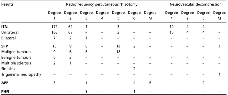
Table 1. Scale for the results of RPR and ND (N=367).
Results Radiofrequency percutaneous rhizotomy Neurovascular decompression Degree Degree Degree Degree Degree Degree Degree Degree Degree Degree Degree
1 2 3 4 5 0 M 1 2 3 M
ITN 172 69 1 – 3 – – 10 4 4 –
Unilateral 165 67 – – 3 – – 10 4 4 –
Bilateral 7 2 1 – – – – – – – –
SFP 16 9 6 – 18 2 – – – – 1
Maligne tumours 9 6 6 – 18 – – – – – –
Benigne tumours 5 2 – – – – – – – – –
Multiple sclerosis 2 1 – – – – – – – – –
Sinusitis – – – – – 2 – – – – –
Trigeminal neuropathy – – – – – – – – – – 1
AFP 5 – 1 – – 4 6 – – 2 –
PHN – – 8 – – 1 – – – – –
m o re than 1 branch (p<0.05), but not associated to the degree of post-operative sensorial deficit: 13.5% of patients with post-operative hypoalgesia, 18.9% with partial analgesia, and 44.5% with complete anal-gesia had re c u rrence. Recurrence and complications a re outlined in Table 3. Transient trigeminal pare s i s o c c u rred in 13 (4.8% - but only 1 discomfort a b l e ) , and facial paresthesia occurred in 36 (13.2%); 2.8% had anaesthesia dolorosa. Numbness sensation was unpleasant in 8 (2.9%) patients; 4 (1.5%) had diff i-culties to adapt their dental prosthesis due to facial hypoesthesia; 10 (3.7%) had ulcerate lesions in the oral mucosa clinically treated. Transient functional deficit of the VI cranial nerve occurred in 3 (1.1%) patients and of the IV nerve in 1 (0.4%); 4 (1.5%) had transient gustatory deficit and 18 (6.6%), transient auditive disturbances; 1 (0.4%) had haematoma of the right temporal lobe due to accidental cere b r a l c o rtex injury. Fort u n a t e l y, he got completely well after surgical treatment. Sensitive deficits are out-lined in Table 4. Hypalgesia/analgesia were transient in 30.4% fo patients (Table 5). Follow-up of patients was up to 65 months (mean 22.6 months).
All patients treated with ND got complete pain relief (Table 1): 1 had still 2 days of paroxysms. Neuro -vascular conflict was present in all of them: 17 ( 9 4 . 4 % ) with the superior cerebellar art e ry, 3 (16.7%) with a n t e r i o r-inferior cerebellar art e ry, 1 (5.6%) vert e b r a l a rt e ry, 1 (5.6%) undetermined vein, and 2 (11.1%)
arachnoiditis. Only 3 (16.7%) had depression of neu-ral surface at the compression area, and compre s s i o n was located in all at the initial 5mm of the trigemi-nal root. Only 1 (5.6%) had re c u rrence, after 30 months ( Table 3). Follow-up was up to 39 months (mean of 25.5 months).
Symptomatic facial pain –The most prevalent gen-der was male (30 patients - 75.8%); all patients were white; ages ranged from 22-77 yo (mean 56.5 yo). B reathing insuff i c i e n c y, hypertension and/or de-nutri-tion were present in 30 cases (Table 2). Recurre n c e rates (9.6%) and complications may be observed on Table 3.
a) Malign tumors (N=39): Tumor location was: cra-nial base, oral cavity, face, oro p h a rynx and lary n x . All patients had RPR as treatment, and all had pain relief (Table 1). Pain distribution was: V3 in 17 ( 4 3 . 6 % ) , V2-3 in 10 (25.6%), V1-2-3 in 6 (15.4%), V1-2 in 3 (7.7%), V2 in 2 (5.1%), V1 in 1 (2.6%). RPR was bilat-eral in 3 (7.7%) patients. Three patients had lesion of adjacent branches during the procedure. Follow-up was Follow-up to 23 months (mean 5.2 months).
b) Benign tumors (N=7): One patient had menin-gioma of Meckel cave, 2 meninmenin-gioma of the medi-um and posterior fossa, 1 clivus meningioma, 1 der-moid cyst of Meckel cave (with previous surg e ry with-out alleviation), and 1 acoustic neurinoma. All were treated with RPR, and all had pain relief (Table 1).
Table 2. Relevant clinical findings of patients (N=367).
Surgical risks Cardiac disease Arterial hypertension
IBI ACA IMI ICI Moderate Severe D DU DB RI
ITN
14 11 17 25 107 40 – 5 14 2
Radiofrequency rhizotomy 14 11 17 25 104 40 – 4 – 2
Neurovascular decompression – – – – 3 – – 1 – –
SFP 7 – – – 2 3 18 – – –
Maligne tumours 6 – – – – 3 18 – – –
Benigne tumours – – – – 2 – – – – –
Multiple sclerosis 1 – – – – – – – 1 –
Sinusitis – – – – – – – – – –
Trigeminal neuropathy – – – – – – – – – –
AFP
2 – – – 3 – – – 1 –
Radiofrequency rhizotomy 3 – – – 3 – – – 1 –
Neurovascular decompression – – – – – – – – – –
PHN 3 – – – 2 2 – 1 – 1
c) Multiple sclerosis (N=3): Facial pain began after 2-20 years from the initial symptoms of the disease, and pain duration ranged from 12 to 39 months (mean 29m); 2 patients had bilateral ITN and 1 uni-lateral AFP; 2 had pain at V3 and 1, V1-2. Neuro l o-gical abnormalities including facial hypoesthesia were p resent in all of them. All underwent RPR, with ini-tial pain relief (Table 1). Maximum follow-up was 56 months (mean 30.7 m).
d) Sinusitis (N=2): Pain was at V1-2 in 1 and at V2 in the other. Both had multiple unsuccessful pre v i-ous treatments. No one had pain alleviation after RPR (Table 1).
e) Trigeminal neuropathy (N=1): Pain was pre s e n t on the left side, associated to moderate hypoesthe-sia. Previous treatments with anticonvulsants and p s y c h o t ropics were unsuccessful. Imaging exams and blood tests were normal. ND was perf o rmed because of arachnoiditis observed on imaging exams, but its coagulation did not relief pain (Table 1).
Atypical facial pain – Half patients were female (8; 50%), and all were white. Pain was on the left facial side in 8 (50%) and bilateral in 1 (6.3%). Ages ranged from 29 to 79 yo (mean 55.6 yo). Pain start-ed mostly between the fifth and sixth life’s decade. It was located at V1 in 2 (12.5%), at V2 in 1 (6.3%), at V3 in 3 (18.8%), at V1-2 in 2 (12.5%), at V2-3 in 5 (31.3%) and at V1-2-3 in 3 (18.8%). Findings were n o rmal in imaging exams. Previous surgeries were : 6 (37.5%) trigeminal neure c t o m y, 6 (37.5%) subtem-poral rh i z o t o m y, and 1 (6.3%) peripheral trigeminal alcoholization. Associated diseases are outlined in Table 2, and post-operative complications in Table 3. All were treated with RPR, but only 37.5% had pain relief (Table 1). Six (37.6%) had lesion of adjacent trigeminal branches during surg e ry. Two patients with re c u rrence had complete pain relief after ND. Patients with degree 0 were treated with trigeminal tractotomy and nucleotomy without impro v e m e n t . Follow-up ranged from 3 to 62 months (mean 33.4m).
Table 3. Pain recurrence and complications (N=367).
Results N Nº % MT CH AC MC K U CP CE OT
ITN 290 29 9.6 14 39 6 13 2 1 36 5 56
Radiofrequency rhizotomy 273 28 10.2 13 39 6 13 2 1 36 4 49
Neurovascular decompression 18 1 5.6 30 – – – – – – 1 7
SFP 52 6 9.6 9.5 2 – 1 1 2 2 1 –
Maligne tumours 39 3 5.1 3 – – 1 1 1 2 1 –
Benigne tumours 7 2 28.5 15 – – – – – – – –
Multiple sclerosis 3 1 33.0 12 1 – – – – – – –
Sinusitis 2 – – – 1 – – – 1 – – –
Trigeminal neuropathy 1 – – – – – – – – – – –
AFP 16 4* 23.5 12 1 – – 1 – 2 1 1
Radiofrequency rhizotomy 16 3 23.5 12 1 – – 1 – 2 – 1
Neurovascular decompression 2 1 50.0 6 – – – – – – 1 –
PHN 9 – – – – – – 1 2 – – 1
ITN, idiopathic trigeminal neuralgia; SFP, symptomatic facial pain; AFP, atypical facial pain; PHN, post-herpetic neuralgia; N, number of patients; No, number of re c u rrences; %, percentage of re c u rrences; MT, mean duration of re c u rrence (months); CH, corneal hyporeflex; AC, abolition of corn e a l reflex; MC, compromise of motor root; K, keratitis; U, corneal ulcer; CP, central pain; CE, compromise of extrinsic motility of the eye; OT, other; *patient that underwent both surgeries.
Table 4. Sensitive post-operative deficits after RPR X trigeminal branch affected (N=290): sensitive deficits occurred in a different trigeminal branch than the pain location in 40 (14.7%).
Area of sensitive deficit
Trigeminal V1 V2 V3 V1-2 V2-3 V1-2-3 Sub-total
branch V1 5 – – – – 1 6
affected V2 – 31 – 3 12 3 49
by pain V3 – – 44 – 7 1 52
V1-2 – 2 – 43 – 9 54
V2-3 – 1 2 2 72 4 81
Post-herpetic neuralgia –Most patients were male (55.6%); 8 (88.8%) were white. Ages ranged from 26 to 88 yo (mean 61.7 yo). Pain location was at V1 in 8 patients and V3 in 1. All had tactile hypoesthesia and allodynia at the affected area. Only 2 had pre v i o u s s u rgical treatment (1 had peripheral neurectomy and the other peripheral V1 alcoholization). After RPR, 88.8% had pain relief (Table 1). Associated diseases a re outlined in Table 2, and post-operative compli-cations in Table 3. During the pro c e d u re, 4 (44.4%) patients had adjacent lesion at V2. Three needed fur-ther trigeminal tractotomy and nucleotomy after re c u rrence, but only one had pain relief. The others u n d e rwent thalamotomy with partial impro v e m e n t . Follow-up ranged from 3 to 60 months (mean 25.1 m).
DISCUSSION
N e u ro s u rgical pro c e d u res have a recognized eff i-cacy in the treatment of facial pain, especially for ITN. The ideal surg e ry should eliminate pain with low risks and post-operative complications, including few cases of recurrence. There are a lot of controversies on the indication of surgical techniques1 7 , 2 1and on hypothetical etiologies and their tre a t m e n t2 2. For ITN, the current techniques include RPR (a perc u t a n e o u s p ro c e d u re )1 4and ND (an open surg e ry )2 1. Perc u t a-neous techniques are very safe, and facial pain often affects elderly people with higher surgical risks.
In this sample, general characteristics of the pa-tients, including gender, age, ethnic group, and pain characteristics (side, branch affected, pain location, duration) are similar to scientific literature1 , 2 , 1 0. Pati-ents selected for ND were usually younger and did
not present clinical contra-indications to an open sur-g e ry. Only 1 patient in this sample had more than 60 years old, but he had association of hemifacial spasm to ITN, which was the reason for ND9.
RPR was indicated for older ITN patients with higher clinical risks for complications, and other pain causes (SFP, AFP, PHN)2 3. For these cases, there is no advantage using more invasive techniques, and the chosen pro c e d u re should be as simpler and safer as possible. All patients had information about types of s u rgeries, their advantages, disadvantages and risks, and could participate during the pro c e d u re ’s choice. Many patients with ND indication chose RPR because of their risks fear.
All complications that occurred in this sample w e re similar to literature1 3 , 1 7 , 1 8. Carotid lesion occurre d in 2.9% of the cases, but did not cause perm a n e n t damage. There was a variable degree of tactile hy-poesthesia after RPR. Corneal sensory deficit was m o re frequent in patients with ITN at V1 (31.4%). Complaints of impossibility of bilateral mastication may occur due to oral mucosa hypoesthesia, and lin-gual hypoesthesia may difficult dental prothesis adap-tation. The low rate of motor complications after RPR may be associated to large nerve fibers pre s e rv a t i o n . Beyond that, we did not observe any association between the degree of hypoesthesia and the recur-rence rate24.
When surgery is indicated to ITN, it usually pres-ents high rate of success1 0 , 1 5 , 2 4. In this study, RPR and ND relieved ITN in 100%, and RPR reduced sympto-matic facial pain in 94.8% immediately after the sur-g e ry. The presence of a primary case, like multiple
Table 5. Pain location X post-operative analgesia (N=221) and hypoalgesia (N=114), after radiofrequency percutaneous rh i z o t o m y.
Location of post-operative analgesia (N=221)
Trigeminal V1 V2 V3 V1-2 V2-3 V1-2-3 V1-3 Sub-total
branch V1 1 – – – – 1 – 2
affected V2 – 32 – 2 6 – – 40
by pain V3 – 1 32 – 5 1 – 43
V1-2 – 21 – 15 1 4 – 41
V2-3 – 6 13 1 47 4 – 71
V1-2-3 – 4 2 – 10 7 1 24
Location of post-operative hypoalgesia (N=114)
Trigeminal V1 V2 V3 V1-2 V2-3 V1-2-3 V1-3 Sub-Total
branch V1 4 – – – – – – 4
affected V2 2 6 2 – 3 – 1 14
by pain V3 1 – 8 – – – – 9 |
V12 15 – 1 13 – – 4 33
V2-3 2 9 5 2 11 1 2 32
s c l e rosis, indicates RPR rather than ND, because of the non evidence of a vascular compression as etiol-o g y1 8. On the other hand, other facial pain causes without a removable cause (eg. AFP, PHN), or tumors impossible to extirpate, do not often present good results with drugs, and surg e ry may be a choice8. In this study, 81.3% AFP and 88.9% PHN patients had p a rtial or complete pain relief, but neuro s u rg e rywas not efficient for inflammatory disease (sinusitis) or trigeminal neuropathy.
Dysesthesias and other sensory complications of these sample were more often after RPR, corre s p o n-ding to other scientific papers, but immediate com-plications after ND are usually more severe1 2 , 1 3 , 2 1. Des-pite that, ND may be a good choice for young pa-tients intending to pre s e rve superficial facial sensi-t i v i sensi-t y, which is an unpleasansensi-t complainsensi-t, and dysessensi-the- dysesthe-sia in these cases is uncommon. The recurrence rate of ND is also lower than RPR21.
In conclusion, both pro c e d u res were useful to treat ITN, and age, clinical conditions, and patients’ opinions are the factors that should be considere d during the surgical choice. RPR was also efficient to t reat symptomatic facial pain and post-herpetic facial pain, but not atypical facial pain and inflammatory pain (sinusitis).
REFERENCES
1. Rothman KJ, Monson RR. Epidemiology of trigeminal neuralgia. J Chron Dis 1973;26:2-12.
2. Siqueira SRDT, Nóbrega JCM, Valle LBS, Teixeira MJ, Siqueira JTT. Idiopathic trigeminal neuralgia: clinical aspects and dental pro c e d u re s . Oral Surg Oral Med Oral Pathol Oral Radiol Endod 2004;98:311-315. 3. G u e r re ro - F i q u e roaAR, Escobar-Juyo A, Caballero - G a rcia G,
Blanco-Castillo IP. Gabapentin effect in orofacial allodynia: experimental cor-relation with trigeminal neuralgia. Rev Neurol 1999;29:1147-1153. 4. Zakrzewska JM, Patsalos PN. Long-term cohort comparing medical
(oxcarbazepine) and surgical management of intractable trigeminal neuralgia. Pain 2002;95:259-266.
5. Benedittis G, Bernasconi V, Ettore G. Tumours of the fifth cranial nerve. Acta Neurochir (Wien) 1977;38:37-64.
6. Zakrzewska J. M. Classification issues related to neuropathic trigemi-nal pain. J Orofac Pain 2004;18:325-331.
7. B rown GR. Herpes zoster: correlation of age, sex, distribution, neural-gia, and associated disorder. South Med 1976;69:576-578
8. Gerson RG. The management of post-herpetic neuralgia. In Murphy JE (ed). Pharmaceutical medicine. London; 1979:177-185.
9. Siegfried J. 500 percutaneous thermocoagulation of the Gasserian gan-glion for trigeminal pain. Surg Neurol 1977;8:126-131.
10. Choudhury BK, Pahari S, Acharyya A, Goswami A, Bhattacharyya MK. P e rcutaneous re t rogasserian radiofrequency thermal rhizotomy for trigeminal neuralgia. J Indian Med Assoc 1991;89:294-296.
11. Mathews ES, Scrivani SJ. Percutaneous stereotactic radiofrequency ther-mal rhizotomy for the treatment of trigeminal neuralgia. Mt Sinai J Med 2000;67:288-299.
12. Taha JM, Tew JM. Comparison of surgical treatments for trigeminal neuralgia: Reevaluation of radiofrequency rh i z o t o m y. Neuro s u rg 1996; 38:865-871.
13. Peters G, Nurmikko TJ. Peripheral and gasserian ganglion-level pre-cedures for the treatment of trigeminal neuralgia. Clin J Pain 2002;18: 28-34.
14. Pollock BE, Foote RL, Staff o rd SL, Link MJ, Gorman DA, Schomberg PJ. Results of repeated gamma knife radiosurgery for medically unre-sponsive trigeminal neuralgia. J Neuro s u rg 2000;93(Suppl 3):S162-S164. 15. Taha JM, Tew JM, Buncher R. A p rospective 15-year follow up of 154 consecutive patients with trigeminal neuralgia treated by perc u t a n e o u s s t e reotactic radiofrequency thermal rh i z o t o m y. J Neuro s u rg 1995;83: 989-993.
16. Bilgin H, Kelebek N, Korfali G, Bekar A, Kerimogl U. A r a re complica-tion of trigeminal nerve stimulacomplica-tion during radiofrequency thermoco-agulation. J Neurosurg Anesthesiol 2002;14:47-49.
17. Kanpolat Y, Savas A, Bekar A, Berk C. Percutaneous controlled radiofre-quency trigeminal rhizotomy for the treatment of idiopathic trigemi-nal neuralgia; 25-years experience with 1600 patients. Neuro s u rg e r y 2001;48:524-534.
18. Pollack IF, Janetta PJ, Bissonette DJ. Bilateral trigeminal neuralgia: a 14-years experience with microvascular decompression. J Neuro s u rg 1988;68:559-565.
19. Merskey H, Bogduk N. Classification of chronic pain. 2ª. Ed. Seattle: IASP Press, 1994.
20. White JC, Sweet WH. Pain and the neuro s u rgeon. Illinois: Charles C Thomas Publisher, 1969.
21. Jannetta PJ. Vascular decompression in trigeminal neuralgia. In Samii M, Jannetta PJ (eds). The cranial nerves. Berlin: Spriger- Verlag, 1981:331-340.
22. Hitchocock H, Teixeira M, Pinto J. Percutaneous trigeminal radiofre-quency rhyzotomy. J Royal Coll Surg Edinburgh 1983;28:74-79. 23. Yoon KB, Wiles JR, Miles JB, Nurmikko TJ. Long-term outcome of
per-cutaneous thermocoagulation for trigeminal neuralgia.A n a e s t h e s i a 1999;54:798-808.