D e p a rtamento de Genética Médica, Faculdade de Ciências Médicas / Universidade Estadual de Campinas, Campinas SP, Brasil (FCM/UNICAMP):1P rofessor Associado, Departamento de Genética Médica;2Doutor em Ciências Médicas com área de concentração
em Genética.
Received 24 February 2006, received in final form 21 June 2006. Accepted 27 July 2006.
Dra. Vera Lúcia Gil-da-Silva-Lopes - Departamento de Genética Médica - FCM / UNICAMP - Caixa Postal 6111 - 13084-971 Campinas SP - Brasil. E-mail: [email protected]
CENTRAL NERVOUS SYSTEM ABNORMALITIES ON
MIDLINE FACIAL DEFECTS WITH HYPERTELORISM
DETECTED BY MAGNETIC RESONANCE IMAGE AND
COMPUTED TOMOGRAPHY
Vera Lúcia Gil-da-Silva-Lopes
1, Silvio David Araújo Giffoni
2ABSTRACT - The aim of this study were to describe and to compare structural central nervous system (CNS) anomalies detected by magnetic resonance image (MRI) and computed tomography (CT) in individuals a ffected by midline facial defects with hypertelorism (MFDH) isolated or associated with multiple congen-ital anomalies (MCA). The investigation protocol included dysmorphological examination, skull and facial X-rays, brain CT and/or MRI. We studied 24 individuals, 12 of them had an isolated form (Group I) and the others, MCA with unknown etiology (Group II). There was no significative diff e rence between Group I and II and the results are presented in set. In addition to the several CNS anomalies previously described, MRI (n=18) was useful for detection of neuronal migration errors. These data suggested that structural CNS anomalies and MFDH seem to have an intrinsic embryological relationship, which should be taken in account during the clinical follow-up.
KEY WORDS: frontonasal dysplasia, hypertelorism, midline, craniofacial abnormalities, facial clefts, CNS abnormalities, magnetic resonance image.
Anomalias de sistema nervoso central em defeitos de linha média facial com hipert e l o r i s m o detectados por ressonância magnética e tomografia computadorizada
RESUMO - Este estudo objetivou descrever e comparar as anomalias estruturais do sistema nervoso central (SNC) detectadas por meio de ressonância magnética (RM) e tomografia computadorizada (TC) de crânio em indivíduos com defeitos de linha média facial com hipertelorismo (DLMFH) isolados ou associados a anomalias congênitas múltiplas (ACM). O protocolo de investigação incluiu exame dismorfológico, RX de crânio e face, CT e RM de crânio. Foram estudados 24 indivíduos, sendo que 12 apresentavam a forma iso-lada (Grupo I) e os demais, DLMFH com ACM de etiologia não esclarecida (Grupo II). Não houve difere n ç a e n t re os dois grupos e os resultados foram agrupados. Além de várias anomalias de SNC já descritas, a RM foi útil para detecção de erros de migração neuronal. Os dados sugerem que as alterações estruturais de SNC e os DLMFH têm relação embriológica, o que deve ser levado em conta durante o seguimento clínico.
PA L AV R A S - C H AVE: displasia frontonasal, hipertelorismo, linha média, malformações craniofaciais, fendas faciais, malformações do SNC, ressonância magnética.
Midline facial defects with hypertelorism (MFDH) is a rare group of disorders characterized by ocular h y p e rtelorism and anomalies of frontonasal pro c e s s . Its isolated form is commonly called frontonasal dys-plasia and it is also detected in several conditions of multiple congenital anomalies (MCA)1 - 3. Its
pathogen-esis is related to a failure during the development of the nasal capsula4 , 5. Diff e rential diagnosis should be
done from some conditions which could lead to a sim-ilar appearance, such as frontal lipoma and encephalo-c e l e5. Several structural abnormalities of central
nerv-ous system (CNS) are often described in this gro u p , such as encephalocele, myelomeningocele, Chiari’s m a l f o rmation and hydro c e p h a l u s6. Corpus callosum
anomalies are re p o rted by several authors3 , 7 - 1 0.
magnet-ic resonance image (MRI) searching CNS anomalies in a large group of individuals with MFDH has not been reported before.
In this article, we describe the neuro rr a d i o l o g i c a l data from 24 patients with MFDH.
METHOD
The casuistry was composed by 24 individuals (8 male and 16 female) affected by MFDH1 0. All of them were
pre-viously evaluated by the same clinical geneticist at Hospital das Clínicas HC/ UNICAMP (Campinas, SP, Brasil). They were selected based upon minimum inclusion criteria, which were ocular hypertelorism and frontonasal process anomalies. Twelve individuals presented an isolated form (Group I) and the others, facial findings were part of a multiple con-genital anomalies condition with unknown etiology (MCA) (Group II). The average age was 12.57 years.
The investigation protocol included dysmorphological evaluation, skull and facial X-rays, computed tomography (CT) of brain and magnetic resonance image. Six individu-als who had metallic clips on their faces were evaluated just by CT.
Brain CT was perf o rmed with parallel slices to the infe-rior orbitomeatal line with 3 mm of thickness and 5 mm of space on the posterior fossa. On the supratentorial fossa, the slices had 10mm of thickness and space from vertex of transversalis axis.
CNS MRI was perf o rmed without contrast at 2.0 T. The sequence was: Axial FSE double eco T2; TE=16/128. TR of 4600, 6 mm of thickness and 2% of space. Axial FSE dou-ble eco DP, TE=16/128. TR=4600, 6 mm of thickness and 2% of space. Sagittal SE T1; TE=10. TR=550, 4 mm of thickness and space of 0%. Axial SE T1; TE=10. TR=550, 6 mm of thick-ness and 2% of space. Coronal SE T1; TE=10. TR=550, 6 mm of thickness and 2% of space. Axial flair inverse re c o v e r TE=90 on CSF. TR=8100, TI=2200, 6 mm of thickness and 2% of space. When necessary, CT scan and MRI were perf o rm e d with anesthesia, according to the American College of E m e rgency Physicians. The statistics evaluation was per-f o rmed by SAS System per-for Windows (statistical analysis sys-tem), version 8.02. It was used Fischer ´s exact test (FET). The rejection level was fixed at 5%11,12.
This study was approved by the Ethics Committee of the University Hospital / UNICAMP (n.057/99) and all indi-viduals consented for their inclusion.
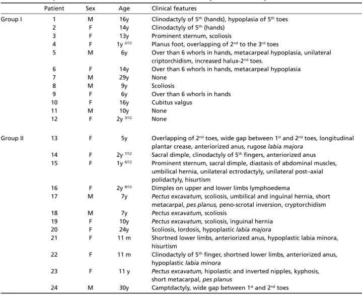
Table 1. General aspects and extra- facial features of individuals affected by MFDH in this study.
Patient Sex Age Clinical features
Group I 1 M 16y Clinodactyly of 5th(hands), hypoplasia of 5thtoes
2 F 14y Clinodactyly of 5th(hands)
3 F 13y Prominent sternum, scoliosis
4 F 1y2/12 Planus foot, overlapping of 2ndto the 3rdtoes
5 M 6y Over than 6 whorls in hands, metacarpeal hypoplasia, unilateral criptorchidism, increased halux-2ndtoes.
6 F 14y Over than 6 whorls in hands, metacarpeal hypoplasia
7 M 29y None
8 M 9y Scoliosis
9 F 6y Over than 6 whorls in hands
10 F 16y Cubitus valgus
11 M 10y None
12 F 2y3/12 None
Group II 13 F 5y Overlapping of 2ndtoes, wide gap between 1stand 2ndtoes, longitudinal
plantar crease, anteriorized anus, rugose labia majora
14 F 2y7/12 Sacral dimple, clinodactyly of 5thfingers, anteriorized anus
15 F 1y6/12 Prominent sternum, sacral dimple, diastasis of abdominal muscles,
umbilical hernia, unilateral ectrodactyly, unilateral post–axial polidactyly, hisurtism
16 F 2y8/12 Dimples on upper and lower limbs lymphoedema
17 M 7y Pectus excavatum, scoliosis, umbilical and inguinal hernia, short
metacarpal,pes planus, peno-scrotal inversion, cryptorchidism
18 M 7y Pectus excavatum, scoliosis
19 F 10y Pectus excavatum, scoliosis, inguinal hernia
20 F 24y Scoliosis, lordosis, hypoplastic labia majora
21 F 11 m Shortned lower limbs, anteriorized anus, hypoplastic labia minora, hisurtism
22 F 11 m Clinodactyly of 5thfinger, shortned lower limbs, anteriorized anus,
hypoplasticlabia minora
23 F 11 y Pectus excavatum, hipolastic and inverted nipples, kyphosis,
short metacarpal, pes planus
RESULTS
Mainly clinical features of Group II are described on Table 1. A considerable heterogeneity of CNS ano-malies was detected (Figs 1A and 1B; 2 and 3). Com-paring the investigation of Groups I and II, no statis-tical diff e rence was found and the results are des-cribed on Table 2.
DISCUSSION
Considering the rarity of the condition and the description by diff e rent specialties, the evaluation of CNS defects in a significant casuistry with MRI had not been perf o rmed before. Until now, the most part of information about the neurological anomalies in MFDH was obtained using CT3 , 7 - 9. These studies
show-ed a large range of CNS anomalies, especially of the corpus callosum, such as lipoma, calcification, and dysgenesis. Some other anomalies were also men-tioned, such as encephalic asymmetry and
encephalo-cele. In this casuistry, the main defects detected by skull and facial X-rays were hyperteleorbitism (24/24), anterior cranium bifidum (8/24) and frontal osseous f a i l u re (4/24). These findings are similar to those pre-viously described.
G u i o n - A l m e i d a9, reviewing 27 patients from the
l i t e r a t u re and personal cases, re p o rted anterior cra-nium bifidum in 12 individuals. Gil-da-Silva-Lopes and M a c i e l - G u e rr a3, evaluating brain CT of 29 personal
patients with MFDH, including isolated form and those with multiple congenital anomalies with un-known etiology, detected anterior cranium bifidum in 6 individuals and frontal osseous failure in 4.
Brain CT in this study showed anomalies of cor-pus callosum (8/21) and several defects, such as osse-ous (7/21), ventricular (3/21), cortical (2/21), fro n t a l lobe atrophies (2/21) and encephaloceles (3/21). Lipo-ma of corpus callosum, a usual feature re p o rted, was detected in just one of our patients. Corpus callosum
Fig 1A and B. MRI shows ence -phalic asymmetry, hypoplasia of corpus callosum, gliosis, fro n t a l perisylvian polymycrogiria and h e t e rotopy of gray matter at left hemisphere.
Fig 2. MRI shows frontal encephalocele, corpus callosum dysgenesis and hypoplasia of cere b e -lar vermis.
Table 2. Radiological findings of individuals affected by MFDH in this study.
Patient Skull and facial X-rays CT scan MRI
Group I 1 Hyperteleorbitism Encephalic and ventricular asymmetry, Corpus callosum agenesis,c o l p o c e p h a l y,
corpus callosum dysgenesis c o l p o c e p h a l y, neuronal migration erro r s 2 Hyperteleorbitism Polips of paranasasal sinus Normal
3 Hyperteleorbitism, Abnormal thickness of ethmoidal, Nodular heterotopia areas anterior bifid cranium, vomeris and cribriform lamina,
median maxilar cleft possible ethmoidal encephalocele
4 Hyperteleorbitism Fronto-parietal atrophy, / ventricular enlargement
5 Hyperteleorbitism Right parietal porencephaly, Corpus callosum dysgenesis, frontal ventricular dilatation micropoligiria
6 Hyperteleorbitism, Sphenoidal hyperostosis Chiari’s anomaly, enlargement
anterior bifid cranium, of 4º ventricle
median nasal cleft
7 Hyperteleorbitism, Normal /
anterior bifid cranium
8 Hyperteleorbitism, / Corpus callosum dysgenesis, cerebellar
anterior bifid cranium dysgenesis, Dandy-Walker anomaly,
nodular heterotopia areas 9 Hyperteleorbitism, Corpus callosum dysgenesis, /
anterior bifid cranium, Tessier (0-14) cleft, auditive partial frontal aplasia channel agenesis
10 Hyperteleorbitism, Lipoma of corpus callosum, / partial frontal aplasia ethmoidal encephalocele
11 Hyperteleorbitism Normal Nodular heterotopia at left
hemispherium, frontal and perisilvian micropoligiria,corpus callosum dysgenesis,cerebellar asymmetry
12 Hyperteleorbitism Normal Normal
Group II 13 Hyperteleorbitism Corpus callosum dysgenesis Nodular heterotopia at frontal left lobe,corpus callosum agenesis,
cerebellar asymmetry
14 Hyperteleorbitism Normal Normal
15 Hyperteleorbitism Atrophy signs at frontal lobes /
16 Hyperteleorbitism, Asymmetry and dilatation of ventricles Cortical displasia at frontal poles, anterior bifid cranium enlargement of 3º and 4º ventricle 17 Hyperteleorbitism, Corpus callosum agenesis Corpus callosum dysgenesis, pineal cyst
anterior bifid cranium
18 Hyperteleorbitism Normal Thin cervical marrow
19 Hyperteleorbitism, Asymmetric skull, corpus callosum Corpus callosum dysgenesis, cranium asymmetric skull dysgenesis deformity that takes to tentorium
verticalization, hemispheres and cerebellar lobes reduction
20 Hyperteleorbitism / Vascular spaces enlargement in the
topography of the basis nucleus
21 Hyperteleorbitism Normal Normal
22 Hyperteleorbitism, Fissure at posterior portion of / anterior bifid cranium corpus callosum, frontal encephalocele,
ventricular system malformation, cerebellar vermis hypoplasia, gray substance ectopic of frontal left lobe
23 Hyperteleorbitism Bilateral fronto parietal atrophy, Compensatory hydrocephalus, corpus
ventricular enlargement, diffuse callosumhypoplasia, signs of subcortical atrophy supratentorium and infratentorium
volumetric reduction
24 Hyperteleorbitism / Enlargement of vascular spaces at basis
is the main anomalous stru c t u redescribed in this con-dition. Fontaine et al.1 3described 3 isolated cases
with MFDH and corpus callosum agenesis. In a clini-cal follow-up of 8 patients, Pascual-Castroviejo et al.7
detected lipoma and calcification of faux in all of them and hypoplasia of the corpus callosum in brain C T. Guion-Almeida9detected 8/100 patients with
cor-pus callosum agenesis. In an extensive review of the l i t e r a t u re, and also adding personal cases, Gil-da-Silva-Lopes and Maciel-Guerr a3o b s e rved agenesis of
this stru c t u re in 5/31 individuals and dysgenesis, in 4/31. These authors also described encephaloceles, ventricular anomalies, cortical atrophy and CNS cal-cifications.
Naidich et al.8re p o rted CNS findings of 11
indi-viduals with MFDH detected, by computed CT of the crania in 10 and MRI in 1 of them. In this study, 5 pa-tients with lipoma of the corpus callosum and 1 with calcification of faux and interhemispheric lipoma w e re observed. Gil-da-Silva-Lopes and Maciel-Guerr a3
detected lipoma of corpus callosum in (2/31) patients.
Two of our patients had errors of migration de-tected by CT scan. This number had an increase when we used MRI (7/18). This kind of investigation also evidenced corpus callosum anomalies (agenesis/dys-genesis) in 8/18 individuals, ventricular anomalies (8/18) and cerebellar hypoplasia in 3/18.
C o rtical anomalies have been described in a gro u p of 10 patients with frontonasal dysplasia, corpus cal-losum agenesis and mental re t a rdation. In this gro u p , MRI was perf o rmed in 8 patients (7M:1F). All the ma-les had periventricular nodular heterotopia (PNH)1 4.
This author suggested that it would be an X-linked defect. PNH was also described by Dobyns et al.1 5, in
3 males affected by cerebellar hypoplasia, corpus cal-losum dysplasia, mental re t a rdation and syndactyly; one of them had distal Xq duplication. Two other u n related males affected by PNH and mental re t a r-dation were described by Guerrini and Dobbyns1 6.
C e rebellar hypoplasia detected in 3 individuals of this c a s u i s t ry, re i n f o rces the clinical suggestion of involve-ment of this structure in MFDH17.
The presence of several anatomic alterations of the CNS was similar in isolated cases and in MCA con-ditions. Considering the rarity of this disorder and the delineation of this study, this casuistry seems to be representative. Spite of it, it may be not enough for a conclusion among isolated and MCA forms of MFDH. In fact, these results could indicate a common e m b ryologic damage or a sequence of events
sec-ondarily to failure during the development of nasal capsule, as well. Thus, structural CNS anomalies and MFDH seem to have an intrinsic embryological rela-tionship. These aspects should be taken into account during the clinical follow-up.
F i n a l l y, the use of MRI in this group of congeni-tal defects would contribute for better characteriza-tion of CNS anatomical subtleness, such as migracharacteriza-tion errors.
Acknowledgements -The authors would thank the exceptional cooperation of the patients and their families, Hospital de Reabilitação Craniofacial (HRAC - “Centrinho” - USP) and Sociedade Brasileira de Pesquisa e Reabilitação de Anomalias Craniofaciais (SOBRAPAR, Campinas, SP). We also thanks Dr. Maria Leine Guion-Almeida (HRAC - “Cen-trinho” - USP) for her clinical opinion in some cases, and Drs. Vanda Maria Gimenes Gonçalves and Verônica A. Za-m a rdi, froZa-m DepartZa-ments of Neurology and Radiology (FCM/UNICAMP) for productive discussions.
REFERENCES
1. Sedano HO, Cohen MM Jr, Jirasek J, Gorlin RJ. Frontonasal dysplasia. J Pediatr 1970;76:906-913.
2. Sedano HO, Gorlin RJ. Frontonasal malformation as a field defect and in syndromic associations. Oral Surg 1988;65:704-710.
3. Gil-da-Silva-Lopes VL, Maciel-Guerra AT. A clinical study of 31 indi-viduals with midline facial defects with hypertelorism (MFDH) and a guideline for follow-up. Clinn Dysmorphol J (2006, submitted) 4. Cohen Jr MM, Sedano HO, Gorlin RJ, Jirasek JE. Frontonasal
dyspla-sia (median cleft face syndrome): comments on etiology and pathogen-esis. Birth Defects. OAS 1971;7:117-119.
5. Cohen MM Jr. Malformations of the craniofacial region: evolutionary, embryonic, genetic and clinical perspectives. Am J Med Genet (Sem Med Genet) 2002;115:245-268.
6. DeMyer W. The median cleft face syndrome. Differential diagnosis of cranium bifidum occultum, hypertelorism, and median cleft nose, lip, and palate. Neurology 1967;17:961- 971.
7. P a s c u a l C a s t roviejo I, PascualPascual SI, PérezHigueras A. Fro n t o -nasal dysplasia and lipoma of the corpus callosum. Eur J Pediatr 1985; 144:66-71.
8. Naidich TP, Osborn RE, Bauer B, Naidich MJ. Median cleft face syn-d rome: MRI ansyn-d CT syn-data from 11 chilsyn-dren. J Comput Assist To m o g r 1988;12:57-64.
9. Guion-Almeida ML. Estudo genético clínico da disostose fro n t o n a s a l . Dissertação. Bauru, 1991.
10. G i ffoni SDA, Gonçalves VMG, Zanardi VA, Gil da Silva Lopes VL. Angular analysis of corpus callosum in 18 patients with fro n t o n a s a l dysplasia. Arq Neuropsiquiatr 2004;62:195-198.
11. C o n o v e r, WJ. Practical nonparametric statistics. New York: John Wi l e y & Sons, 1971.
12. Fleiss JL. Statistical methods for rates and proportions. New York: John Wiley and Sons, 2nded., 1981.
13. Fontaine G, Walbaum R, Poupard B, et al. La dysplasie fro n t o - n a s a l e (a propos de quatre observations). J Génét Hum 1983;31:351-365. 14. Guion-Almeida ML. Defeito de linha média facial e hipertelorismo.
Tese. Campinas, 2000.
15. Dobyns WB, Guerrini R, Czapansky-Beilman DK, et al. Bilateral peri-ventricular nodular heterotopia with mental re t a rdation and syndacty-ly in boys: a new X-linked mental re t a rdation syndrome. Neuro l o g y 1997;49:1042-1047.
16. Guerrini R, Dobyns WB. Bilateral periventricular nodular hetero t o p i a with mental re t a rdation and frontonasal malformation. Neuro l o g y 1998;51:499-503.