Arq Neuropsiquiatr 2004;62(3-B):789-792
Department of Radiology State University of Campinas (UNICAMP), Campinas SP, Brazil. Received 9 December 2003, received in final form 25 March 2004. Accepted 13 May 2004.
Dra. Andréia Vasconcellos Faria - Departamento de Radiologia - UNICAMP - Caixa Postal 6111 - 13083-970 Campinas SP - Brasil. E-mail: [email protected]
COMPUTERIZED TOMOGRAPHY FINDINGS
IN FAHR’S SYNDROME
Andréia V. Faria, Inês C. Pereira, Livio Nanni
ABSTRACT - We analyzed computerized tomography (CT) findings in six patients with Fahr’s syndrome. They presented calcifications in basal ganglia, dentate nucleus, subcortical region and semioval center, due to alteration in calcium metabolism or due to senile relative hypoxemic state. The image pattern was not strict-ly related with etiology, although some differences in dystrophic senile calcifications (the onstrict-ly one pres-ent in semioval cpres-enter and abspres-ent in subcortical region). CT is an easy exam, has maximum sensitivity and allows diagnosis, contributing to early treatment of many etiologies of Fahr’s syndrome.
KEY WORDS: calcifications, computerized tomoghaph, Fahr’s syndrome.
Achados da tomografia computadorizada na síndrome de Fahr
RESUMO - Analisamos os achados de tomografia computadorizada (TC) de seis pacientes com síndrome de Fahr. Eles apresentaram calcificações nos gânglios da base, núcleo denteado, região subcortical e centro semi-oval, devidas a distúrbios no metabolismo do cálcio ou a estado de hipóxia relativa, por senilidade. O padrão de imagem não apresenta relação clara com a etiologia, apesar de algumas diferenças no caso das calcificações distróficas senis (as únicas presentes nos centros semi-ovais e ausentes na região subcor-tical). TC é um exame de fácil realização, máxima sensibilidade e permite o diagnóstico, contribuindo para o tratamento precoce de muitas das etiologias da síndrome de Fahr.
PALAVRAS-CHAVE: calcificações, sindrome de Fahr, tomografia computadorizada.
Fahr’s disease refers to idiopathic calcification of the basal ganglia. This condition has been known since 1800s, many years before Fahr’s description, in 1930, of a man with seizures and diffuse calcifi-cations of the brain vessels and basal ganglia possi-bly due to hypoparathyroidism1. So, Fahr’s disease
might be considered a misnomer2, because Fahr’s
report was not the first one and does not describe idiopathic calcifications. Although this, the more general term Fahr’s syndrome has been applied un-til today to describe a variety of pathological situa-tions of many etiologies, that occur with basal ganglia calcification (BGC) without specific charac-terization by neuroradiology.
Clinical features are important because BGC may be viewed as an incidental finding. Headache, vertigo, movement disorders, paresis, stroke like events, cognitive impairment, psychiatric disor-ders, pyramidal signals and seizures are the most common manifestations3-6. Additionally, it has been
stated that “pathological calcifications” exist when regions other than globus pallidus are involved7.
This study was carried out to correlate patholo-gical BGC, dentate nucleus and subcortical calcifica-tions on computerized tomography (CT) with disea-se that caudisea-sed them and to attempt to define sys-temic metabolic mechanisms of calcification for-mation.
METHOD
We selected and analyzed CT images of six patients with BGC and dentate nucleus, subcortical region and semioval center calcifications, performed with 2.5 mm slice in skull base and 5 mm slice in cerebrum.
RESULTS
panhypoptuita-790 Arq Neuropsiquiatr 2004;62(3-B)
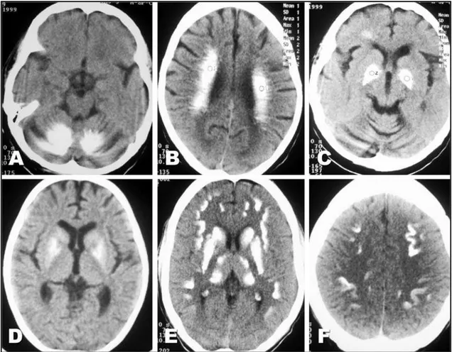
rism secondary to Shehan’s syndrome in one). The symptoms were variable as motor deficits, seizures, comportment disturbances, headache and others. All patients presented calcifications in globus palli-dus, five (with calcium disturbances) in caudate, len-tiform nucleus and subcortical region, two in den-tate nucleus, one (with senile dystrophic calcifica-tions) in semioval center, and one (with hypopara-thyroidism) in thalamus (Fig 1).
DISCUSSION
We present patients with pathological BGC, den-tate nucleus and white matter calcifications of dis-tinct etiologies and clinical features. This kind of ab-normality is related with many etiologies that can be classified as inflammatory (CMV infection, neu-rocysticercosis, toxoplasmosis, neurobrucellosis, tuberculosis, HIV infection), tumoral (astrocytomas), hypoxic and vascular (arteriovenous malforma-tions calcified infarct, ischemic encephalophaty), endocrine (hypoparathyroidsm, pseuso and pseudo-hypoparathyroidism, hyperparathyroidism), toxic (CO and Pb intoxication, hypervitaminosis D, radio-therapy), metabolic and degenerative (senility, mi-thocondrial encephalopaties, leukodistrophic disea-ses, idiopathic familial, motor neuron disease, myo-tonic muscular dystrophy, carbonic anidrase deficit, biopterin deficit) and other (malabsorption, Down syndrome, lupus, tuberous sclerosis, arthrogripo-sis)8-13.
Except by familial cause, with unknown
mecha-nism, genetically determined, all other causes appear to result of calcium deposits by serum abnormali-ties (with a change in vascular permeability relat-ed to local calcium concentration)14or of
dystroph-ic calcifdystroph-ications, with physdystroph-ical abnormalities in small vessel, focal circulatory disabilities and metabolic disorders (hypoxia, hypoglycemia, and abnormal-ities in the acid-base balance or electrolytes)15.
Re-duced blood flow to calcified regions is confirmed in SPECT of the brain with 99m
Tc-hexamethhylpropi-lenamine (99mTc-HMPAO)16.
We report patients with pathological calcifica-tions attributed to different etiologies: dystroph-ic (probable senile in one patient) and secondary to abnormal calcium metabolism (in two patients with hypoparathyroidsm). Both etiologies might be associated in patients with AIDS (because abnor-mal calcium levels are commonly associated with the disease17and because they presented hypoxic
epi-sodes caused by pulmonary disease) and in the pa-tient with panhypopitutarism (secondary to abnor-mal calcium levels and hypoglicemic crisis). The only difference noted in the calcifications pattern was their absence in subcortical region and basal nucleus and their presence in semioval center, in the patient with senile calcifications. Moreover, the pa-tient with senile calcifications presented seizures and headache. Usually, the dystrophic form is silent or occur with extrapyramidal symptoms18. Since no
associated disease was identified as responsible
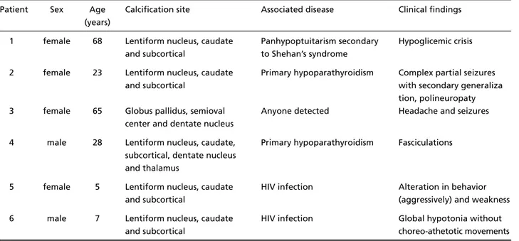
Table 1. Clinical and radiological aspects of patients.
Patient Sex Age Calcification site Associated disease Clinical findings (years)
1 female 68 Lentiform nucleus, caudate Panhypoptuitarism secondary Hypoglicemic crisis and subcortical to Shehan’s syndrome
2 female 23 Lentiform nucleus, caudate Primary hypoparathyroidism Complex partial seizures
and subcortical with secondary generaliza
tion, polineuropaty 3 female 65 Globus pallidus, semioval Anyone detected Headache and seizures
center and dentate nucleus
4 male 28 Lentiform nucleus, caudate, Primary hypoparathyroidism Fasciculations subcortical, dentate nucleus
and thalamus
5 female 5 Lentiform nucleus, caudate HIV infection Alteration in behavior
and subcortical (aggressively) and weakness
6 male 7 Lentiform nucleus, caudate HIV infection Global hypotonia without
by these clinical features, in agreement with pre-vious studies7we suppose that dystrophic
calcifica-tions in other places than globus pallidus (for exam-ple, dentate nucleus and semioval center as occurs in this patient), are related to other symptoms in adults (particularly to psychiatric disorders)19-21.
Because it involves many etiologies, it is diffi-cult attribute neurological deficits as cause or con-sequence of Fahr’s syndrome. Classical and recent anatomical studies about the role of basal ganglia in movement coordination22, subcortical region in
cognitive association and mood determination, and hypoxia in epileptic activity23, points to correlation
between clinical features and calcifications site and consequently hypoperfused area7,10.
Recognition of the intracranial calcifications in Fahr’s syndrome has been made easier by the high resolving capability of CT. Calcifications consist of hydroxyapatite of a nature similar to that found
Arq Neuropsiquiatr 2004;62(3-B) 791
in bones. Other elements include zinc, iron and magnesium24. Because this material composition,
they are always hyperdense on CT. On magnetic re-sonance imaging (MRI), however, their signal is va-riable. On T1 weighted images, low signal is due to the low proton density of calcium and other min-eral ions present in higher concentration. Howe-ver, they might present hyperintense signal, due to proteins and mucopolysaccharides binding the mineral ions. They might also to be undetected on MRI when are in a intermediary stage25.
In conclusion, Fahr’s syndrome is manytimes a treatable entity. Etiology is not directly correlat-ed with image calcification pattern, except for so-me differences noticed in calcifications site in dystrophic senile ones. Topographic image studies are promising to predict neurological deficits. Their recognition by CT is easy, has maximum sensitivi-ty and may be responsible by early treatment.
REFERENCES
1. Klein C, Vieregge P. Fahr’s disease: far from a disease. Mov Disord 1998;13:620-621.
2. Manyam BV, Walters AS, Keller IA, Ghobrial M. Parkinsonism associ-ated with autosomal dominant bilateral striopallidodentate calcinosis. Parkinsonism Relat Disord 2001,7:289-295.
3. Konig P. Psychopathological alterations in cases of symmetrical basal ganglia sclerosis. Biol. Psychiatry 1989;25:459-468.
4. Vega MG, Souza AA, de Lucca F, Purich S, Tenassi ML. Síndrome extra-piramidal e hipoparatireoidismo: acerca da identidade da doença de Fahr. Arq Neuropsiquiatr, 1994,52:419-423.
5. Cavalcanti CE. Cisticercose calcificada em gânglios da base e síndrome parkinsoniana. Arq Neuropsiquiatr 1984;42:183-186.
6. Levy G. Basal ganglia calcification (striopallidodentate calcification) and cognitive impairment. Arq Neuropsiquiatr, 1999:57:148.
7. Harrington MG, Macpherson P, Mcintosh WB, Allam BF, Boné I. The sig-nificance of the incidental finding of basal ganglia calcification on com-puted tomography. J Neurol Neurosurg. Psychiatry. 1981;44:1168-1170. 8. Perez MA, Martin PE, Sarmiento GE, et al. Farh’s disease and hypocalcemic
syndromes: presentation of a clinical case. An Med Interna 1992;9:495-497. 9. Diament AJ, Machado LR, Cypel S, Ramos JLA. Síndrome de calcifica-ções dos gânglios da base, leucodistrofia e pleocitose linfomonocitária crôni-ca de líquido cefalorraquidiano. Arq Neuropsiquiatr 1986,44:185-190. 10. Delgado-Rodrigues RN. Neurocisticercose associada a
hipoparatireoidis-mo e doença de Fahr. Arq Neuropsiquiatr. 1984,42:388-391, 11. Rocha C, Gonfinitti NV, Pelucci LA. Aspectos clínicos e radiológicos
do pseudo-hipoparatireoidismo. Arq Neuropsiquiatr 1997;55:139-143. 12. Guerreiro MM, Scotoni AE. Calcificações dos gânglios da base na
infân-cia. Arq Neuropsiquiatr, 1992;50:513-518.
13. Queiroz AC, Malboisson AMB. Calcificações nos núcleos da base do cérebro. Arq Neuropsiquiatr. 1981;39:321-326.
14. Brannan TS, Burger AA, Chaudhary MY. Bilateral basal ganglia
calcifi-792 Arq Neuropsiquiatr 2004;62(3-B)
cations visualised on CT scan. J Neurol Neurosurg Psychiatry. 1980; 43:403-406.
15. Ogi S, Fukumitsu N, Tsuchida D, Uchiyama M, Mori Y, Matsui K. Ima-ging of bilateral striopallidodentate calcinosis. Clin Nucl Med 2002; 27:721-724.
16. Uygur GA, Liu Y, Hellman RS, Tikofsky RS, Collier BD. Evaluation of regional cerebral blood flow in massive intracerebral calcifications. J Nucl Med 1995;36:610-611.
17. Fenelon G, Gray F, Paillard F, et al. A prospective study of patients with CT detected pallidal calcifications. J Neurol Neurosurg Psychiatry 1993;56:622-625.
18. Barbosa ER, Culchebachi MDB, Navarro JM, Scaff M, Canelas HM. Le-sões hipoatenuantes dos gânglios da base associadas a quadro pirâmi-do-extrapiramidal. Arq Neuropsiquiatr 1986;44:55-59.
19. Ostling S, Andreasson LA, Skoog I. Basal ganglia calcification and psy-chotic symptoms in the very old. Int J Geriatr Psychiatry 2003;18:983-987. 20. Cartier L, Passig C, Gormaz A, Lopez J. Neuropsychological and neuro-physiological features of Fahr’s disease. Rev Med Chil 2002;130:1383-1390. 21. Warren JD, Mummery CJ, Al-Din AS, Brown P, Wood NW. Corticobasal degeneration syndrome with basal ganglia calcification: Fahr’s dis-ease as a corticobasal look-alike? Mov Disord 2002;17:563-567. 22. Hempel A, Henze M, Berghoff C, Garcia N, Ody R, Schroder J. PET
find-ings and neuropsycological déficits in a case of Fahr’s disease. Psych Res 2001;108:133-140.
23. Petroff OAC, Errante LD, Kim JH, Spencer DD. N-acetyl-aspartate, total creatine, and myo-inositol in the epileptogenic human hippocam-pus. Neurology 2003;60:1646-1651.
24. Smeyers-Verbeke J, Michottey Y, Pelsmoeckers J, et al. The chemical com-position of idiopathic non ateriosclerotic cerebral calcification. Neurology 1975;25:48-57.