Antigenic and Genetic Characterization of Twenty-six Strains
of Human Respiratory Syncytial Virus (Subgroup A) Isolated
During Three Consecutive Outbreaks in Havana City, Cuba
Angel Valdivia/
+, Grehete González, Danay Chacón, Clara Savón,
Anselmo Otero, Odalys Váldes, Reynel Cancio, Suset Oropesa,
José A Melero*, Blanca García-Barreno*, Angel Goyenechea
Instituto de Medicina Tropical “Pedro Kouri”, Apartado 601, Marianao 13, La Habana, Cuba *Centro Nacional de Biología Fundamental, Madrid, España
Twenty-six human respiratory syncytial virus strains (subgroup A) isolated from three outbreaks in Havana City during the period 1994/95, 1995/96 and 1996/97 were analyzed to determine their anti-genic and genetic relationships. Analyses were performed by monoclonal antibodies and restriction mapping (N gene) following amplification of the select region of the virus genome by polymerase chain reaction. All isolated strains were classified as subgroup A by monoclonal antibodies and they showed a restriction pattern NP4 that belonged to subgroup A. Thus the results obtained in this work, showed a close relation (100%) between antigenic and genetic characterization of the isolated strains in our laboratory. These methods permit the examination of large numbers of isolates by molecular techniques, simplifying the researchs into the molecular epidemiology of the virus.
Key words : respiratory syncytial virus - molecular characterization - restriction mapping - monoclonals antibodies
Human respiratory syncytial virus (HRSV) is the major cause of lower respiratory tract infec-tions in infants and young children (McIntosh & Chanock 1990). This virus is a member of the Pneumovirus genus of the family Paramixoviridae. It has two surface glycoproteins (G and F), which are the main targets of neutralizing and protective antibodies (Taylor et al. 1984, Walsh et al. 1984). Variability of HRSV isolates was first demon-strated with hyperimmune serum (Coates et al. 1966). Different panels of monoclonal antibodies were later used to subdivide HRSV isolates into two antigenic groups, A and B (Anderson et al. 1985, Mufson et al. 1985), that correlate with ge-netically distinct viruses (Cristina et al. 1990). These monoclonals have identified three types of epitopes in the G molecule (García-Barreno et al. 1989, García et al. 1994): (i) variables epitopes, (ii) subgroup specific epitopes, and (iii) conserved
This work was funded in part by a grant (BIO95-2066-E) from Comisión Interministerial de Ciencia y Tecnología for Spanish-Cuban cooperation.
+Corresponding author. Fax: 246051 and
+53-7-220633. E-mail: [email protected] Received 26 May 1998
Accepted 9 December 1998
epitopes shared by subgroups A and B. The level of antigenic and genetic divergence among the G glycoproteins of HRSV isolates is the highest re-ported for an RNA virus gene product (García et al. 1994).
On the other hand the isolates of subgroup A and B of HRSV can be further subdivided into dis-tinct groupings or lineages on the basis of the fol-lowing criteria: (a) through monoclonal antibod-ies, (b) restriction mapping of part of the nucleo-capsid (N Gene), (c) nucleotide sequencing of part of the small hidrophobic (SH) gene and (d) nucle-otide sequencing of the attachment (G) protein gene (Cane & Pringle 1991, 1992, Cane et al. 1992, García et al. 1994, Cane & Pringle 1995b). These methods allow the speedy analysis of large num-bers of samples without the necessity of nucleotide sequencing (Cane & Pringle 1992).
These lineages appear to be distributed world-wide but as yet their significance in terms of de-gree of virulence and immunity at the individual and community level has not been determined (Cane & Pringle 1992).
Molecular Epidemiology of RSV in Havana City, Cuba • Angel Valdivia et al.
MATERIALS AND METHODS
Viruses, cells, patients and clinical samples -Twenty six nasopharyngeal exudates were obtained from children admitted to the respiratory ward to some of the main pediatric hospitals of Havana City (William Soler, Juan Manuel Márquez and Centro Habana). All samples were suspended in a final volume of 2 ml of minimun essential medium (MEM) containing antibiotics, (100 U/ ml penicil-lin and 100 µg/ml streptomycin sulfate). For PCR analysis, 500 µl were used in a 1.5 ml tube, and the samples stored at -70oC until being tested. Cell culture and indirect immunofluorescence were con-ducted according to previously reported procedures (Parrott et al. 1979, Valdivia et al. 1997).
The samples used in this study were from the 15 municipalities forming Havana City.
All RS virus-positive cultures (confirmed by immunofluorescence) were kept at -70oC until fur-ther use. Viruses from original isolates (Table) were subcultured two times in HEp-2 cells, until a cy-topathic effect (CPE) was observed within 48 hr after inoculation. At this time, the cells were re-suspended in the virus-containing supernatant and were kept as a working seed at -70oC.
Dot-Elisa - Characterization of antibody reac-tivity in Dot-Elisa was done as described by García et al. (1994). Briefly, one plastic flasks (Costar) with a surface area of 75 cm2, containing 20 ml of MEM containing 10% fetal calf serum, 1% glutamine, 100 U/ml penicillin and 100 µg/ml streptomycin sulfate was infected with each of the viruses used in this study. When an extensive CPE was evident by the formation of syncytia, the cells were scraped off with a rubber policeman, pelleted by low-speed centrifugation (3,000 x g, 5 min), and washed with phosphate-buffered saline (PBS). The cell pellets were then resuspended in 300 µl of extraction buffer (10 mM Tris-HCl pH 7.6, 5 mM EDTA, 140 mM NaCl, 1% Triton X-100, and 1% sodium deoxycholate). The extracts were clari-fied by centrifugation at 10,000 x g for 5 min and then were diluted with PBS to 10 µg of protein per ml, and 5 µl were spotted onto strips of Immobilon paper (Millipore). After air drying, the paper was satured with 5% skimmed milk in PBS and was revealed with the Mabs indicated in Fig. 1, by us-ing biotinylated antimouse immunoglobulin, streptavidin-peroxidase, and 4-chloro-1-naphthol according to the recommendations of the manu-facturer (Amersham Corp). The monoclonal
anti-TABLE
Cuban isolates of human respiratory syncytial virus used in the present study
Virus Date of isolation Age of patient Disease Cub/54/94 18/10/94 6 years URI (ashtma)
Cub/60/94 20/10/94 2 months URI
Cub/67/94 25/10/94 3 months Laringitis
Cub/69/94 25/10/94 1 month URI
Cub/81/94 01/11/94 3 months URI
Cub/82/94 01/11/94 4 months Bronchiolitis Cub/83/94 01/11/94 6 months Bronchiolitis Cub/97/94 08/11/94 6 months Bronchiolitis Cub/105/94 15/11/94 2 months Bronchiolitis Cub/106/94 15/11/94 5 months Bronchiolitis Cub/111/94 15/11/94 5 months Bronchiolitis
Cub/115/94 17/11/94 not known URI
Cub/128/94 01/12/94 10 months Bronchiolitis
Cub/134/94 06/12/94 3 months URI
Cub/140/94 13/12/94 22 days URI
Cub/141/94 13/12/94 4 months Bronchiolitis Cub/151/94 20/12/94 5 months Bronchiolitis
Cub/5/95 17/01/95 5 months URI
Cub/8/95 17/01/95 9 months Bronchiolitis Cub/10/95 19/01/95 5 months Bronchiolitis Cub/11/95 19/01/95 6 months Bronchiolitis
Cub/104/96 02/04/96 5 years URI
Mem Inst Oswaldo Cruz, Rio de Janeiro, Vol. 94(4), Jul./Aug. 1999
bodies were used at a dilution of 1:100. These monoclonals were 021/1G and 021/21G (con-served), 021/19G and 021/2G (subgroup specific), and 63G, 25G, 68G, 58G, 59G, 61G, 62G, 64G and 74G (“Long” strains specific)(García-Barreno et al. 1989, García et al. 1994) and they were pro-vided by Prof. JA Melero, Deparment of Molecu-lar Biology, National Center of Microbiology, Vi-rology and Sanitary Immunology, Madrid, Spain. One reference strain of group A (Long) and one of group B (CH 18537) were included as negative and positive controls in this work. One HEp-2 ex-tract was also included as negative control.
Nucleic acid extraction from clinical specimens - All small-scale RNA extractions were carried out in 1.5-ml microfuge tubes without any attemp at separating RNA from DNA, using an adaptation of the method of Kumar and Lindberg (1972). Nasopharyngeal exudates samples were centri-fuged in a microfuge for 2 min. The cell pellet was resuspended in 0.5 ml of 3.5 M urea, 200 mM NaCl, 10 mM Tris-HCl pH 7.8, 5 mM EDTA, 0.75 mM MgCl2, 0.5% SDS and 0.35% NP40. 0.5 ml of phenol-chloroform (1:1)(equilibrated with 150 mM NaCl, 10 mM Tris-HCl pH 7.8, 1 mM EDTA) was added, the mixture vortexed for about 5 sec and then centrifuged for 10 min. The aqueous layer was extracted again with phenol-chloroform and then added to 1 ml of ethanol; the nucleic acids were precipitated at -20oC for 20 hr, pelleted, washed with 0.5 ml 70% ethanol, vacuum-dried and re-suspended directly into 30 ml reverse transcriptase mix (see below).
RNA extraction from tissue cultures - Virus iso-lates were cultured in HEp-2 cells in plastic flasks (Costar) with a surface area of 25 cm2 , containing 5 ml of MEM with glutamine, penicillin, strepto-mycin, and 5% foetal calf serum. When extensive CPE was present, the cells were detached into the tissue culture medium by shaking with sterile glass beads, and the suspension aliquotted and stored at -70oC. Approximately 0.5 ml of these infected cell suspensions were extracted as for the nasopharyn-geal exudates. The resulting nucleic acids were resuspended in 20 µl H2O and 3 µl used for each cDNA synthesis.
cDNA synthesis and PCR - The preparation of cDNA was carried out with approximate 20 µg of total RNA (spectrophotometrically quantified) in a 20 µl volume containing 100 ng of each primer. First, the target RNA was mixed with both prim-ers and placed at 65oC. After 15 min the solution was placed on ice and completed with 100 mM Tris-HCl pH 8.3, 500 mM KCl, 25 mM MgCl2 , 25 mM (each) dATP, dCTP, dGTP and dTTP, 20 U Rnasin (Boehringer Mannhein GmbH, Germany) and three units of AMV reverse transcriptase
(Boehringer Mannhein GmbH, Germany). Incuba-tion was at 42oC for 30 min. Finally, the reaction mixture was placed at 95oC for 5 min and kept on ice until the PCR was carried out. PCR mix was made up to a volume of 100 µl, which made a re-action mix containing 100 mM Tris-HCl pH 8.3, 500 mM KCl, 25 mM MgCl2, distilled water and 2.5 µ Taq DNA polymerase (Boehringer Mannhein GmbH, Germany). The amplification was carrried out in 30 cycles in a Perkin-Elmer Cetus Thermal cycler. Each cycle consisted of denaturation at 93oC for 1.5 min, annealing of the primer at 55oC for 1.5 min, and chain elongation at 72oC for 1.5 min. After the last cycle of amplification, 10 µl of the amplified products were analyzed by electrophore-sis on 2% agarose gels with Tris-borate buffer.
Analysis of the amplified products - The remain-ing products were diluted with 100 µl H2O, ex-tracted with 150 µl phenol-chloroform and etha-nol precipitated. Samples were then restricted with Pst I, Hae III, Hind III, and Bgl II. All this was performed according to previous reports (Cane & Pringle 1991, 1992). In these experiments the strain 021, isolated in Uruguay (Montevideo) with an NP5 know pattern was always included as control. This strain was provided by Prof. JA Melero.
Primers - The primers originally designed to amplify between nucleotides 858 and 1135 of the HRSV (N gene)(Collins et al. 1985) have been previously described (Cane & Pringle 1991):
N1: 5’GGAACAAGTTGTTGAGGTTTATGAATATGC 3' N2: 5' CTTCTGCTGTCAAGTCTAGTACACTGTAGT 3'
Controls - Distilled water, mixed buffer solu-tions, full-time open vial with final buffer mixture, and HEp-2 RNA were included as negative con-trols (Stoker 1990, Wright & Wynford-Thomas 1990, Kitchin & Bootman 1993). The cDNA reac-tion and PCR were performed using the recom-mended strict protocol, with all precautions to pre-vent contamination (Kwok & Higuchi 1989).
RESULTS
Molecular Epidemiology of RSV in Havana City, Cuba • Angel Valdivia et al.
A B C
021/1G 021/21G 021/19G 021/2G 63 G 25 G 68 G 58 G 59 G 61 G 62G 64 G 74 G - - - - - - - Hep-2 + + + + + + + + + + + + + Long + + - - - - - CH18537 + + + + - + + + + + + + + 54/94 + + + + - + + + + + + + + 60/94 + + + + - + + + + + + + + 67/94 + + + + - + + + + + + + + 69/94 + + + + - + + + + + + + + 81/94 + + + + - + + + + + + + + 82/94 + + + + - + + + + + + + + 83/94 + + + + - + + + + + + + + 97/94 + + + + - + + + + + + + + 105/94 + + + + - + + + + + + + + 106/94 + + + + - + + + + + + + + 111/94 1
+ + + + - + + + + + + + + 115/94
+ + + + - + + + + + + + + 128/94 + + + + - + + + + + + + + 134/94 + + + + - + + + + + + + + 140/94 + + + + - + + + + + + + + 141/94 + + + + - + + + + + + + + 151/94 + + + + - + + + + + + + + 5/95 + + + + - + + + + + + + + 8/95 + + + + - + + + + + + + + 10/95 + + + + - + + + + + + + + 11/95 + + + + - + + + + + + + + 104/96 2 + + + + - + + + + + + + + 195/96 + + + + - + + + + + + + + 201/96 + + + + - + + + + + + + + 202/96 3 + + + + - + + + + + + + + 220/96
Fig. 1-1: strains from 94/95(21); 2: strain from 95/96(01); 3: strains from 96/97(04); A: conserved monoclonal antibodies; B: group-specific monoclonal antibodies; C: antibodies specific for the Long strain; +: positive; -: negative.
variable monoclonals from strain Long, with the exception of monoclonal 63G, which was the only one that did not react against the strains isolated in our laboratory. With the results obtained with this monoclonal panel it would be inferred that the strains isolated in our laboratory are closely related to Long strain.
Fig. 2 shows the restriction analysis of the PCR products obtained from each of the strains belong-ing to the first outbreak. The restriction analysis of strains 105/94, 106/94 and 128/94 were carried out in another gel (data not shown). The digestion
was only made with enzymes BgI II and Hind III, since they had already been classified as members of the subgroup A, by means of monoclonals cor-respondent.
Mem Inst Oswaldo Cruz, Rio de Janeiro, Vol. 94(4), Jul./Aug. 1999
in this study were obtained directly from clinical samples as well as from cell culture.
DISCUSSION
Sequence analysis of numerous HRSV isolates, mostly group A, have shown extensive variability of the G glycoprotein (Cane & Pringle 1991, Sullender et al. 1991, García et al. 1994, Cane & Pringle 1995b ). Several features about HRSV evo-lution emerged from these studies: (a) viruses from antigenic group A belong to different lineages that correlate with previously identified genotypes (Cane & Pringle 1992, García et al. 1994). Most epidemics are produced by viruses classified into more than one genotype. At a local level, replace-ment of predominant genotypes was observed dur-ing consecutive years (Cane & Prdur-ingle 1995a); (b) HRSV genotypes have a worldwide distribu-tion and viruses isolated in distant places and in slightly different years may be more closely re-lated than viruses isore-lated in the same place on two consecutive days (Melero et al. 1997); (c) antigenic changes, detected with a panel of anti-G mono-clonal antibodies, correlated with the position of viruses in the phylogenetic tree (García et al. 1994, Cane & Pringle 1995b). However, the viruses used in those studies were isolated mainly in places of temperate climate (where annual outbreaks of HRSV occur in winter months) and in most cases in countries of high economic status (Melero et al. 1997). Very limited epidemiological data is
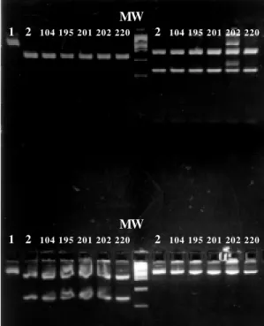
avail-Fig. 3: restriction analysis of the polymerase chain reaction prod-ucts obtained from each of the strains belonging to the second and third outbreaks. Lane 1: reference strain Long without di-gesting; Lane 2: reference strain Long digested; Lane 3 to 7: strains belonging of these outbreaks. Upper left square: diges-tion with Hae III. Upper right square: digestion with Rsa I. Lower left square: digestion with Bgl II. Lower right square: digestion with Hind III.
80/ 81/ 82/ 5/ 54/ 83/ 10/ 8/ 115/
021MVV 11/ 111/ 134/ 151/ 07/ 69/ 97/ 140/141/ 0 4 0 4 0 4 0 6 0 4 0 4 0 5 0 5 0 4 0 5 0 4 0 4 0 4 04 0 4 0 4 0 4 0 4
278 bp A
B
Fig. 2: restriction analysis of the polymerase chain reaction products obtained from each of the strains belonging to the first outbreak. A: digestion with Hind III; B: digestion with Bgl II; strain from Montevideo with known pattern NP5 (021); MW: PBR 322/Hae III.
MW
1 2 104 195 201 202 220 2 104 195 201 202 220
MW
Molecular Epidemiology of RSV in Havana City, Cuba • Angel Valdivia et al.
able from tropical countries where HRSV infec-tions may follow a different pattern (Heirholzer et al. 1994).
With regard to the above considerations and the reports made in previous studies by other au-thors, the most remarkable facts of our results are: (1) the strains analized in three consecutive out-breaks are identical using both methods; (2) the pattern obtained from the analyzed strains and by the two methods used, established a very closed relation between these strains and the standard Long strains, which is one of the oldest around the world (ends of the 50’s).
In this work we combined the use of mono-clonal antibodies and restriction analysis of PCR products for the select region of N gene to make a molecular characterization of three outbreaks of HRSV in Havana City.
All the isolated strains were identified as RS subgroup A by means of monoclonal antibodies and gave N gene fragment restriction pattern NP4. In our opinion, many factors have contributed to the peculiar behavior of the molecular epidemiol-ogy of this virus in Cuba.
After the end of the 50’s, the exchange of people with others countries was extremely reduced due to economic, social and political reasons. This situ-ation has continued for 30 years, reducing the prob-ability of entrance of new strains. On the other hand, Cuba, as an island, does not share borders with any other country, which limits the entrance of people from other neighboring countries. Nev-ertheless, in the 90’s, an outburst of the tourist in-dustry took place in Cuba, but the most important fact is that this increase was observed in grown-ups and not in young children who constitute the natural reservoir of the virus.
All these geographical, social and economic factors could be the main cause of such peculiar behavior for the molecular epidemiology of this virus in our country.
It will be interesting to examine strains from Havana City and other Cuban regions in the same country in subsequent years to determine when and where a new pattern appear or reappear. In this sense it would be interesting to extend such mo-lecular approach to other tropical countries even in distant geographical areas.
ACNOWLEDGEMENTS
To Dr German Roges and Orquidea Biart for critical review of the manuscript.
REFERENCES
Anderson LJ, Heirholzer JC, Tsou C, Hendry RM, Fernie BN, Stone Y, McIntosh K 1985. Antigenic charac-terization of respiratory syncytial virus strains with
monoclonal antibodies. J Infect Dis 151: 626-633. Cane PA, Pringle CR 1991. Respiratory syncytial virus heterogeneity during an epidemic: analysis by lim-ited nucleotide sequencing (SH gene) and restric-tion mapping (N gene). J Gen Virol 72: 349-357. Cane PA, Pringle CR 1992. Molecular epidemiology of
respiratory syncytial virus: Rapid identification of subgroup A lineages. J Virol Methods 40: 297-306. Cane PA, Pringle CR 1995a. Molecular epidemiology of human respiratory syncytial virus. Seminars Virol 6: 371-378.
Cane PA, Pringle CR 1995b. Evolution of subgroup A respiratory syncytial virus: evidence for progressive accumulation of aminoacid changes in the attach-ment protein. J Virol 69: 2918-2925.
Cane PA, Matthews DA, Pringle CR 1992. Analysis of relatedness of subgroup A respiratory syncytial vi-ruses isolated worldwide. Virus Research 25: 1522. Coates HV, Alling DW, Chanock RM 1966. An anti-genic analysis of respiratory syncytial virus isolates by a plaque reduction netralization test. Am J Epidemiol 89: 299-313.
Collins PL, Anderson K, Langer SJ, Wertz GW 1985. Correct sequence for the major nuclecapsid protein rnRNA of respiratory syncytial virus. Virology 146: 69-77.
Cristina J, López JA, Albo C, García-Barreno B, García J, Melero JA, Portela A 1990. Analysis of genetic variability in human respiratory syncytial virus by the Rnase A mismatch cleavage method: subtype diver-gence and heterogeneity. Virology 174: 126-134. Garcia O, Martin M, Dopazo J, Arbiza J, Frabasile S,
Russi J, Hortal M, Perez-Brena P, Martinez I, Garcia Barreno B, Melero JA 1994. Evolutionary pattern of human respiratory syncytial virus (Subgroup A): Cocirculating lineages and correlation of genetic and antigenic changes in the G glycoprotein. J Virol 68: 5448-5459.
Garcia-Barreno B, Palomo C, Penas C, Delgado T, Perez-Brena P, Melero JA 1989. Marked differences in the antigenic structure of human respiratory syncytial virus F and G glycoprotein. J Virol 63: 925-932. Heirholzer JC, Tannock GA, Heiholzer CM, Coombs
RA, Kennett ML, Phillips PA, Gust ID 1994. Subgrouping of respiratory syncytial virus strains from Australia and Papua New Guinea by biologi-cal and antigenic characteristic. Arch Virol 136: 133-147.
Kitchin PA, Bootman JS 1993. Quality control of the polymerase chain reaction. Med Virol 3: 107-114. Kumar A, Lindberg N 1972. Characterization of
messanger ribonucleoprotein and messanger RNA from KB cells. Proc Natl Acad Sci 69: 681-685. Kwok S, Higuchi R 1989. Avoiding false positives with
PCR. Nature 339: 237-238.
McInstosh K, Chanock RM 1990. Respiratory syncytial virus, p.1313-1351. In BN Fields, DM Knipe, PM Howley (eds) Fields Virology, Lippincot-Raven, Philadelphia.
vi-Mem Inst Oswaldo Cruz, Rio de Janeiro, Vol. 94(4), Jul./Aug. 1999
rus attachment (G) protein. J Gen Virol 78: 2411-2418.
Mufson MA, Orvell C, Rafnar B, Norrby E 1985. Two distinct subtypes of human respiratory syncitial vi-rus. J Gen Virol 66: 2111-2124.
Parrott RH, Kim HW, Brandt CD, Beem MO, Richardson L, Gerin JL, Chanock RM 1979. Respiratory syncy-tial virus, p. 695-708. In EH Lennette & NJ Schmidt (eds), Diagnostic Procedure for Viral, Rickettsial and Chlamydial Infection, Amer Pub Health Ass, Wash-ington, D.C.
Stoker NG 1990. The polymerase chain reaction and infectious diseases: hopes and realities. Trans R Soc Trop Med Hyg 84: 775-758.
Sullender WM, Mufson MA, Anderson LJ, Wertz GM 1991. Genetic diversity of the attachment protein of subgroup B respiratory syncytial viruses. J Virol 65: 5425-5434.
Taylor G, Stott EJ, Bew M, Fernie BF, Cote PJ, Collins AP, Hughes M, Jebbett J 1984. Monoclonal anti-bodies protect against respiratory syncytial virus infection in mice. Immunology 52: 137-142. Valdivia A, Savon C, Chacón D, Sarmiento L, Morier
L, Otero A, Soto Y, Oropesa S, Goyenechea A 1997. Analysis of respiratory syncytial virus in clinical samples by reverse transcriptase-polymerase chain reaction restriction mapping. Mem Inst Oswaldo Cruz 92: 389-393
Walsh EE, Schlesinger JJ, Brandis MW 1984. Protec-tion from respiratory syncytial virus infecProtec-tion in cotton rats by passive tranfer of monoclonal anti-bodies. Infect Immun 43: 756-758.
Wright PA, Wynford-Thomas D 1990. The polymerase chain reaction: miracle or mirage? A critical rewiew of its uses and limitations in diagnosis and research.